대한정형외과학회지:제 43 권 제 3 호 2008 J Korean Orthop Assoc 2008; 43: 400-403
400
통신저자:이 정 섭
부산시 서구 아미동 1-10 부산대학교 의과대학 정형외과학교실 TEL: 051-240-7248ㆍFAX: 051-247-8395 E-mail: [email protected]
*이 논문은 부산대학교 자유과제 학술연구비(2년)에 의하여 연구되었음.
Address reprint requests to Jung Sub Lee, M.D.
Department of Orthopaedic Surgery, Pusan National University School of Medicine, 1-10, Ami-dong, Seo-gu, Busan 602-739, Korea
Tel: +82.51-240-7248, Fax: +82.51-247-8395 E-mail: [email protected]
Lumbar Artery Injury Combined with a Transverse Process Fracture of the Lumbar Spine Presenting
with Hypovolemic Shock after a Fall
- A Case Report -
Jung Sub Lee, M.D., Chang Won Kim, M.D.*, and Kuen Tak Suh, M.D.
Departments of Orthopaedic Surgery and Radiology*, Pusan National University School of Medicine, Pusan, Korea
낙상 사고 후 요추 횡돌기 골절과 동반된 요추 동맥 손상에 의한 혈류 감소성 쇼크 - 증례 보고-
이정섭ㆍ김창원*ㆍ서근택
부산대학교 의과대학 정형외과학교실, 영상의학교실*
There are many reports on lumbar artery injuries. However, there are only a few case reports of a lumbar artery injury presenting with hypovolemic shock from either a blunt or penetrating trauma. We described a 47-year-old man with a retroperitoneal hemorrhage secondary to a lumbar artery injury presenting as hypovolemic shock after a 3 m fall.
Key Words: Lumbar artery injury, Hypovolemic shock, Fall
An injury to the lumbar arteries is relatively rare.1-9) Nevertheless, once damaged they can be- come a significant source of hemorrhage, par- ticularly because these injuries can be easily over- looked. A lumbar artery injury can cause a poten- tially fatal hemorrhage if not diagnosed and treated promptly. We report a patient with a retro- peritoneal hemorrhage secondary to a lumbar artery injury who presented with hypovolemic shock after a 3 m fall.
CASE REPORT
A 47-year-old man was transferred by ambu- lance after sustaining pain to the lower back and
left wrist injury after a 3m fall. The patient was awake and alert and denied any loss of conscious- ness. He had no medical history of any bleeding disorders. The initial evaluation at a local clinic revealed obvious hemodynamic instability with resuscitation being performed. The patient was then transferred to the authors’ institution 18 hours after his initial injury for definitive care. The vital signs upon arrival to the authors’ institution are as follows: a heart rate of 160 beats per mi- nutes and a systolic blood pressure of 80 mmHg.
The laboratory data showed a low hemoglobin level (6.9 g/dl). The patient was intubated and further resuscitated with lactated Ringer’s solution and
Lumbar Artery Injury Combined with a Transverse Process Fracture of the Lumbar Spine Presenting with Hypovolemic Shock after a Fall 401
Fig. 1. Anteroposterior radiograph of the lumbar spine shows a fracture of the left second, third and fourth lumbar transverse processes (arrows).
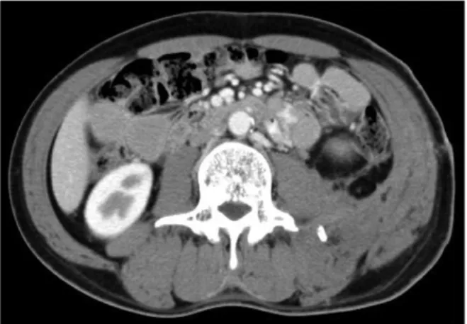
Fig. 2. Enhanced abdominopelvic computed tomographic scan at the level of the third lumbar vertebrae shows a large retroperitoneal hematoma.
Fig. 4. Arteriography shows the cessation of bleeding after embolization (arrow).
Fig. 3. Arteriography shows an actively bleeding second lumbar artery (arrow).
packed red blood cells.
The physical examination revealed abdominal pain and distention, low back pain, and an open fracture of the left wrist. The radiographic survey documented a fracture of the left second, third and fourth lumbar transverse processes (Fig. 1) in addition to a comminuted fracture of the left wrist.
A computed tomography (CT) scan of the abdomen and pelvis revealed a retroperitoneal hematoma extending from the diaphragm to the pelvis (Fig. 2).
The patient required an additional 7 units of
packed red blood cells before his vital signs had stabilized. Emergent arteriography was performed because he showed signs of continuous hemor- rhage. No pelvic arterial hemorrhage was noted.
However, arteriography revealed a second lumbar artery bleeding actively on the left (Fig. 3). The lumbar artery was embolized successfully using Gelfoam (SpongostanⓇ, Johnson & Johnson Medi-
402 Jung Sub Lee, Chang Won Kim, Kuen Tak Suh
Fig. 5. Control computed tomography scan at the level of the third lumbar vertebrae 3 months after embolization shows a markedly decreased retroperitoneal hematoma.
cal Limited, Gargrave, Skipton, UK) with a ces- sation of the hemorrhage (Fig. 4). After emboli- zation, the tachycardia had resolved, the hemor- rhage ceased and no further transfusions were required. He underwent successful closed reduction and percutaneous pinning of his wrist fracture. His transverse processes fractures were treated by wearing a corset for 6 weeks. He was eventually discharged after a 4 week hospital stay. The CT scan performed 3 months after embolization showed a marked decrease in the retroperitoneal hematoma (Fig. 5).
DISCUSSION
An early diagnosis of a lumbar artery injury is important because complications can occur, such as aneurysmal expansion, rupture, hemorrhage, thrombosis, and ischemia1-9). Unfortunately, a diag- nosis of a pseudoaneurysm in the lumbar artery can be difficult for many reasons. These include nonspecific symptomatology and signs, an una- wareness of this condition, delayed presentation, and neglecting the condition in the differential diagnosis.
The plain radiographs can reveal fractures of the pelvis, lumbar vertebrae, or lower ribs, and can
suggest an associated retroperitoneal hematoma5). A suspicion of a lumbar artery pseudoaneurysm should be heightened in patients with these frac- tures who are symptomatic. An enhanced CT scan is useful for diagnosing retroperitoneal bleeding in patients with abdominal or pelvic injuries and suspected vascular injures1-9). A hemodynami- cally unstable patient with a large retroperitoneal hematoma and no obvious intra- abdominal injury should undergo emergency arteriography.
A lumbar arterial hemorrhage is a relatively rare cause of retroperitoneal hemorrhage and shock in patients who have fallen from a height. The possi- bility of a lumbar artery injury must be consi- dered in all such patients.
REFERENCES
1. Armstrong NN, Zarvon NP, Sproat IA, Schurr MJ:
Lumbar artery hemorrhage: unusual cause of shock treated by angiographic embolization. J Trauma, 42: 544-545, 1997.
2. Haydu P, Chang J, Knox G, Nealon TF Jr: Transcatheter arterial embolization of a traumatic lumbar artery false aneurysm. Surgery, 84: 288-291, 1978.
3. Hulnick DH, Naidich DP, Balthazar EJ, Megibow AJ, Bosniak MA: Lumbar artery pseudoaneurysm: CT demon- stration. J Comput Assist Tomogr, 8: 570-572, 1984.
4. Ikubo A, Komura M, Matoba N, et al: Lumbar artery pseudoaneurysm: an unusual cause of a retroperitoneal hematoma: report of a case. Surg Today, 23: 635-638, 1993.
5. Kalangos A, Walder B, Faidutti B: Ruptured lumbar artery pseudoaneruysm: a diagnostic dilemma in retroperitoneal hemorrhage after abdominal trauma. J Trauma, 45: 829-832, 1998.
6. Kornberg E: Lumbar artery aneurysm with acute aortic occlusion resulting from chiropractic manipulation: a case report. Surgery, 103: 122-124, 1988.
7. Marty B, Sanchez LA, Wain RA, et al: Endovascular treatment of a ruptured lumbar artery aneurysm: case report and review of the literature. Ann Vasc Surg, 12: 379-383, 1998.
Lumbar Artery Injury Combined with a Transverse Process Fracture of the Lumbar Spine Presenting with Hypovolemic Shock after a Fall 403
8. Peters M, Henny CP, ten Cate JW, Marsman JW, Breederveld C: Lumbar arterial rupture secondary to iliopsoas hemorrhage in a hemophiliac patient. Acta Haematol, 71: 128-129, 1984.
9. Sclafani SJ, Florence LO, Phillips TF, et al: Lumbar arterial injury: radiologic diagnosis and management. Radio- logy, 165: 709-714, 1987.
= 국문초록=
요추 동맥 손상에 대한 여러 보고가 있으나 둔상이나 관통상에 의한 요추 동맥 손상으로 발생한 혈류 감소성 쇼크에 대한 보고는 드물다. 이에 저자들은 3 m 높이에서 낙상하여 2번째 요추 동맥 손상으로 후복막 출혈을 보인 47세 남자 환자를 보고하고자 한다.
색인 단어: 요추 동맥 손상, 혈류 감소성 쇼크, 낙상