www.jkfas.org pISSN 1738-3757 eISSN 2288-8551 J Korean Foot Ankle Soc 2018;22(3):127-130 https://doi.org/10.14193/jkfas.2018.22.3.127
대퇴부 피판술의 장점은 적절한 크기의 혈관 직경을 가진 혈관 경을 제공할 수 있고 수혜부의 혈관의 조건에 맞게 중요 혈관의 재 건이나 보존에도 적용할 수 있으며 넓은 피판이 가능하면서도 동 시에 근육, 건, 근막 등의 다른 조직을 동시에 이용할 수 있는 복합 (composition) 피판술이 가능하다는 점이다.
저자들은 화상에 의하여 생긴 족관절 부위 피부괴사에 대하여 동측 대퇴부 유리 피판술과 함께 아킬레스건의 동종건 및 동종골 을 이용한 재건을 동시에 시행하였으며, 좋은 임상적 결과를 얻었 기에 이를 보고하고자 한다.
본 연구는 본원 윤리위원회(Institutional Review Board)의 승인 을 받아 진행되었다.
증례 보고
43세 여자 환자가 연탄가스로 인한 일산화탄소 중독으로 생긴 아킬레스건의 종골 부착부는 외상에 취약하며 피부괴사 혹은 연
조직 손상을 쉽게 입을 수 있는 조직이다. 이러한 연부조직이 외부 에 노출될 경우 근육이 적고 감염에 취약하여 치료하기 어렵다.1,2) 특히 화상의 경우, 피부 및 건 결손이 함께 동반된다면 조직의 회 복이 어려운 경우가 많아 절단이나 괴사제거 등 극단적인 치료방 법을 선택하게 된다. 아킬레스건 부위의 화상 및 손상에 관한 재건 및 치료를 위하여 여러 방법들이 시도되고 있다.3-10)
Case Report
This is an Open Access article distributed under the terms of the Creative Commons Attribution Non-Commercial License (http://creativecommons.org/licenses/CC
by-nc/4.0) which permits unrestricted non-commercial use, distribution, and reproduction in any medium, provided the original work is properly cited.
Copyright 2018 Korean Foot and Ankle Society. All rights reserved.ⓒ
A 3rd degree burn on the heel including the Achilles tendon is vulnerable and requires active treatment to improve the functional out- comes. Previously, there have been a few treatments on severe burns, such as amputation, debridement or simple skin graft. The coop- erative technique of an anterior lateral thigh flap with Achilles tendon reconstruction can be an innovative procedure that preserves the major arteries. The authors review a case and report the clinical outcome.
Key Words: Achilles tendon, Burns, Reconstructive surgical procedures, Surgical flaps
아킬레스건을 포함한 뒷발굽 접촉성 피부 화상에 대해 시행한 전외측 대퇴피부 피판술 및 아킬레스건 재건술: 증례 보고
-기능적 회복을 위한 수술적 치료법-
박준식, 백승하, 김갑래
강동성심병원 정형외과
Anterior Lateral Thigh Free Flap and Achilles Tendon Reconstruction Surgery for Contact Dermal Burn of Heel
Including Achilles Tendon: A Case Report -Surgical Treatment for Functional Recovery-
Jun-Sik Park, Seung-Ha Baek, Gab-Lae Kim
Department of Orthopedic Surgery, Kangdong Sacred Heart Hospital, Seoul, Korea
Received June 20, 2018 Revised August 15, 2018 Accepted August 19, 2018 Corresponding Author: Gab-Lae Kim
Department of Orthopedic Surgery, Kangdong Sacred Heart Hospital, 150 Seongan-ro, Gangdong-gu, Seoul 05355, Korea
Tel: 82-2-2224-2230, Fax: 82-2-489-4391, E-mail: [email protected] ORCID: https://orcid.org/0000-0002-0282-1721
The main point of this paper was presented at the Fall Meeting of the Korean Foot and Ankle Society in 2018.
Financial support: None.
Conflict of interest: None.
128 Vol. 22 No. 3, September 2018
의식저하로 응급실에 내원하였다. 환자는 키 162 cm, 몸무게 51 kg 이었고 기저질환 및 약물 복용력은 없었다. 본원 내과에서 일산화 탄소 중독에 대하여 산소 호흡기 치료를 받았고, 호흡기 치료가 끝 난 후 본원 정형외과로 의뢰되었다. 당시 근처에 있던 연탄통에 좌 측 아킬레스건 부위를 닿아 족관절 후방부위에 11 cm×23 cm 크 기의 3도 이상의 화상을 입었다. 좌측 아킬레스건을 포함한 피부 괴사가 진행된 상태였고(Fig. 1), 화상에 의한 아킬레스건 완전 파 열이 동반되었음을 자기공명영상을 통해 확인하였다(Fig. 2). 이에 저자들은 피부 복원과 아킬레스건 복원을 동시에 하는 것을 목표 로 전외측 대퇴 피판술과 동종건 이식술 및 동종골 이식술을 함께 시행하였다.
일차적으로 20 cm 정도의 괴사된 피부의 제거를 시행하였다. 그 후 피부층 아래에 있는 건의 화상으로 인한 손상 범위 및 정도를 확인하였다. 화상으로 인하여 건은 완전 파열됐으며 건강하지 않 은 육아조직이 확인되었다. 아킬레스건은 종골 도입부부터 손상 부위까지 전반적인 제거를 시행하였다(Fig. 3).
건재건술은 동종 아킬레스건을 이용한 ‘turn down’ 기법을 이용 하였으며 고정을 위하여 고정나사 및 일반 봉합사를 이용하였다.
상전장골극에서 우측 슬개골까지 일직선상의 선을 내려 도플러 초 음파를 이용하여 이식판 및 혈관 부위를 확인하였다. 11 cm×26 cm의 피부 공여부를 디자인한 후, 외측대퇴회선동맥의 하위 가지 를 줄기로 하는 피판술을 시행하였다(Fig. 4). 이후 미세현미경을
Figure 1. Photograph taken before the surgery shows a large soft tis- sue defect of posteromedial side of ankle and heel area.
A B
Figure 2. The sagittal images of magnetic resonance imaging show Achilles tendon defect (A) and damaged soft tissue (B).
A B
Figure 3. (A) Photograph of necrotic tis- sues and nonfunction of Achilles tendon.
(B) The damaged Achilles tendon was removed from the calcaneus to one third of distal lower leg.
www.jkfas.org 129 Jun-Sik Park, et al. Thigh Free Flap and Achilles Tendon Reconstruction
이용하여 미세혈관 및 신경 문합술을 시행하였다. 이식 후 피판 부 위의 생존을 확인하였다.
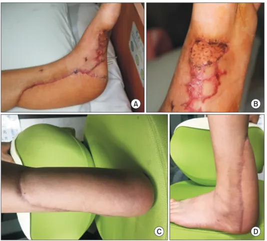
수술 후 괴사나 또 다른 재건술이 필요한 합병증이 발생하지 않 았고 피판의 생존을 성공적으로 확인할 수 있었다. 국소 부위의 발 적, 또는 발열 등의 감염 소견은 없었다. 3개월 후 환자는 체중부하 및 보행이 가능하였다. 술 후 족관절의 외측 부위 및 이식 부위의 감각 소실이 일부 확인되었으나 최종 추시 동안 기능적으로 문제
가 없었으며, 공여부 역시 합병증 없이 치유되었다(Fig. 5, 6).
고 찰
아킬레스건 인대 재건술 시 중요한 핵심은 기능적 회복과 함께 감염과 같은 합병증을 줄이는 것이다. 아킬레스건 인대 수술의 합 병증은 크게 3가지로, 창상 부위의 감염, 창상 부위의 피열, 그리고
A B C
Figure 4. (A) An allotendon of Achilles tendon and allobone used in reconstruction. (B) An intraoperative lateral image of allotendon and allobone. (C) Flap donor site of anterior surface of thigh.
A B
C D
Figure 5. Gross photography (A) and plan- tar area (B) of foot flap site of 1 month after the surgery shows no complications.
Posteroanterior view (C) and laterial view (D) of lower leg gross photography of 3 months after the surgery shows good healing and no complications.
130 Vol. 22 No. 3, September 2018
구축이다. 재건술 시 이러한 합병증 없이 연부조직의 회복을 함께 할 수 있어야 한다. 강한 에너지 및 심한 손상이 동반될 경우 복합 적인 연조직 결손은 이러한 아킬레스건 손상 치료에 큰 걸림돌이 된다.
연조직의 손상은 혈관에 대한 접근을 필요로 하며, 변연절제술 을 필요로 할지 판단이 필요하다.9) 최종적인 연조직의 치료는 대 개 표피피부 이식으로도 가능하나 유리 피판술, 피판 성형술, 근막 피부판 이식술 등 여러 가지 방법을 도입하고 있다.10)
본 증례의 환자는 특별한 기저질환이 없었으나 당뇨, 말초혈관 질환, 정맥부전 등의 동반질환이 있는 환자의 경우 피판괴사의 확 률이 높아질 수 있다. 따라서 환자의 상태가 피판 및 재건술의 영 향을 줄 수 있는 경우, 수술 계획 전 혈관의 상태에 대한 판단이 우 선되어야 한다.
재건술과 피판술을 동시에 시행한 술기는 기존 치료에 비해 기 능적으로 탁월한 결과를 확인할 수 있었다. 환자의 체중부하 및 보 행은 수술 후 3개월 이후 가능하였으며, 통증은 없었다.
결론으로, 이에 저자들은 이러한 피판술과 함께 아킬레스건 재 건술을 동시에 시행할 경우 환자의 기능적인 예후가 매우 좋은 것 으로 생각하며, 적극적인 재건술을 통해 유용한 임상적 결과를 가 져올 수 있을 것이라 기대한다.
REFERENCES
1. Leppilahti J, Orava S. Total Achilles tendon rupture. Sports Med.
1998;25:79-100.
2. Marchesi A, Parodi PC, Brioschi M, Riccio M, Perrotta RE, Co- lombo M et al. Soft-tissue defects of the Achilles tendon region:
management and reconstructive ladder: review of the literature.
Injury. 2016;47 Suppl 4:147-53.
3. Bullocks JM, Hickey RM, Basu CB, Hollier LH, Kim JY. Single- stage reconstruction of Achilles tendon injuries and distal lower extremity soft tissue defects with the reverse sural fasciocutane- ous flap. J Plast Reconstr Aesthet Surg. 2008;61:566-72.
4. Deiler S, Pfadenhauer A, Widmann J, Stützle H, Kanz KG, Stock W. Tensor fasciae latae perforator flap for reconstruction of composite Achilles tendon defects with skin and vascularized fascia. Plast Reconstr Surg. 2000;106:342-9.
5. Lee JW, Yu JC, Shieh SJ, Liu C, Pai JJ. Reconstruction of the Achilles tendon and overlying soft tissue using antero-lateral thigh free flap. Br J Plast Surg. 2000;53:574-7.
6. Smith PJ, Foley B, McGregor IA, Jackson IT. The anatomical ba- sis of the groin flap. Plast Reconstr Surg. 1972;49:41-7.
7. Baumeister SP, Spierer R, Erdmann D, Sweis R, Levin LS, Ger- mann GK. A realistic complication analysis of 70 sural artery flaps in a multimorbid patient group. Plast Reconstr Surg.
2003;112:129-40; discussion 141-2.
8. Kim GC, Chung CI, Kim SE, Kim HS, Rhyou IH. Reconstruction of soft tissue defect of lower extremity with anterolateral thigh perforator flap. J Korean Soc Micorsurg. 2006;15:70-6.
9. Clark N, Sherman R. Soft-tissue reconstruction of the foot and ankle. Orthop Clin North Am. 1993;24:489-503.
10. Choi YR, Lee SY, Lee SC, Lee HJ, Han SH. Reverse superficial sural artery flap for the reconstruction of soft tissue defect on posterior side of heel exposing Achilles tendon. J Korean Soc Micorsurg. 2012;21:159-64.
A B
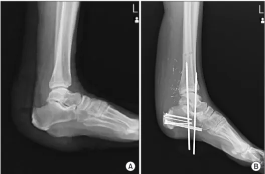
Figure 6. (A) Preoperative simple lateral radiography shows soft tissue damage.
(B) Postoperative simple lateral radiog- raphy shows fixation of allobone to cal- caneus with screws. And ankle joint was immobilized with Steinmann pins.