경추 추간판 탈출증 치료에서 경추 추간판전치환술과 전방고정술의 임상적 결과 및 방사선학적 결과분석
울산대학교 의과대학 서울아산병원 신경외과학교실
박은석·노성우·박진훈·전상룡·임승철·김창진
Clinical and Radiological Analysis of Cervical Arthroplasty Compared to Anterior Cervical Discectomy and Fusion in Cervical Disc Disease
Eun-Suk Park, MD, Sung-Woo Roh, MD, Jin-Hoon Park, MD,
Sang-Ryong Jeon, MD, Seung-Chul Rhim, MD and Chang-Jin Kim, MD
Department of Neurological Surgery, Asan Medical Center, University of Ulsan College of Medicine, Seoul, Korea
Objective: Although anterior cervical discectomy and fusion (ACDF) is the most common treatment for degenerative cervical disc disease, concerns about adjacent level degeneration and loss of motion have led to suggestions that total disc replacement may be a better alternative. Methods: Since April 2006, 35 cases of cervical arthroplasty have been perform- ed at our institute. Here we compare clinical and radiological results in patients who have cervical disc herniations treated with arthroplasty or with ACDF. We evaluated 67 patients treated for cervical disc herniations with radiculopathy and neck pain, of whom 35 underwent cervical arthroplasty using the Mobi-C® (LDR medical, Troyes, France) implant and 32 underwent ACDF using the Solis® cage (Stryker Spine, Allendale, NJ). Clinical measurements of outcome included the numeric rating scale (NRS) score for radiculopathy and neck pain, neck disability index (NDI) score, duration of hospital stay and convalescence time. All patients were assessed radiologically by measuring overall cervical lordosis (Cobb’s angle), segmental lordosis and segmental range-of-movement (ROM) of operated disc levels and adjacent disc levels.
Results: Mean hospital stay (5.52 vs. 6.26 days, p<0.05) and interval between surgery and return to work (1.15 vs. 2.92 months, p<0.05) were significantly shorter in the arthroplasty than in the ACDF group. After 12 months, mean NDI and neck and extremity NRS scores had improved in both groups. Patients in the arthroplasty group, but not in the ACDF group, maintained their baseline overall preoperative cervical and segmental lordosis scores after surgery. Segmental ROM of adjacent levels were higher in the ACDF group than in the arthroplasty group, and segmental motion of operated level scores in the arthroplasty group were maintained at the last follow-up assessment. The ROM of adjacent segment were smaller in the arthroplasty group than in the ACDF group, but the difference was not statistically significant (p>
0.05). In addition, segmental motion of operated level in the arthroplasty group were maintained at the last follow-up assessment. In two cases of arthroplasty group, new bony growth at the treated level, indicating heterotrophic ossification, was suspected based on radiographic (film) results. Conclusion: Although clinical results were similar in the two groups, postoperative recovery was significantly shorter in the arthroplasty group. Postoperative overall cervical and segmental lordosis were reduced in the ACDF group compared with preoperative levels, but not in the arthroplasty group. (J Kor Neurotraumatol Soc 2009;5:83-88)
KEY WORDS: Anterior cervical discectomy and fusion·Cervical arthroplasty·Outcome.
Received: September 22, 2009 / Revised: September 22, 2009 / Accepted: September 30, 2009 Address for correspondence: Sung-Woo Roh, MD
Department of Neurological Surgery, Asan Medical Center, University of Ulsan College of Medicine, 86 Asanbyeongwon-gil, Songpa-gu, Seoul 138-736, Korea
Tel: +82-2-3010-3555, Fax: +82-2-476-6738, E-mail: [email protected]
서 론
경추 전방고정술(anterior cervical discectomy and fusion)는 경추 신경근병증과 척수병증 환자 치료의 한 방법으로 가장 널리 이용되는 방법이다. 그러나 수술 후 인접분절의 퇴행성 변화(adjacent segment degenera- tion: ASD) 같은 합병증의 발생은 경추 추간판전치환술 (cervical arthroplasty)의 개발을 촉진하였다.1,3,4,9) 경 추 추간판전치환술에 대한 가장 중요한 이론적 근거 역 시 움직임을 유지하여 인접분절의 퇴행성 변화를 감소시 키는 것에 있으며,15) 생리적 굴곡 및 운동의 보존은, 환 자가 빠른 속도로 회복하게 하고 빨리 일상활동을 수행 할 수 있게 하는 장점이 있다. 경추 추간판전치환술의 또 다른 이점은 뼈 이식에 의한 이환율의 감소 및 전방 금 속판 고정술과 관련된 합병증을 감소시키는 것이다.16,17)
이 논문에서는 Mobi-C®(LDR medical, Troyes, Fran- ce)를 이용한 경추 추간판전치환술과 Solis® cage(Stry- ker Spine, Allendale, NJ)를 이용한 경추 전방고정술 을 수술적 방법으로 사용하였으며, 경추 추간판전치환술 과 경추 전방고정술의 임상적 결과 및 방사선학적 결과 에 대해 후향적으로 연구하였다. 특히 이환부 운동의 보 존 및 인접부 운동의 보존 여부에 대해 방사선학적으로 평가하였다.
대상 및 방법
본원에서 수술받은 상지의 경추 신경근병증을 동반한 단일 분절의 연성 디스크(soft disc) 환자를 대상으로 하 였으며, 복수 분절의 디스크 환자 및 방사선학적으로 심 한 변성을 보인 환자, 이전에 수술을 받았던 병력이 있는 환자는 제외하였다. 이에 속하는 경추 전방고정술 환자군 은 2005년 2월부터 2006년 12월까지 치료받은 총 32 명 (남자 20명, 여자 12명) 환자들로, 평균 나이는 47세
(범위는 26~63세)였으며, 평균 추적관찰기간은 35개월 (범위는 24~45개월)이었다. 경추 추간판전치환술 환자군 은 2006년 4월부터 2008년 12월까지 치료받은 총 35 명 (남자 18명, 여자 17명) 환자들로 평균 나이는 45.3세 (범위는 31~61세)였으며, 평균 추적관찰기간은 20개월 (범위는 6~33개월)이었다. 수술부위는 경추 전방고정술 환자군에서는 제4-5경추간이 1명, 제5-6경추간이 21 명, 제6-7경추간이 10명이었으며, 경추 추간판전치환술 환자군에서는 제3-4경추간이 2명, 제4-5경추간이 3명, 제5-6경추간이 20명, 제6-7경추간이 10명이었다. 두 환자군 간에 연령 및 성별, 수술분절에는 통계학적 차이 는 없었다 (p>0.05)(Table 1).
저자들은 전례에서 우측 접근법을 사용하여 디스크 제 거술을 시행하였으며, 시험용 삽입물(trial implant)을 디 스크 공간에 넣어 적절한 크기, 높이, 위치를 확인 후 삽입 물(Mobi-C® 혹은 Solis® cage)을 디스크공간에 삽입하 였다. 삽입물을 디스크공간에 삽입 후 투시장치(fluoro- scopic guidance)를 이용하여 정렬 및 위치를 확인하고 교정하였다. Mobi-C®를 이용하여 경추 추간판전치환술 을 시행하였고, Polybone®(Kyungwon medical, Seoul, Korea)과 Solis® cage를 이용한 경추 전방고정술을 시행 하였다.
임상적 결과(clinical outcomes)는 neck disability in- dex (NDI)와 numerical rating scale (NRS)을 이용하 여 평가하였다.
전반적인 경추 전만도(lordosis), 수술한 분절의 전만도 및 운동범위(range of motion) 및 인접분절의 운동범위 를 방사선학적으로 평가하였다. 전반적 경추 전만도는 제 2경추의 하종판과 제7경추의 하종판이 이루는 각으로 정 의하였으며, 분절의 전만도는 측정분절의 상위 척추의 하 종판과 하위 척추의 상종판이 이루는 각으로 정의하여 측 정하였다. 경추 운동범위는 측면 방사선사진을 통해 측 정되는 경추의 굴곡과 신전시 발생하는 각의 차이로 정의
TABLE 1. Baseline demographic and clinical characteristics
Arthroplasty group (n=35) ACDF group (N=32)
Age range (mean), years 31-61 (45.3) 26-63 (47)
Sex (male : female) 18 : 17 20 : 12
Operation period April 2006- September 2008 February 2005-December 2006
Followed-up period (mean), months 6-35 (20) 27-49 (35)
Operation level
C3-4 02 00
C4-5 03 01
C5-6 20 21
C6-7 10 10
ACDF: anterior cervical discectomy and fusion
하였다. 분절의 운동범위는 역동 방사선사진(dynamic X- ray)을 통해 굴곡과 신전시 발생하는 분절의 각의 차이 로 정의하고 측정하였다. 수술 후 방사선사진을 통해 새 로 발생한 이소성 골화증(heterotrophic ossification)에 대해 평가하였다.
통계적 분석 방법으로는 Mann-Whitney test과 Re- peated Measures Analysis of Variance를 이용하였으 며, p-value가 0.05 이하시 통계적 의의가 있는 것으로 평가하였다.
결 과
임상적 결과
평균 수술시간은 경추 추간판전치환술 환자군에서 160 분, 경추 전방고정술 환자군에서 153분으로 비슷하였으 며 (p>0.05), 재원일은 경추 추간판전치환술 환자군이 5.52일, 경추 전방고정술 환자군이 6.26일이었으며, 회복 기는 경추 추간판전치환술 환자군이 1.15개월, 경추 전방 고정술 환자군이 2.93개월로 경추 추간판전치환술 환자 군에 비해 긴 것으로 확인됐다 (p>0.05). 환자의 만족도 는 마지막 외래 경과관찰에서 평가되었으며, 두 환자군 간에 차이가 없는 것으로 평가됐다 (p>0.05)(Table 2).
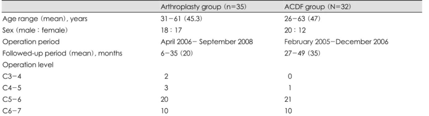
NDI 점수는 수술 후 12개월 후부터 두 환자군 모두 감 소를 보였다. 경추 추간판전치환술 환자군은 22.9에서 10.05로, 경추 전방고정술 환자군은 23.43에서 8.36으 로 감소하였다 (Figure 1A). 상지의 NRS 점수도 수술 후 12개월 후 평가상 두 환자군 모두에서 감소를 보였으 나, 두 환자군 간의 큰 차이는 없었다 (Figure 1B).
방사선학적 결과
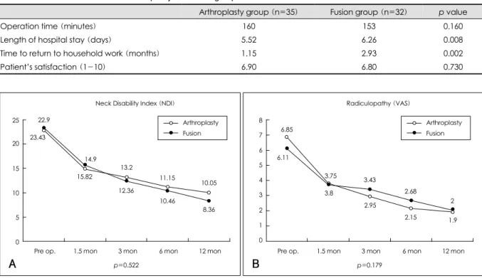
수술 후 12개월에서 검사한 경추 전만도는 경추 추간판 전치환술 환자군에서 29.79도에서 28.59도로 감소하였 으며, 경추 전방고정술 환자군에서는 24.27도에서 17.69 도로 감소하였다. 경추 추간판전치환술 환자군의 경우 경 추 전만도가 수술 직후에는 34.75도로 증가를 보이다가 점차 감소하였으며, 경추 전방고정술 환자군에서는 수술 직후부터 점차적으로 감소하는 양상을 확인할 수 있었다 (Figure 2A). 분절 전만도의 경우 경추 추간판전치환술 환자군에서는 4.78도에서 8.28도로 증가를 보인 반면, 경 추 전방고정술 환자군에서는 3.93도에서 2.82도로 감소 를 보였다 (Figure 2B).
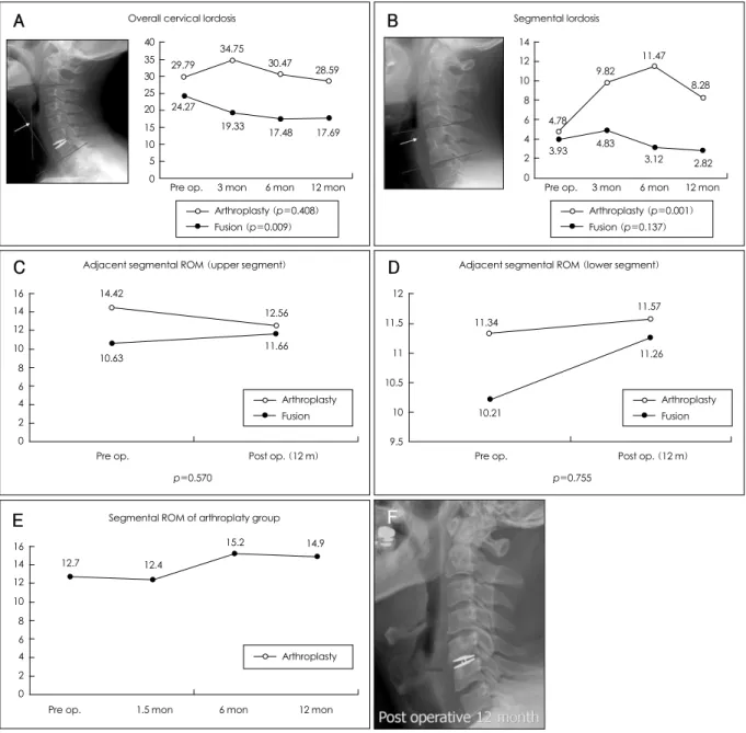
상위 분절의 운동범위는 경추 추간판전치환술 환자군 에서 14.42도에서 12.56으로 감소를 보였으나, 경추 전 방고정술 환자군에서는 10.63도에서 11.66도로 증가를
TABLE 2. Clinical outcomes in the arthroplasty and fusion groups
Arthroplasty group (n=35) Fusion group (n=32) p value
Operation time (minutes) 160 153 0.160
Length of hospital stay (days) 5.52 6.26 0.008
Time to return to household work (months) 1.15 2.93 0.002
Patient’s satisfaction (1-10) 6.90 6.80 0.730
FIGURE 1. A: Neck disability index (NDI) scores in the arthroplasty and fusion groups (p>0.05). The scores of both groups de- creased gradually; however, the greatest decrease was seen in the immediate postoperative period. B: NRS scores of radicul- opathy in the arthroplasty and fusion groups (p>0.05). The scores of both groups decreased gradually, but the decreases were greatest in the immediate postoperative period. NRS: nume ical rating scale, VAS: visual analogue scale.
Arthroplasty Fusion
A
Neck Disability Index (NDI)
Pre op. 1.5 mon 3 mon 6 mon 12 mon 25
20
15
10
5
0
p=0.522
8.36 10.46
12.36 15.82
23.43 22.9
14.9
13.2
11.15
10.05
Arthroplasty Fusion
B
Radiculopathy (VAS)
Pre op. 1.5 mon 3 mon 6 mon 12 mon 8
7 6 5 4 3 2 1 0
p=0.179 2.15 1.9 2.95
3.8 6.11
6.85
3.75 3.43
2.68
2
보였다. 그러나, 이러한 결과는 통계적 의의는 없었다 (p>
0.05)(Figure 2C). 하위 분절의 운동범위의 경우는 두 환자군 모두에서 증가를 보였다 (p>0.05)(Figure 2D).
수술부위 분절의 운동범위의 경우, 경추 추간판전치환술 환자군에서는 수술 직후 잠시 감소를 보이다가 수술 후
12개월까지 12.7도에서 14.9도로 점차적으로 증가를 보 였다 (Figure 2E).
경추 추간판전치환술 환자군 중 2예에서 이소성 골화 증이 수술분절에서 관찰되었다 (Figure 2F).
Arthroplasty (p=0.408) Fusion (p=0.009)
A Overall cervical lordosis
Pre op. 3 mon 6 mon 12 mon 40
35 30 25 20 15 10 5 0
29.79
24.27 19.33 34.75
30.47
17.48 17.69 28.59
Arthroplasty (p=0.001) Fusion (p=0.137) Segmental lordosis
Pre op. 3 mon 6 mon 12 mon 14
12 10 8 6 4 2 0
3.93 4.78
4.83 9.82
11.47
3.12 2.82 8.28
B
Adjacent segmental ROM (upper segment)
16 14 12 10 8 6 4 2 0
Pre op. Post op. (12 m) p=0.570
Arthroplasty Fusion 14.42
10.63 11.66
12.56
C Adjacent segmental ROM (lower segment)
12
11.5
11
10.5
10
9.5
Pre op. Post op. (12 m) p=0.755
Arthroplasty Fusion 11.34
10.21
11.26 11.57
D
FIGURE 2. A: Overall cervical lordosis in the arthroplasty and fusion groups. Measurement was performed as shown in the left panel. In the arthroplasty group, cervical lordosis increased immediately after surgery, then decreased to preoperative levels.
Cervical lordosis in the fusion group gradually decreased after surgery and did not return to preoperative levels. B: Segmental lordosis in the arthroplasty and fusion groups. Measurement was performed as shown in the left panel. In the arthroplasty group, segmental lordosis score increased immediately after surgery, but then decreased after 6 months and returned to preoperative levels. In the fusion group, however, segmental lordosis increased immediately after surgery, then decreased but did not return to preoperative levels. C: Adjacent ROM of the upper level in the arthroplasty and fusion groups. After surgery, upper ROM decreased in the arthroplasty group but increased in the fusion group (p>0.05). D: Adjacent ROM of the lower level in the arthroplasty and fusion groups. After surgery, lower ROM increased in both groups, but the increase was greater in the fusion group (p>0.05). E:
Segmental ROM in the arthroplasty group decreased immediately after surgery before increasing to a level greater than the preoperative level. F: In two cases of arthroplasty group, new bony growth at the treated level, indicating heterotrophic ossification, was suspected based on postoperative X-ray. ROM: range of movement.
Segmental ROM of arthroplaty group
16 14 12 10 8 6 4 2 0
Pre op. 1.5 mon 6 mon 12 mon Arthroplasty
12.7 12.4
15.2 14.9
E F
고 찰
현재 경추 전방고정술은 퇴행성 경추질환 환자들에서 가장 많이 쓰이는 수술적 치료이다.3) 비록 경추 전방고 정술이 수술적인 측면에서 여러 장점이 있지만, 인접 분 절 디스크의 이상 유발이나, 목 움직임 제한 등과 같은 부 작용이 따를 수 있다. 수술 후의 운동 장애는 인접한 척 추분절에 대한 과도한 운동부화와 관련이 있으며, 이러 한 운동부화는 인접분절의 퇴행성 변화를 유발하거나 악 화시킬 수 있다.2,9,12) 척추 운동 장애의 부작용이 없는 경 추 추간판전치환술은 인접분절의 퇴행성 변화와 같은 부 작용을 감소시킬 수 있다. 지금까지의 연구들은 인접분절 의 운동 장애 증가와 병적인 퇴행(pathogenic degenera- tion) 간에 뚜렷한 관련성이 있음을 보여주고 있으며,19) 같은 맥락으로 우리 연구 역시 수술 후 인접분절 운동 장 애의 증가를 보여주고 있다.
경추 추간판전치환술에 대해서, 우리는 상대적으로 조 작이 쉬우며 경추 전방고정술의 술기와 유사점이 있는 Mobi-C®를 사용하고 있다. Mobi-C®의 수술적 삽입은 삽입의 위치, 각도, 깊이의 조정을 편리하게 해주는 이식 물 고정장치(implant holder)로 인하여, 쉽고, 안전하고, 재현이 가능하다는 특징이 있다. 마찬가지로, 우리가 사 용하는 Solis® cage 역시 간단한 장치이며, 유일한 차이 점인 장골이식을 제외한다면, Mobi-C®만큼이나 간편한 이식방법이다.
이 논문에서 저자들은 두 환자군 간의 평균 수술시간 은 비슷하고, NDI와 NRS 점수로 평가된 임상결과 역시 차이가 없음을 확인하였으며, 이는 과거의 몇몇 연구들 의 결과와 같이 병변의 감압과 고정이 증상의 호전과 유 사한 관계가 있음을 알 수 있다.10,11) 이러한 과거 연구들 은, 또한 두 환자군 모두에서 큰 차이 없이 목과 팔의 통 증이 현저하게 감소하였다고 보고하였다.11) 다른 연구들 에서 볼 수 있듯이, 유발 병변을 제거하는 수술후에 즉각 적으로 NDI와 NRS 점수가 향상되었다. 우리 연구에서 가장 빠른 경우는 수술 후 한 달 반만에 통증과 증상의 감소를 보인 경우였다. 입원기간과 회복 시간으로 평가 되는 수술 후 회복(postoperative recovery)은 경추 전 방고정술 환자군보다 경추 추간판전치환술 환자군에서 훨 씬 더 짧은 것으로 나타났다. 우리는 수술 후 보조기 착용 이나 장골이식이 경추 전방고정술 환자에서 수술 후 회 복에 영향을 미친다고 결론을 내렸다.
우리는 경추 전방고정술 환자군에서 수술 후의 전체 혹 은 부분적인 경추 전만도가 수술 전의 수준으로까지 회복
되지 못함을 발견하였다. 이 결과는 경추 후만증(kypho- sis)을 시사한다. 몇몇 연구들은 경추 전방고정술 후 이 식물의 침전을 보고하고 있다. 예를 들어, 경추의 전방 고 정판이 없는 환자의 약 10% 이상에서 경추 고정술 수술 후 비대칭적 cage 침전(subsidence)을 보이게 된다.18) 실 제로, 현재의 연구에서 전방경유 추간판절제술 및 골유 합술 환자군의 몇몇 환자들은 수술 후 cage 침전을 의미 하는 경추 후만증이 더 심해진 소견을 보이고 있다. 그러 나, 경추 추간판전치환술 환자군에서는 환자들의 전체 혹은 부분 경추 전만증이 수술 이전 수준을 유지하고 있 었다. 또한, 척추 배열의 유지가 인접분절들의 퇴행(de- generation)을 막아주는 것으로 보고되고 있다.5-8)
다른 연구들에서도 경추 추간판전치환술을 받은 환자 들이 치료받은 분절의 운동성이 훨씬 더 유지가 잘되어, 인접분절의 운동성에 영향을 적게 미친다는 것을 보여주
었다.10,11,14,21) 본 연구 역시 수술 후 분절의 운동범위가
보존됨을 보여주었다. 우리는 상위 인접분절에서의 운동 범위가 경추 추간판전치환술 환자군에서는 감소하는 반면, 경추 전방고정술 환자군에서는 증가함을 발견하였다. 게 다가, 하위 분절의 운동범위 증가는 경추 추간판전치환 술 환자군 보다 경추 전방고정술 환자군에서 더 컸다. 이 러한 결과들을 종합해 보면, 경추 전방고정술에 비해 경 추 추간판전치환술이 인접분절의 과운동성 예방에 더 큰 장점을 가진다고 결론을 내릴 수 있다.
최근 경추 추간판전치환술 후 발생한 이소성 골화증에 대해 발표되고 있으며, Wenger 등20)의 연구에서는 경추 추간판전치환술 후 약 6%에서 이소성 골화증이 수술분 절에 발생하였다고 발표하였다. 이소성 골화증의 발생 이 유에 대해서는 아직 명확히 밝혀져 있진 않으나, 수술부위 의 해면골(cancellous bone)의 노출에 의한 것으로 설명 하고 있다.13) 우리 연구에서도 35예의 경추 추간판전치 환술 환자 중 약 2예 (5.7%)에서 이소성 골화증이 발생 하였다. Mobi-C®를 이용한 경추 추간판전치환술의 경우 수술분절의 종판(endplate)에 손상없이 비교적 쉽게 삽입 이 가능하므로, 이소성 골화증의 발생을 줄이는 데 도움 이 될 수 있을 것으로 생각되며, 이에 대한 연구를 위해 좀 더 많은 증례와 추적관찰이 필요할 것으로 생각된다.
결 론
두 환자군을 비교할 때 NDI 점수, NRS 점수와 같은 임상적 결과에 있어서 유의한 차이는 없었다. 그러나, 수 술 후 회복 기간에 있어서, 경추 추간판전치환술 환자군의
회복 시간이 빨랐는데, 그것은 수술 후 보조기 착용이나 장골이식이 없었던 점과 관련이 있는 것으로 추정된다.
경추 전방고정술을 시행한 환자군에서, 경추 추간판전 치환술을 시행한 환자군에 비해서 수술 후 후만증이 더 많이 발생하는 경향을 보였는데, 이것은 몇몇의 증례에 서 수술 후 이식물 침전(postoperative graft subsidence) 과 연관이 되어 있는 것으로 판단된다. 마지막 외래 경과 관찰에서 경추 추간판전치환술 환자군에서는 분절 운동 (segmental motion)이 유지되었으며, 비록 통계적으로 유의하지는 않지만, 인접한 분절의 운동범위가 경추 전 방고정술 환자군에 비해 경추 추간판전치환술 환자군에 서 더 작았다. 그러나 우리가 시행한 연구는 짧은 경과관 찰 기간과 대상 환자 수가 적다는 점에서 한계점을 지닌 다. 두 수술 간에 차이점을 명확히 밝히기 위해서는 좀 더 많은 환자와 장기간의 경과관찰기간이 필요하다.
중심 단어: 경추 전방고정술·경추 추간판전치환술·결과.
REFERENCES
1) Cummins BH, Robertson JT, Gill SS. Surgical experience with an implanted artificial cervical joint. J Neurosurg 88:943-948, 1998 2) Eck JC, Humphreys SC, Lim TH, Jeong ST, Kim JG, Hodges SD,
et al. Biomechanical study on the effect of cervical spine fusion on adjacent-level intradiscal pressure and segmental motion. Spine (Phila Pa 1976) 27:2431-2434, 2002
3) Goffin J, Geusens E, Vantomme N, Quintens E, Waerzeggers Y, Depreitere B, et al. Long-term follow-up after interbody fusion of the cervical spine. J Spinal Disord Tech 17:79-85, 2004 4) Hilibrand AS, Carlson GD, Palumbo MA, Jones PK, Bohlman
HH. Radiculopathy and myelopathy at segments adjacent to the site of a previous anterior cervical arthrodesis. J Bone Joint Surg Am 81:519-528, 1999
5) Katsuura A, Hukuda S, Saruhashi Y, Mori K. Kyphotic malalign- ment after anterior cervical fusion is one of the factors promoting the degenerative process in adjacent intervertebral levels. Eur Spine J 10:320-324, 2001
6) Kim SH, Shin HC, Shin DA, Kim KN, Yoon do H. Early clinical experience with the mobi-C disc prosthesis. Yonsei Med J 48:457- 464, 2007
7) Kim SW, Limson MA, Kim SB, Arbatin JJ, Chang KY, Park MS, et al. Comparison of radiographic changes after ACDF versus Bryan
disc arthroplasty in single and bi-level cases. Eur Spine J 18:218- 231, 2009
8) Kim SW, Shin JH, Arbatin JJ, Park MS, Chung YK, McAfee PC.
Effects of a cervical disc prosthesis on maintaining sagittal align- ment of the functional spinal unit and overall sagittal balance of the cervical spine. Eur Spine J 17:20-29, 2008
9) Kulkarni V, Rajshekhar V, Raghuram L. Accelerated spondylotic changes adjacent to the fused segment following central cervical corpectomy: magnetic resonance imaging study evidence. J Neu- rosurg 100 (1 Suppl Spine):2-6, 2004
10) Mummaneni PV, Burkus JK, Haid RW, Traynelis VC, Zdeblick TA. Clinical and radiographic analysis of cervical disc arthroplasty compared with allograft fusion: a randomized controlled clinical trial. J Neurosurg Spine 6:198-209, 2007
11) Nabhan A, Ahlhelm F, Pitzen T, Steudel WI, Jung J, Shariat K, et al. Disc replacement using Pro-Disc C versus fusion: a prospective randomised and controlled radiographic and clinical study. Eur Spine J 16:423-430, 2007
12) Pickett GE, Rouleau JP, Duggal N. Kinematic analysis of the cer- vical spine following implantation of an artificial cervical disc.
Spine (Phila Pa 1976) 30:1949-1954, 2005
13) Sasso RC, Best NM, Metcalf NH, Anderson PA. Motion analysis of bryan cervical disc arthroplasty versus anterior discectomy and fusion: results from a prospective, randomized, multicenter, clinical trial. J Spinal Disord Tech 21:393-399, 2008
14) Sasso RC, Smucker JD, Hacker RJ, Heller JG. Artificial disc versus fusion: a prospective, randomized study with 2-year follow-up on 99 patients. Spine (Phila Pa 1976) 32:2933-2942, 2007
15) Shim CS, Lee SH, Park HJ, Kang HS, Hwang JH. Early clinical and radiologic outcomes of cervical arthroplasty with Bryan Cer- vical Disc prosthesis. J Spinal Disord Tech 19:465-470, 2006 16) Silber JS, Anderson DG, Daffner SD, Brislin BT, Leland JM,
Hilibrand AS, et al. Donor site morbidity after anterior iliac crest bone harvest for single-level anterior cervical discectomy and fu- sion. Spine (Phila Pa 1976) 28:134-139, 2003
17) St John TA, Vaccaro AR, Sah AP, Schaefer M, Berta SC, Albert T, et al. Physical and monetary costs associated with autogenous bone graft harvesting. Am J Orthop 32:18-23, 2003
18) van Jonbergen HP, Spruit M, Anderson PG, Pavlov PW. Anterior cervical interbody fusion with a titanium box cage: early radiolo- gical assessment of fusion and subsidence. Spine J 5:645-649, 2005 19) Weinhoffer SL, Guyer RD, Herbert M, Griffith SL. Intradiscal pres- sure measurements above an instrumented fusion. A cadaveric study.
Spine (Phila Pa 1976) 20:526-531, 1995
20) Wenger M, Hoonacker P, Zachee B, Lange R, Markwalder TM.
Bryan cervical disc prostheses: preservation of function over time.
J Clin Neurosci 16:220-225, 2009
21) Yoon DH, Yi S, Shin HC, Kim KN, Kim SH. Clinical and radio- logical results following cervical arthroplasty. Acta Neurochir (Wien) 148:943-950, 2006