Comparison of Positional and Non-Positional Obstructive Sleep Apnea Patients by Nocturnal Polysomnography
Min-Woo Park, D.D.S., Jung-Hwan Cho, D.D.S., Won-Kyu Park, D.D.S.,M.S.D.,Ph.D.,
Jin-Woo Nam, D.D.S.,M.S.D.,Ph.D., Chong-Il Yun, D.D.S.,M.S.D.,Ph.D., Jin-Woo Chung, D.D.S.,M.S.D.,Ph.D.
Department of Oral Medicine and Oral Diagnosis
School of Dentistry and Dental Research Institute, Seoul National University
Objectives: The aim of this study was to evaluate the differences in the polysomnography data between positional and non-positional obstructive sleep apnea (OSA) patients.
Methods: Forty-seven patients diagnosed with OSA were evaluated using full night polysomnography. According to the criteria of Cartwright et al., the patients were classified into two groups with 37 positional (supine apnea-hypopnea index [AHI] ≥ 2x's the lateral AHI) and 10 non-positional (supine AHI < 2x's the lateral AHI) OSA patients, and the differences of polysomnography data between the two groups were evaluated.
Results: There were no significant differences in demographic variables (age, gender, and BMI), daytime sleepiness, overall AHI, total arousal index, and percent time of snoring between two groups. However, AHI, arousal index, and mean oxygen saturation (SpO2) of the REM sleep stage were significantly more severe in the positional OSA group than the non-positional OSA group. Mean SpO2 and the lowest SpO2 during overall sleep stage were also significantly lower in the positional OSA group than the non-positional OSA group.
Conclusions: Our results of differences in the polysomnography data of REM sleep stage suggest that non-positional OSA patients may have higher collapsibility of the oropharyngeal airway during sleep than positional OSA patients.
Key words: Sleep apnea, Positional OSA, Polysomnography, REM sleep, Oxygen saturation
Corresponding author : Jin-Woo Chung
Department of Oral Medicine and Oral Diagnosis School of Dentistry and Dental Research Institute Seoul National University
28 Yunkeun-Dong, Chongro-Ku, Seoul 110-749, Korea Tel: +82-2-2072-3021, Fax: +82-2-744-9135
E-mail: [email protected] Received: 2009-09-22
Revised: 2009-10-20 Accepted: 2009-11-10
* This Study was supported by the Seoul National University Dental Hospital Research Fund (Grant number 04-2008-0027).
Ⅰ. INTRODUCTION
Obstructive sleep apnea (OSA) is a commonly occurring disorder affecting between 2 and 4 % of general population
1,2)and a potentially life- threatening condition in which periodic cessation of breathing occurs during sleep inspite of the presence of inspiratory effort. This disorder affects not only the quality of life but also causes a significant increase in morbidity. The reduction in blood oxygen saturation may give rise to hypertension, cardiac arrhythmias, nocturnal angina, and myocardial ischemia.
3)Many studies suggest that body position plays an
important role in breathing functions during sleep.
4-6)In the majority of patients with obstructive apnea and hypopnea the severity decreased surely in the lateral position but in certain patients, apnea and hypopnea decreased only slightly in the lateral position. Cartwright defined positional OSA patients as those whom the respiratory disturbance index (RDI) or the apnea-hypopnea index (AHI) was at least twice as high while sleeping in the supine position as in the lateral position. Those cases in which the RDI in the supine position was less than twice that in the lateral position were defined non-positional OSA patients.
5)Previous studies on the differences in the clinical characteristics of patient and risk factors between positional and non-positional OSA are few and report conflicting results. Some investigators have reported that positional OSA patients are younger, and less obese, therefore they have less severe respiratory disturbance than non-positional patients.
5,6)Others have found no differences in the body mass index (BMI) between positional and non-positional patients.
7)Although several studies have shown less severe respiratory disturbance in positional OSA patients,
5,6)the anatomical and physiological mechanisms for this phenomenon have not been clarified yet. We hypothesized that there may be factors other than age and BMI that influence the differences in disordered breathing between positional and non-positional OSA patients.
The aim of this study was to evaluate the differences found in the polysomnography data between positional and non-positional obstructive sleep apnea (OSA) patients.
Ⅱ. MATERIALS AND METHODS 1. Subjects
Subjects were the consecutive patients who visited the Snoring and Sleep Apnea Clinic,
Department of Oral Medicine in Seoul National University Dental Hospital with the complaint of snoring, sleep apnea, or daytime sleepiness, from August, 2007 to October, 2009. The subjects were diagnosed with OSA (RDI>5) based on full night polysomnography results. The subject group consisted 47 (37 men and 10 women) patients and age ranged from 16 to 70 years. The daytime sleepiness of each subject was evaluated with the Epworth Sleepiness Scale (ESS) questionnaire.
The subjects were categorized into positional and non-positional OSA patients following the criteria suggested by Cartwright.
5)Specifically these criteria state that positional OSA patients have a supine AHI at least two times higher than their lateral AHI, and non-positional patients will have their supine AHI less than two times higher than their lateral AHI.
2. Polysomnography evaluation
Multi-channel recordings of electroencephalo- gram (EEG), submental and leg electromyogram (EMG), electrocardiogram (ECG), nasal thermistor, nasal pressure transducer, thoracic and abdominal piezoelectric belts, and oxygen saturation were conducted using Alice 4 (Respironics, Pittsburgh, USA) polysomnography. Body position was also confirmed through direct observation of the patient by the technician using a low light camera and simultaneously digital recording with a posture tag at the thoracic piezoelectric belt was also carried out.
Sleep was staged by an eligible sleep physician
and respiratory events were scored using the
standard criteria of the American Academy of Sleep
Medicine.
8)Briefly, obstructive apnea was defined
as a reduction in airflow greater than 90% with a
duration of at least 10 sec in which there was
persistent respiratory effort, whereas hypopnea was
defined as a reduction of airflow by 30% for more
than 10 sec accompanied by oxygen desaturation ≥
3%.
3. Statistical analysis
The differences in demographic variables and polysomnography data between positional and non-positional OSA patients were analyzed using independent t-test. P-values less than 0.05 were considered statistically significant. The statistical analysis was performed by SPSS Version 12.0 (Chicago, IL, USA) software.
Ⅲ. RESULTS 1. Demographic comparison
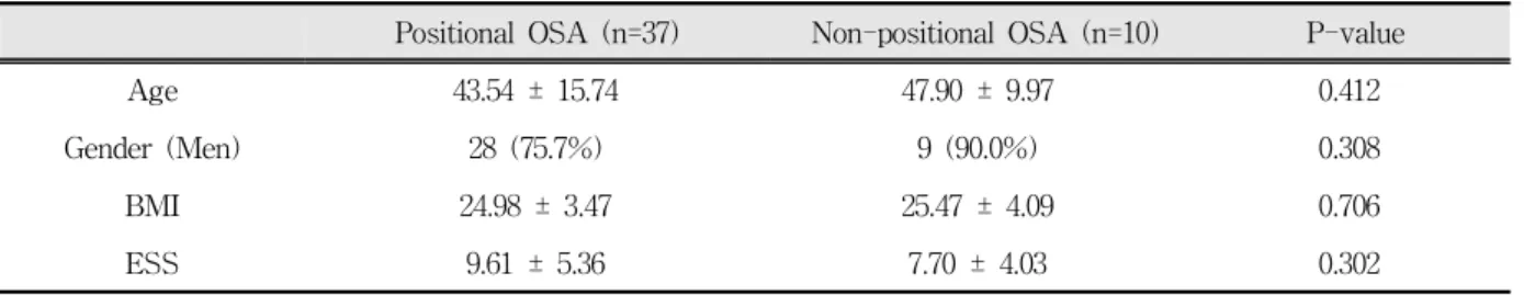
From the 47 subjects 10 positional OSA and 37 non-positional OSA patients were classified. Table 1 shows the demographic data of the subjects.
There were no significant differences in age, gender, BMI, and ESS between positional and non-positional OSA patients.
Positional OSA (n=37) Non-positional OSA (n=10) P-value
Age 43.54 ± 15.74 47.90 ± 9.97 0.412
Gender (Men) 28 (75.7%) 9 (90.0%) 0.308
BMI 24.98 ± 3.47 25.47 ± 4.09 0.706
ESS 9.61 ± 5.36 7.70 ± 4.03 0.302
P-values were obtained from independent t-test and Chi-square test Table 1. Demographic comparison of positional and on-positional OSA patients
Positional OSA (n=37) Non-positional OSA (n=10) P-value
Overall AHI 25.44 ± 14.33 39.18 ± 25.97 0.139
Supine AHI 35.00 ± 20.79 38.27 ± 30.29 0.753
Lateral AHI 5.96 ± 7.83 32.91 ± 22.18 0.004
% time of snoring 50.4 ± 16.28 44.80 ± 23.54 0.486
% time in supine 70.53 ± 23.22 66.69 ± 30.98 0.668
P-values were obtained from independent t-test.
Table 2. Apnea-hypopnea indices (AHIs), percent time of snoring, and percent time in supine position of positional and non-positional OSA patients
2. Apnea and hypopnea indices (AHIs), percent time of snoring, and percent time in supine position
There were no significant differences in overall AHI and supine AHI between positional and non-positional OSA groups. Lateral AHI was significantly higher in the non-positional OSA group than the positional OSA patients (p<0.01).
There were no significant differences in percent time of snoring and time in supine position between positional and non-positional OSA patients (Table 2).
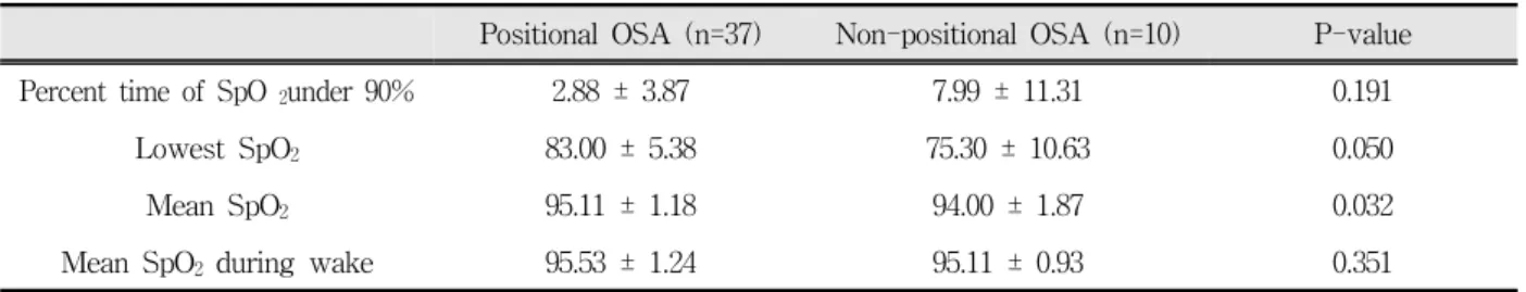
3. Oxygen desaturation
Non-positional OSA patients showed signifi- cantly lower mean SpO
2(p<0.05) and lowest SpO
2(p=0.05) than positional OSA patients (Table 3).
There were no significant differences in percent
Positional OSA (n=37) Non-positional OSA (n=10) P-value
Percent time of SpO 2under 90% 2.88 ± 3.87 7.99 ± 11.31 0.191
Lowest SpO2 83.00 ± 5.38 75.30 ± 10.63 0.050
Mean SpO2 95.11 ± 1.18 94.00 ± 1.87 0.032
Mean SpO2 during wake 95.53 ± 1.24 95.11 ± 0.93 0.351
P-values were obtained from independent t-test.
Table 3. Oxygen saturation comparison of positional and non-positional OSA patients
Positional OSA (n=37) Non-positional OSA (n=10) P-value Apnea & hypopnea arousal index 13.87 ± 12.14 23.10 ± 19.78 0.187
Snoring arousal index 4.85 ± 4.79 4.78 ± 4.19 0.967
Spontaneous arousal index 4.697 ± 4.67 3.00 ± 2.59 0.278
Arousal index 28.40 ± 17.26 33.40 ± 22.43 0.450
P-values were obtained from independent t-test.
Table 4. Comparisons of arousal indices between positional and non-positional OSA patients.
time of SpO
2under 90% and mean SpO
2during wake between positional and non-positional OSA patients.
4. Arousals
There were no significant differences in apnea and hypopnea arousal index, snoring arousal index, spontaneous arousal index, and overall arousal index between positional and non-positional OSA groups (Table 4).
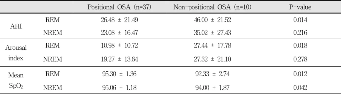
5. Polysomnography data according to REM and NREM sleep stages
Table 5 shows the comparisons of polyso- mnography data according to REM and NREM sleep stages between the two groups. REM AHI and REM arousal index were significantly higher in the non-positional OSA group than the positional OSA group. AHI and arousal index during NREM sleep stage did not show significant differences between the two groups. Both REM and NREM
mean SpO
2were significantly lower in the non-positional OSA group than the positional OSA group.
Ⅲ. DISCUSSION
In this study there were no significant differences in age, gender, BMI, ESS, and overall AHI between positional and non-positional OSA patients composed of Korean adults. Oksenberg et al.
reported that positional OSA patients were younger
and less obese than non-positional OSA patients.
6)They also reported that positional OSA patients had
better sleep quality and were less sleepy during
daytime hours. They speculated that age and BMI
might explain the difference between the two
groups and they also suggested that positional and
non-positional OSA are part of the same disorder
with the only difference being a progression in
severity. However, our data are contradictory to
their results and we suggest that some factors
beyond gender, and BMI changes influence being
positional or non-positional OSA patients.
Positional OSA (n=37) Non-positional OSA (n=10) P-value
AHI REM 26.48 ± 21.49 46.00 ± 21.52 0.014
NREM 23.08 ± 16.47 35.02 ± 27.43 0.216
Arousal index
REM 10.98 ± 10.72 27.44 ± 17.78 0.018
NREM 19.27 ± 13.64 27.32 ± 21.10 0.278
Mean SpO2
REM 95.30 ± 1.36 92.33 ± 2.74 0.012
NREM 95.06 ± 1.18 94.00 ± 1.87 0.042
P-values were obtained from independent t-test.
Table 5. Comparisons of AHI, arousal index, and mean SpO2 of REM and NREM stages between positional and non-positional OSA patients
Interestingly, AHI, arousal index, and mean SpO
2during REM sleep stage were significantly higher in the non-positional OSA group than the positional OSA group in this study. REM sleep is often identified as the stage of sleep in which people are most vulnerable for having sleep-disordered breathing.
9,10)Tone of the upper airway including the genioglossus muscle decreases from wakefulness to non-rapid eye movement (NREM) sleep and still further in REM sleep.
11,12)During REM sleep, generalized muscle atonia increase pharyngeal collapsibility and decrease air ventilation. Arousal responses to hypoxia are likely responsible for the worsening of sleep apnea during REM sleep. Many OSA patients have more respiratory events in REM sleep which are often associated with the greatest degree of oxygen desaturation.
13)We suspect that the most current explainable hypothesis with evidence for differences in breathing function between positional and non-positional OSA patients could be pharyngeal collapsibility. The non-positional group must have more inherent pharyngeal airway collapsibility than the positional group (i.e. they collapse even when they are on their side), and therefore, AHI, arousal index, and mean SpO
2should be more severe in REM sleep especially for the non-positional OSA patients which was the case
aintained during wakefulness through the activation of dilator muscles by protective reflex mechanisms.
14)These protective reflex mechanisms are diminished during sleep, leading to collapse of the pharyngeal airway in anatomically predisposed individuals. During the REM sleep stage, the muscles are changed to the atonia state and the pharyngeal space becomes more affected by the collapsibility of the pharyngeal airway. The main mechanism for increasing apnea severity in the supine position compared to the lateral position is most probably related to the effect of gravity on the pharyngeal airway.
15,16)In the supine position, the gravity forces the tongue and soft palate to fall back into the throat narrowing the pharyngeal space.
Therefore the risk for pharyngeal airway obstruction is higher which would lead to a large number of apneic events.
17)If the pharyngeal collapsibility is higher, airway obstruction would be affected regardless of body position. We did not measure the pharyngeal collapsibility in this study, therefore future studies should be performed to seek further evidence that will explain the differences between positional and non-positional OSA patients.
Two limitations should be mentioned about this
study. First, we did not consider the anatomical
characteristics of the two groups in this study.
wall parallel to the Frankfurt horizontal measurement was narrower in non-positional OSA patients compared to positional OSA patients.
18)We could suspect that the anatomical variability such as hyoid bone position, tongue size, relationships between maxilla and mandible, and soft palate size affected the airway space in both supine and lateral sleep position. Further imaging analysis including cephalometric evaluation should be needed to obtain more convincing evidence on the pharyngeal airway and sleep position. Second, the sample size for each group was not relatively modest and the vast majority of patients were composed of middle aged or older men. To investigate the effect of gender and aging on the risk factors for the positional and non-positional OSA patients, a larger sample with a wider age range should be studied for both genders.
In summary, we have shown that the non- positional OSA patients have more severe respiratory disturbance during the REM sleep stage than age-, gender- and BMI-matched positional OSA patients. These results suggest that other factors beyond gender and BMI are contributory to OSA in these subjects. Additional studies, including evaluation of dynamic pharyngeal airway collapsibility, will be required to identify the contributing factors that cause the differences in positional and non-positional OSA patients and to evaluate substantial differences in treatment efficacy for these two patient groups.
V. CONCLUSION
The mechanisms underlying positional and non-positional obstructive sleep apnea (OSA) have not been clarified. In this study, there were no significant differences in demographic variables (age, gender, and BMI), daytime sleepiness, overall AHI, total arousal index, and percent time of snoring between the two groups. However, AHI, arousal index, and mean SpO
2of REM sleep stage were significantly more severe in positional OSA group than non-positional OSA group. The
differences in the polysomnography data of REM sleep stage found in our results suggest that non-positional OSA patients may have higher collapsibility of the oropharyngeal airway during sleep than positional OSA patients.
REFERENCES
1. O’Connor C, Thornley K, Hanly P. Gender differences in the polysomnographic features of obstructive sleep apnea. Am J Respir Crit Care Med 2000;161:
1465-1472.
2. Resta O, Carpanano G, Lacedonia D, DiGioia G, Gilberti T, Stefano A, Bonfitto P. Gender difference in sleep profile of severely obese patients with obstructive sleep apnea. Respir Med 2005;99:91-96.
3. Klitzman D, Miller A. Obstructive sleep apnea syndrome: complications and sequelae. Mt Sinai J Med 1994;61:113-121.
4. Gastaut H, Tassinari CA, Duron B. Polygraphic study of the episodic diurnal and nocturnal (hypnic and respiratory) manifestations of the Pickwickian syndrome. Brain Res 1966;2:167-186.
5. Cartwright RD. Effect of sleep position on sleep apnea severity. Sleep 1984;7:110-114.
6. Oksenberg A, Silverberg DS, Arons E, et al. positional vs nonpositional obstructive sleep apnea patients.
Anthropomophic, nocturnal polysomnographic and multiple sleep latency test data. Chest 1997;112:
629-639.
7. Pevernagie DA, Shepard JW. Relations between sleep stage, posture and effective nasal CPAP levels in OSA. Sleep 1992;15:162-167.
8. American Academy of Sleep Medicine Task Force.
Sleep-related breathing disorders in adults:
recommendations for syndrome definition and measurement techniques in clinical research. Sleep 1999;22:667-669.
9. Shea SA, Edwards JK, White DP. Effect of wake- sleep transitions and rapid eye movement sleep on pharyngeal muscle response to negative pressure in humans. J Physiol 1999;520 Pt 3:897-908.
10. Series F, Cormier Y, La Forge J. Influence of apnea type and sleep stage on nocturnal postapneic desaturation. Am Rev Respir Dis 1990;141:1522-1526.
11. Eckert DJ, Malhotra A, Lo YL, White DP, Jordan AS.
The influence of obstructive sleep apnea and gender on genioglossus activity during rapid eye movement sleep. Chest 2009;135:957-964.
12. Siddiqui F, Walters AS, Goldstein D, LaheyM, Desai H. Half of patients with obstructive sleep apnea have a higher NREM AHI than REM AHI. Sleep Med 2006;7:281-285.
13. Findley LJ, Wilhoit SC, Suratt PM. Apnea duration and hypoxemia during REM sleep in patients with obstructive sleep apnea. Chest 1985;87:432-436.
14. Pierce R, White D, Malhotra A, et al. Upper airway collapsibility, dilator muscle activation and resistance in sleep apnoea. Eur Respir J 2007;30:345-353.
15. Oksenberg A, Khamaysi I, Silverberg DS, Tarasiuk A. Association of body position with severity of apneic events in patients with severe nonpositional obstructive sleep apnea. Chest 2000;118;1018-1024.
국문요약
야간수면다원검사를 이용한 자세성 및 비자세성 수면무호흡증 환자의 비교 연구
서울대학교 치의학대학원 구강내과진단학교실, 치학연구소
박민우․조정환․박원규․남진우․윤종일․정진우
본 연구의 목적은 자세성 수면무호흡증 환자군과 비자세성 수면무호흡증 환자군의 수면다원검사 결과를 비교 분석하여 그 원인 요소를 알아보는데 있다. 서울대학교치과병원에 코골이 및 수면무호흡증을 주소로 내원한 환자들 중 수면다원검사 결과 수면무호흡증으로 진단된 (무호흡-저호흡 지수 5이상) 환자 47명을 Cartwright 등의 분류에 따라 37명의 자세성 수면무호흡 증 환자 (앙와위에서의 수면무호흡-저호흡 지수가 측와위에서의 수면무호흡-저호흡 지수의 2배 이상)와 10명의 비자세성 수면무호흡증 환자 (앙와위에서의 수면무호흡-저호흡 지수가 측와위에서의 수면무호흡-저호흡 지수의 2배 미만)로 분류하 여 각 군간의 수면다원검사 지수들을 비교 분석 하였다. 연구 결과, 나이, 성별, 체질량지수, 주간졸리움 지수, 전체 수면무호 흡-저호흡 지수, 전체 각성 지수, 코골이 시간에서 두 군간의 유의한 차이를 보이지 않았다. 그러나 비자세성 수면무호흡증 환자군에서 자세성 수면무호흡증 환자군보다 높은 REM 수면에서의 수면무호흡-저호흡 지수 및 각성지수와 낮은 REM 수면 평균혈중산소포화도를 나타내었다. 결론적으로 본 연구의 결과는 비자세성 수면무호흡환자가 자세성 수면무호흡증 환자보다 구인두 기도에서의 더 높은 협착도를 갖고 있을 가능성을 제시하여 준다.
주제어: 수면무호흡, 자세성 수면무호흡, 수면다원검사, REM 수면, 혈중산소포화도
16. Pevernagie DA, Stanson AW, Sheedy PF 2nd, Daniels BK, Shepard JW Jr. Effects of body position on the upper airway of patients with obstructive sleep apnea. Am J Respir Crit Care Med 1995;152:179-185.
17. Oksenberg A, Khamaysi I, Silverberg DS. Apnoea characteristics across the night in severe the night in severe obstructive sleep apnoea: influence of body posture. Eur Respir J 2001:18:340-346.
18. Chang ET, Shiao GM. Craniofacial abnormalities in Chinese patients with obstructive and positional sleep apnea. Sleep Med 2008;9:403-410.