大 韓 不 妊 學 會 誌 : 第 31 卷 第 1號 2004 Kor. J. Fertil. Steril., Vol. 31, No. 1, 2004, 3
A New Efficient Cryopreservation of Human Embryonic Stem Cells by a Minimum Volume Cooling Method
Eun Young Kim
1, Sae Young Park
1, Ji Yeon Yoon
1, Gwang Su Ghil
1, Chang Hyun Lee
1, Gun Soup Lee
1, Jin Cheol Tae
1, Nam Hyung Kim
2,
Won Don Lee
3, Kil Saeng Chung
4Sepill Park
1*, Jin Ho Lim
31
Maria Infertility Hospital Medical Institute/Maria Biotech,
2Chungbuk National University,
3
Maria Infertility Hospital,
4Kon Kuk University ‐
방법을 이용한 효율높은 인간배아줄기세포동결 Minimum Volume Cooling
마리아 기초의학연구소 마리아바이오텍/ 1, 충북대학교2, 마리아불임병원3, 건국대학교4
김은영
1․ 박세영
1․ 윤지연
1․ 길광수
1․ 이창현
1․ 이건섭
1․ 태진철
1김남형
2․ 이원돈
3․ 정길생
4․ 박세필
1*․ 임진호
3연구 목적: 본 연구는 인간배아줄기세포 동결에minimum volume cooling (MVC) 초자화 동결방법 이 유용하게 이용될 수 있는지의 여부를 조사하고자 실시하였다.
연구재료 및 방법: 인간배아줄기세포 콜로니는0.05% collagenase 처리와 기계적 처리에 의해 작은
로 자른 다음 동결 방법에 따른 효율을 비교 검토하고자 대략 개의 를
clumps , i) 40-50 clumps 10%
가 들어있는 동결액에
DMSO 5~10분간 처리하여 1ml cryo-vial에 넣고 slow-cooling용 cryo-module에 장착하고 -80C에서 overnight한 후 LN2에 침지하여 완만동결을 실시하거나 ii) 10% ethylene glycol (EG)이들어 있는 동결액에서 5~ 분 처리하고10 30% EG과 0.5 mol sucrose가 들어 있는 동결액에서
초간 처리하여 본 연구를 위해 개발된 에 씩 적재한 다음
30 MVC straw 10 clumps 4~5 MVC straw를
LN2가 들어있는 cryo-vial에 넣어 MVC 초자화동결을 실시하였다 융해 후 생존율을 조사하였고 배. 아줄기세포의 특성을 유지하고 있는지의 여부를 조사하였다.
결 과: 융해 후 인간배아줄기세포의 생존율은 완만동결을 실시했던 군, (20.0%) 보다 MVC 초자 화동결을 실시했던 군에서 (76.0%) 유의하게 높게 나타났다 또한 회복과정에서 완만동결을 실시했.
던 군에서는 세포성장이 매우 더디고 안 좋은 반면, MVC 초자화동결을 실시했던 군은 배아줄기세
포의 증식이 동결을 실시하지 않은 세포와 같은 상태로 2주 이후부터 빠르게 전환되고 회복되는
것을 확인할 수 있었다 이와 더불어. MVC 초자화동결-융해 후 회복된 배아줄기세포에서 정상 핵
형, alkaline phosphatase acitivity, SSEA-4와 TRA-1-60 염색 및 Oct-4 발현을 확인하였으며 체외분화 의 특성도 확인하였다.
결 론: 새로이 개발된 MVC 초자화동결을 이용하면 인간배아줄기세포는 고유의 특성을 잃지 않
주관책임자 박세필 우: , ) 130-812서울특별시 동대문구 신설동103-11,마리아 기초의학연구소 마리아 생명공학연구소/ Tel: (02) 2250-5653, Fax: (02) 2250-5669, e-mail: [email protected]
*본 연구는 보건복지부 바이오보건기술개발사업 우수 핵심사업연구비(01-PJ10-PG8-01EC01-0010)의 지원에 의하여 이루 어진 것임.
*This study was supported by a grant (01-PJ10-PG8-01EC01-0010) of the Korea Health 21 R&D Project, Ministry of Health &
Welfare, Republic of Korea.
고 성공적으로 동결될 수 있다.
Key Words: Human embryonic stem cell, Vitrification, Minimum volume cooling (MVC)
Established human embryonic stem (hES) cell lines have become powerful tools for in vitro research on early human embryogenesis. Further- more, they have a large clinical application potential and serve as an unlimited source of cells for transplantation and tissue generation therapies (Thomson et al. 1998). The hES cell lines derived from blastocyst stage embryos have normal karyotypes and exhibit remarkable long-term proliferative potential in an undifferen- tiated state (Reubinoff et al. 2000). Moreover, hES cells can differentiate into derivatives of all three embryonic germ layers when trans- ferred to in vitro differentiation culture conditions or an in vivo environment. Recently, studies of the in vitro differentiation characteristics of hES have been performed by many groups (Kaufman et al. 2001; Kehat et al. 2001; Lumelsky et at.
2001; Xu et al. 2002; Zwaka and Thomson, 2003). Thus, the optimization of efficient cryo- preservation methods for hES cells is very important to store limited hES cell lines and to transfer the cells between different laboratories.
In one report (Reubinoff et al. 2001), hES cells were successfully cryopreserved by open- pulled straw (OPS) vitrification. Vitrification has been widely used and is now regarded as a potential alternative to traditional slow-rate freezing methods. Vitrification is particularly useful for cryopreserving sensitive mammalian embryos (Lane et al. 1999). The hES cells indi- cate colony shaped growing pattern and they are very highly chilling sensitive. In present study, we introduced a new vitrification method for freezing hES cells using minimum volume cooling (MVC) straw. This method is very simple, easy, cost-effective, and reliable. In preliminary tests, we confirmed the efficiency
of MVC vitrification by the results that live calves were produced from transfer in vitro developed blastocysts of frozen-thawed bovine oocytes into recipient cows (Kim et al. 2001).
In this study, we compared the freezing effi- ciency between slow-cooling methods using a cryo-vial and freezing-module (0.4~0.6 ℃ /min ) and MVC vitrification method using a modified 0.5-ml French mini-straw and cryovial (< 20000℃
/min) for cryopreservation of hES cells.
MATERIALS AND METHODS 1. Derivation and culture of hES cells
We used the MB01-MB03 hES cell lines,
which have been established at our institute
(MB01-09, Maria Biotech), for cryopreservation
experiments. These cell lines have been register-
ed with the National Institutes of Health (NIH),
USA. All hES cells were derived from 5-year-
old frozen-thawed blastocysts that were destined
to be discarded according to patient consent in
a routine human IVF-ET program. Establishment
of our hES cell lines was described in Park et
al. (2004). Briefly, donated and frozen-thawed
blastocysts were cultured in modified CR1aa
medium supplemented with 20% human follicular
fluid for 24 h to ensure embryo survival. After
zona pellucida digestion by 0.25% pronase (Sigma,
Saint Louis, MO), the inner cell mass (ICM)
was isolated by immunosurgery (Solter and
Knowles, 1975) using our developed rabbit
anti-human cell antibody (1:10, 15 min) followed
by exposure to guinea pig complement (10%,
1.5 min, Sigma) diluted in 0.05% BSA in
DPBS. Subsequently, the recovered ICM was
plated on a 10 µg/ml mitomycin C (Sigma) mi-
totically inactivated STO cell (ATCC CRL-1503,
250,000 cells/ 1.77 cm
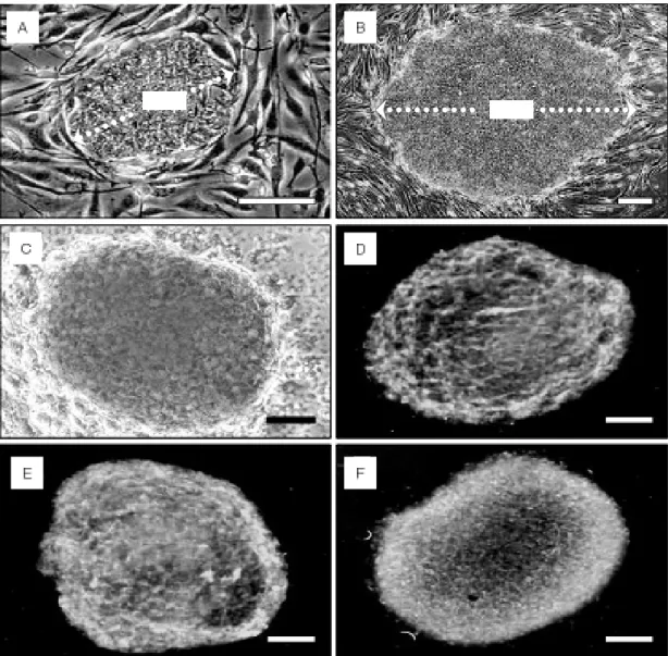
2, #3653, Becton Dickinson, NJ) feeder layer. STO cells are ready-made immortal mouse embryonic fibroblast cells that are maintained as easily as other cell lines. ICM colonies produced at 5 to 8 days (Figure 1A) were replated on a fresh STO cell feeder layer and expanded colonies were propagated further in clumps of about 100~200 hES cells about every 7 days. The ES cell culture medium consisted of Knockout-Dulbecco's modified Eagle's medium (KO-DMEM, Gibco, Grand Island, NY) supple- mented with 20% fetal bovine serum (FBS;
Hyclone, Logan UT), 1 mM glutamine, 0.1 mM -mercaptoethanol, 1% ribonucleosides, 1% none
β -
ssential amino acids (NEAAs) and 4 ng/ml human basic fibroblast growth factor (b-FGF; KOMA Biotech, Inc).
2. Freezing and thawing of hES cells
The hES cell colonies were cryopreserved by either slow-cooling using a freezing module (StrataCooler, Stratagene, USA) or MVC vitrifi- cation using a modified 0.5 ml French mini- straw (IMV; designated as MVC straw). Large hES cell colonies were subjected to a 0.05%
collagenase (Sigma) treatment for 10~15 min
and then mechanically dissected into several
small clumps using a 28 gauge needle (Figure1
C-D). For slow-cooling, about 40~50 clumps
of hES cells were transferred into a cryo-vial
containing 1 ml freezing medium (10% DMSO
in ES culture medium). The vials were transferred
into the pre-chilled (4 ℃ ) cryo-module, which
was then placed in an -80 ℃ freezer. This cryo-
module was designed to freeze mammalian cells
Figure 1. Human embryonic stem (hES) cells and dissected hES cell clumps before freezing. (A) Growing
ICM cells on STO cell feeder layer at day 7 after immunosurgery. (B) Magnification of hES cells. (C) Recovered hES cell colonies after collagenase treatment. (D) Mechanically dissected hES cell clumps. Scale bars: (A-B) 100 µm; (B) 300 µm; (D) 400 µm.at a controlled rate of 0.4~0.6 ℃ /min. After an overnight incubation at -80 ℃ , the vials were plunged into liquid nitrogen (LN
2). Vials were rapidly thawed in a water bath at 36 ℃ and their contents were poured into a petri-dish containing ES culture medium. The hES cell clumps were recovered and transferred into fresh ES culture medium. After incubating for 20~30 min, recovered hES cell clumps were plated onto a STO feeder cell layer. During MVC vitrification, hES cell clumps were frozen using a modified French mini-straw (the MVC straw; total length 2.5~3.0 cm) that was prepared by cutting off the end piece of one side (1.5~
2.0 cm). This vitrification method allows direct contact between the hES cell clump-containing medium and LN
2, which increases cooling and warming rates (>20,000 ℃ /min). Ten hES cell clumps were loaded individually onto open place (1.5~2.0 cm) of MVC straw followed by serial exposure to two vitrification solutions (VS):
(1) 10% ethylene glycol (EG) and 10% FBS in D-PBS for 5 min, and (2) 30% EG + 0.5 mol sucrose (MS) + 10% FBS in D-PBS for 30 sec.
The MVC straw was plunged directly into LN
2and about 4~5 straws were put into the pre- chilled cryo-vial, which was held in freezing cane and then plunged into LN
2. Thawing was rapidly performed in five steps (1 MS, 0.5 MS, 0.25 MS, 0.125 MS, and/or 10% FBS in D-PBS) for 1 min each. Recovered hES cell clumps were then plated onto a fresh STO feeder cell layer.
3. Assessment of hES cell survival
Survival of frozen-thawed hES cells was assessed along three criteria: hES cell clump- recovery rate after thawing, expansion of hES cell colony on STO cell feeder layer and complete survival which spend the same number of days as growing condition of non-frozen hES cells on STO cell as described in Park et al. (2004,
Figure 2A-B).
4. Stem cell marker staining
To assess AP activity, frozen-thawed hES cell colonies were fixed in 4% formaldehyde for 15 min and then stained using Fast red TR/
Naphthol AS-MX for 15 min (Figure 2C). To detect human stem cell markers, frozen-thawed ES colonies were fixed with 90% acetone in H
2O and 100% ethanol at RT for 15 min and stained with stage-specific embryonic antigen (SSEA)-4 (MC-813-70; Santa Cruz, CA) and tumor rejection antigen (TRA)-1-60 (Santa Cruz).
Antibody localization (Figure 2D-E) was detected using a rabbit anti-mouse antibody conjugated to fluorescein isothiocyanate (FITC).
5. Oct4 expression by indirect immunos- taining
To confirm that frozen-thawed and surviving hES cells were undifferentiated, Oct4 expression was assessed using an H-134 antibody (Santa Cruz). ES colonies were fixed in a 4% para- formaldehyde solution at 4 ℃ for 10 min. Anti - body localization (Figure 2F) was determined using a goat anti-rabbit antibody labeled with FITC.
6. Chromosome analysis
For chromosome analysis, control or frozen -thawed hES cells were cultured in Matrigel coated plates for 4 to 6 days. After treatment in 5% colcemid (v/v, Gibco), harvested hES cells were stained using a standard G-banding tech- nique. Karyotyping was performed using the Cytovision program (Applied Imaging Co.).
7. in vitro differentiation
For in vitro differentiation, embryoid bodies
(Figure 4A) were prepared in a bacteriological
dish (#1007, Falcon) for 5 days following colla-
genase treatment and mechanical dissection of control or frozen-thawed hES cell colonies. To initiate differentiation, EBs were treated with 1 M retinoic acid (RA) for 1 week and plated μ
onto a 0.1% gelatin-coated dish in differentia- tion medium (KO-DMEM containing 1 mM glu- tamine, 0.1 mM β -mercaptoethanol, 1% NEA A, and 10% FBS) for 3 weeks. in vitro differentia-
tion was assessed by indirect immunocytoche- mistry. For muscle-specific actin staining, cells were fixed in methanol/acetone (1:1) at 4 ℃ for 15 min, and for neuronal cell and alpha-feto protein (AFP) staining, cells were fixed in 4%
paraformaldehyde at 4 ℃ for 15 min. Monoclona l antibodies against muscle actin (1:20, Figure 4C), β -tubulin (1:100, Figure 4B, green), AFP Figure 3. Normal karyotype (46, XX) of vitrified-thawed human ES cell line (MB03).
Figure 2. in vitro survival and positive marker expression of surviving hES cells after MVC vitrification
and thawing. hES cell colony at day 3 after thawing (A). Expanded hES cell colony showing a normal growth pattern at day 9 after thawing (B). Growing speed is indicated as doubling over the dotted line(A-B). hES cell colony stained histochemically for alkaline phosphatase (C). hES cell colony immunostained
with anti-SSEA-4 (D), anti-TRA-1-60 (E), and anti-Oct-4 (F). Scale bars: 100 µm.(1:50, Figure 4D), and polyclonal antibody to glial fibrillary acid protein (GFAP; Figure 4B, red) (1:200) were supplied from Sigma (USA).
Primary antibody reactions were performed at 4℃
overnight. Antibody localization was determined by staining with a rabbit anti-mouse antibody conjugated to FITC or a goat anti-rabbit anti- body labeled with tetramethyl rhodamine iso- thiocyanate (TRITC, 1:200, Jackson Immuno Re- search, USA) for 1 h.
RESULTS
Three hES cell lines (MB01, 52 passages;
MB02, 39 passages; MB03, 31 passages) were applied for freezing test. Before freezing, hES
cells were mechanically dissected into small clumps of about 100~200 cells. During this procedure, a reasonable number of cells were discarded as single cells (Figure 1D). Survival at day 3 after thawing (Figure 2 A) of hES cells frozen by slow-cooling (20.0%, 50/250) was significantly low compared to those frozen by MVC vitrification (76.0%, 190/250) (p<0.01), suggesting that hES cells may be sensitive to chilling during cryopreservation even though they are small and have a relatively high nucleus:
cytoplasm ratio. Furthermore, almost all of the
surviving slow-cooled hES cells had very poor
growth potential, while all of the surviving
MVC-vitrified hES cells indicated rapid recovery
and demonstrated a proliferation speed similar
Figure 3. Normal karyotype (46, XX) of vitrified-thawed human ES cell line (MB03).
to that of non-frozen hES cells at day 14 after thawing. From the 3 passages, doubling time of vitrified-thawed hES cells was ~36 h. However, the frequency of spontaneous differentiation was higher in the freezing groups (MVC vitrifi- cation, 25%; slow cooling, 40%) than in the control group (15%). After 5 passages, MVC- vitrified hES cells were AP positive, SSEA-4 positive, TRA-1-60 positive, and express Oct-4.
When compared to control, the intensity of immunostaining in MVC-vitrified hES cell colony was not diminished. Karyotyping of MVC- vitrified hES cells was done in 6 passages after thawing and they were normal (MB01, 46XY;
MB02, 46XX; MB03, 46 XX). The MVC-vitrified cells also indicated in vitro differentiation chara- cteristics of three embryonic germ layers. Em- bryoid body formation is the start of differ-
entiation of hES cells into a wide variety of cell types. In vitro differentiation into some specific cell types (neuron, muscle, and AFP) was detected by indirect immunocytochemistry (Figure 4).
DISCUSSION
Efficient cryopreservation should be useful for improving the handling of hES cell lines, storing of cell stocks, and establishing ES cell banks (Reubinoff et al., 2001). Cryopreservation of hES cells may also allow limited hES cell lines to be shared between laboratories. In this study, hES cells were successfully cryopreserved by a vitrification method using a MVC straw.
These results demonstrate that hES cells are
sensitive to chilling and that vitrification is an
Figure 4. in vitro differentiation of MVC vitrified hES cells. (A) Embryoid body with characteristics of
three embryonic layers. Indirect immunocytochemistry using β-tubulin III and glial fibrillary acidic protein was used to show neuronal (green) and astrocyte (red) differentiation, respectively, indicating the presence of ectoderm (B), and expression of muscle actin (C) and α-fetoprotein(D) indicated the presence of mesoderm
(C) and endoderm (D). Scale bars: (A) 200 µm; (B) 1000 µm; (C, D) 100 µm.
effective method for their cryopreservation.
Vitrification is fundamentally different from freezing by slow cooling. Slow cooling methods attempt to maintain a delicate balance between various factors to prevent damage such as ice crystal formation, osmotic injury, and toxic effect of cryoprotectants, concentrated intracellular elec- trolytes, chilling injury, cell fracture, and altera- tions of intracellular organelles, the cytoskeleton, and cell-to-cell contacts (Massip et al., 1995).
While vitrification totally eliminates ice crystal formation, it carries an increased risk of nearly all other types of injury according to use high percent of cryoprotectants. Thus, different pro- tocols have been developed that minimize toxic, osmotic, and other injuries; use less toxic che- micals (such as ethylene glycol or/and glycerol) as cryoprotectants; and incorporate stepwise addition and/or exposure of cells to pre-cooled concentrated solutions (Rall, 1987; Kasai et al., 1990). However, the increased cooling rate decreases the potential for chilling injuries such as damage of intracellular lipid droplets, lipid- containing membranes, and the cytoskeleton by passing rapidly through the dangerous +15 to -5 ℃ zone (Zeron et al., 1999). Moreover, vitri- fication does not require expensive coolers or any special skills and can be performed re- latively quickly.
Our MVC vitrification method is very simple, easy, cost-effective, and reliable. Unlike OPS straw of Reubinoff’s method (2001), our MVC straw can be easily prepared by simple cutting- off end place (1.5~2.0 cm) of sterilized 0.5 ml French mini straw (total length 2.5~3.0 cm) with sharp scissors. Also 4~5 straws can be safely storaged in a cryo-vial before plunged into LN
2. Time to spend for freezing and thawing procedures is very short, 6~11 min and 5 min, respectively. Recovery and in vitro survival of MVC-vitrified hES cell clumps were efficient
and these results were reliable. Therefore, it suggested that MVC method could be useful freezing method to cryopreserve for chilling sensitive hES cells compared to slow-cooling method indicated very poor developmental po- tential. In this study, frozen-thawed hES cells were assessed according to three criteria: hES clump-recovery rate after thawing, expansion of hES cell colony on an STO cell feeder layer and complete survival which spend the same number of days as growing condition of non- frozen hES cells on STO cell as described in Park et al (2004). After the early day of thawing, survival of frozen-thawed hES cells by slow cooling was much lower than that for cells frozen by vitrification. Furthermore, almost all of the surviving slow-cooled hES cells had poor growth potential; while the entire surviving MVC-vitrified hES cells demonstrated a pro- liferation speed similar to that of non-frozen hES cells at day 14 after thawing. We cultured MVC vitrified cells until 22 passages and freezed them again.
Furthermore, after 5 passages post-MVC vitrification, hES cells were AP positive, SSEA -4 positive, TRA-1-60 positive, and they ex- pressed high levels of Oct-4. Further, 8 passage cultured cells had a normal karyotype. In vitro differentiation, which was initiated with RA, into certain cell types representing the three embryonic germ layers, was detected by indirect immunocytochemistry. Among growth factors, RA has been identified as a morphogenic and teratogenic compound and as a signaling mo- lecule that influences gene expression in a complex manner via a family of RA receptors (Rohwedel et al., 1999). In this study, in vitro differentiation of human ES cells into muscle cells, neuronal, and glial cells was confirmed using either monoclonal or polyclonal antibodies.
Moreover, AFP staining was positive, indicating
endodermal differentiation of ES cells.
These results demonstrate that hES cells can be successfully cryopreserved without losing their pluripotent characteristics using a newly developed MVC vitrification method. This method may help in future studies of hES cell charac- teristics and their applications.
Acknowledgement
This study was supported by a grant (01-PJ10 -PG8-01EC01-0010) of the Korea Health 21 R&D Project, Ministry of Health & Welfare Republic of Korea.
REFERENCES
1. Kasai M, Komi JH, Takakamo K, Isudera H, Sakurai T, Machida T. A simple method for mouse embryo cryopreservation in a low toxicity vitrification solution, without appreciable loss of viability. J Reprod Fertil 1990; 89: 91-7.
2. Kaufman DSD, Hanson ET, Lewis RL, Auerbach RA, Thomson JA. Hematopoietic colony-forming cells derived from human embryonic stem cells.
Proc. Natl. Acad. Sci. USA. 2001; 98: 10716- 21.
3. Kehat I, Kenyagin-Karsenti D, Snir M, Segev H, Amit M, Gepstein A, Livne E, Binah O, Itskovitz-Eldor J, Gepstein L. Human embryonic stem cells can differentiate into myocytes with structural and functional properties of cardomyo- cytes. The J. Clinic. Invest. 2001; 108(3): 407- 14.
4. Kim EY, Kim DI, Lee MG, Weon YS, Nam HK, Lee KS, Park SY, Park EM, Yoon JY, Heo YT, Cho HJ, Park SP, Chung KS, Lim JH. Establishment of bovine ovum bank: I.
Full term development of vitrified in vitro matured Hanwoo (Korean cattle) oocytes by minimum volume cooling (MVC) method. Kor.
J. Anim. Reprod. 2001; 25(1): 1-7.
5. Lane M, Forest KT, Lyons EA, Bavister BD.
Live births following vitrification of hamster embryos using a novel containerless technique.
Theriogenology 1999; 51: 167.
6. Lumelsky N, Blondel O, Laeng P, Velasco I, Ravin R, Mckay R. Differentiation of embryonic stem cells to insulin-secreting structures similar to pancreatic islets. Science 2001; 292: 1389- 94.
7. Massip A, Mermillod P, Dinnyes A. Morphology and biochemistry of in-vitro produced bovine embryos: implications for their cryopreservation.
Hum. Reprod. 1995; 10: 3004-11.
8. Park SP, Lee YJ, Lee KS, Shin HY, Cho HW, Chung KS, Kim EY, Lim JH Establishment of human embryonic stem cell lines from frozen- thawed blastocysts using STO cell feeder layers.
Hum. Reprod. 2004, in press.
9. Rall WF. Factors affecting the survival of mouse embryos cryopreserved by vitrification. Cryo- biolog,y 1987; 24: 387-402.
10. Reubinoff BE, Pera MF, Fong CY, Trounson A, Bongso A. Embryonic stem cell lines from human blastocysts: somatic differentiation in vitro. Nat.Biotech. 2000; 18: 399-404.
11. Reubinoff BE, Pera MF, Vajta G, Trounson AO. Effective cryopreservation of human em- bryonic stem cells by the open pulled straw vitrification method. Hum. Reprod. 2001; 16(10):
2187-94.
12. Rohwedel J, Guan K, Wobus AM. Induction of cellular differentiation by retinoic acid in vitro.
Cells tissues Organs 1999; 165(3-4): 190-202.
13. Solter D, Knowles B. (1975) Immunosurgery of mouse blastocyst. Proc. Natl Acad. Sci. USA.
1975; 72: 5099-102.
14. Thomson JA, Itskovitz-Eldor J, Shapiro SS, Waknitz MA, Swiergiel JJ, Marshall VS, Jones JM. Embryonic stem cell lines derived from human blastocysts. Science 1998; 282: 1145-7.
15. Xu RH, Chen X, Li DS, Li R, Addicks GC,
Glennon C, Zwaka TP, Thomson JA. BMP4 initiates human embryonic stem cell differen- tiation to trophoblast. Nat.Biotech. 2002; 20:
1261-4.
16. Zeron Y, Pearl M, Borochov A, Arav A. Kinetic and temporal factors influence chilling injury
to germinal vesicle and mature bovine oocytes.
Cryobiology 1999; 38: 35-42.
17. Zwaka TP, Thomson JA. Homologous recombi- nation in human embryonic stem cells. Nat.
Biotech. 2003; 10: 1038.