Received: May 31, 2016, Revised: July 4, 2016, Accepted: July 4, 2016, Published online: August 5, 2016
Corresponding author: Hisayoshi Suma, Suma Heart Clinic, Suma Square, Hillside West B-002, 13-4 Hachiyamacho, Shibuya-ku, Tokyo 150-0035, Japan
(Tel) 81-3-6416-4567 (Fax) 81-3-6416-4567 (E-mail) [email protected]
© The Korean Society for Thoracic and Cardiovascular Surgery. 2016. All right reserved.
This is an open access article distributed under the terms of the Creative Commons Attribution Non-Commercial License (http://creativecommons.org/
licenses/by-nc/4.0) which permits unrestricted non-commercial use, distribution, and reproduction in any medium, provided the original work is properly cited.
Throughout its 30-year history, the right gastroepiploic artery (GEA) has been useful for in situ grafts in cor- onary artery bypass grafting (CABG). The early graft patency rate is high, and the late patency rate has im- proved by using the skeletonized GEA graft and proper target selection, which involves having a target coro- nary artery with a tight >90% stenosis. Total arterial revascularization with the internal thoracic artery and GEA grafts is an option for achieving better outcomes from CABG procedures.
Key words: 1. Coronary artery disease 2. Ischemic heart disease 3. Coronary artery bypass graft 4. Myocardial revascularization 5. Gastroepiploic artery
Introduction
The history of coronary artery bypass grafting (CABG) exceeds 50 years. The saphenous vein and various arterial grafts have been used and evaluated for their functional efficacy; however, the choice of conduit has been a major concern for cardiac sur- geons desiring a good surgical outcome [1]. It is clear that the internal thoracic artery (ITA) is superi- or to the saphenous vein in terms of the patency rate and patients’ long-term outcomes [2]. Conse- quently, intensive investigations were undertaken to find a new arterial conduit for CABG.
Before CABG, a procedure in which the graft is di- rectly anastomosed to the coronary artery, was in- stituted, the gastroepiploic artery (GEA) was used for indirect myocardial revascularization (Vineberg pro- cedure) for the posterior or inferior wall of the heart [3]. The aorta-coronary bypass using the saphenous
vein graft was successfully introduced by Favaloro [4] in the Cleveland Clinic in 1967. Afterwards, anas- tomosis of the GEA to the right coronary artery was attempted by Edwards in the early 1970s [5]. The GEA graft then emerged as an alternative arterial conduit for CABG, and pioneering articles were pub- lished in 1987 with successful clinical results by Pym et al. [6] in Canada and Suma et al. [7] in Japan.
Since that time, the GEA graft gained attention as an attractive arterial conduit for CABG.
Anatomy, Histology, and Physiology
The right GEA is one of four arteries that supply blood to the stomach. It is the largest terminal branch of the gastroduodenal artery, which originates from the common hepatic artery (Fig. 1).
It is important to know that the GEA arises from the superior mesenteric artery in rare occasions [8].
Fig. 1. (A) Schematic and (B) angio- gram of the gastroepiploic artery (GEA).
If a cardiologist was not aware of this, hepatic or se- lective gastroduodenal angiography to seek the GEA graft patency after the operation can fail to find a patent GEA graft. The right GEA reaches more than half of the greater curvature of the stomach in about 90% of people [7,9], and terminates in a v arying fa- shion with or without communication to the left GEA [10].
Histologically, the wall thickness of the GEA is sim- ilar to the ITA [11], but has different characteristics from the ITA in terms of spasmogeneity. The GEA contains many smooth muscle cells in the media, whereas the ITA has rich elastic fibers in media.
Therefore, the GEA is considered a muscular artery and the ITA is an elastic artery. This difference is quite important in clinical situations during and after surgery, because GEA spasm readily arises from sur- gical manipulation whereas the ITA hardly becomes spastic.
Arteriosclerosis of the GEA was found to be less frequent than in the coronary artery [12]. In our previous study, the GEA and ITA used in CABG were examined pathologically and moderate to severe arte- riosclerosis was noted in 8% of GEA grafts and 1%
of ITA grafts [13]. Although the GEA had arterio- sclerosis more frequently than the ITA, the GEA may be an arteriosclerosis-resistant artery, as all patients included in the study had severe coronary artery diseases.
Various functional studies have been published as the GEA has been increasingly used for CABG. In re- sponse to vasoactive agents, the GEA contracts with ergonovine, serotonin, and phenylephrine, and the ITA reacted similarly [14]. Another study demon-
strated that the GEA contractions by potassium chlor- ide, serotonin, and norepinephrine were stronger than the ITA contractions [15]. These results suggest that prevention of spasm provoked by platelet aggrega- tors, adrenergic stimulation, or depolarizing agents is important in GEA grafting [16].
Interestingly, the responses of the GEA to hista- mine were different than in the ITA. Histamine di- lates the GEA and contracts the ITA [17,18]. Our group introduced an implantable ultrasonic Doppler miniprobe for the patients undergoing CABG, which was attached to the GEA and other grafts at the end of the operation. Then the skin was closed and the patient started postoperative rehabilitation. During exercise, drug administration, and meals, the graft blood flow was monitored. A week postoperativ ely, the probe was removed without injury. We found that the GEA flow increased after meals [19]. This finding is compatible with the previously mentioned GEA dilatation by histamine. We also found that the GEA was more capable of prostacyclin production than the saphenous vein [20].
Surgical Procedure
The midline incision was about 5 cm longer than the usual sternal splitting incision of ordinary CABG.
Following a pericardiotomy, the peritoneum was open and the stomach was pulled out to the surgical field.
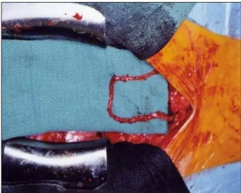
The GEA was detected by palpation to determine size and length. It is important to touch the GEA softly because it readily contracts under mechanical stimu- lation. Then the GEA was mobilized from the greater curvature of the stomach using electric cautery or an
Fig. 2. Detachment of the gastroepiploic artery from the stomach.
Fig. 3. Skeletonized gastroepiploic artery graft.
ultrasonic scalpel as a pedicle (Fig. 2), or in a ske- letonized fashion without any surrounding tissue (Fig. 3).
The skeletonized GEA can be used as a free graft if necessary [21]. The skeletonized GEA produces a lon- ger graft length and is easier to use for sequential anastomoses. Also, Y- or I-composite grafts using a free GEA graft in combination with the in situ left or right ITA graft further increase the possibility of total arterial complete myocardial revascularization.
There was a concern about gastric ischemia due to GEA detachment from the stomach in the early peri- od when GEA grafting was first instituted. Therefore, we measured gastric mucosal blood flow during sur- gery. The endoscopic Doppler flow study clearly de- monstrated that no gastric ischemia occurred in any part of the stomach following GEA detachment [22].
After mobilizing the GEA from the pylorus to more than half of the greater curvature, its distal end was cut and a diluted papaverine solution was slowly in- jected into the GEA lumen to relieve spasm. The in- traluminal papaverine procedure is effective to obtain a good caliber and maximum flow of the GEA for successful anastomosis. Accordingly, perioperative use of diltiazem is essential.
The GEA and the coronary artery anastomosis are performed under a cardioplegic arrest or an off-pump beating heart. Finally, the GEA is fixed to the epicar- dium properly to avoid kinking or twisting. Fig. 4 shows a complete arterial CABG procedure with bi- lateral ITA and GEA in situ grafts.
Indication For Gastroepiploic Artery Grafting
The GEA is most suitable for grafting the distal right coronary artery and the posterior descending artery because this site is the nearest for the in situ GEA graft and the most distant from the in situ right ITA graft.
The distal circumflex artery is also a possible tar- get for the GEA graft. The anterior descending artery is a common site for the left ITA, but it is an option for the GEA grafting target when the ITA is un- available (Fig. 5).
The GEA is advantageous for the patient with athe- rosclerotic ascending aorta because the in situ arte- rial grafts are necessary for the aortic no-touch tech- nique [23]. In cases of redo CABG, the GEA is useful because the abdomen is a v irgin area, which makes GEA preparation easy before re-sternotomy.
GEA grafts are not recommended in very elderly patients or those with extreme obesity. In addition, unstable hemodynamics during emergency surgery or proposed future abdominal operations are a contra- indication for the in situ GEA graft.
Clinical Results
Since 1987 when Pym et al. [6] and Suma et al.
[7] reported their successful clinical applications of GEA grafting, international investigators have started using the GEA graft, and clinical and basic research
Fig. 4. (A, B) Coronary artery by- pass grafting with in situ bilateral internal thoracic artery and gastro- epiploic artery graft.
Fig. 5. The in situ GEA graft anasto- mosed to various sites of the coro- nary artery. (A) Main RCA. (B) Posterior descending artery. (C) Left anterior descending artery. (D) Circumflex. RCA, right coronary ar- tery; GEA, gastroepiploic artery.
Ejection fraction (%) 51.6
No. of grafts 3.4
Concomitant grafts
Internal thoracic artery 1,458 (97)
Radial artery 133 (9)
Saphenous vein 823 (55)
Site of GEA anastomosis
Left anterior descending 70
Diagonal 10
Left circumflex 319
Right coronary artery 1,238
Mode of GEA graft
In situ single 1,346
Free single 52
Free composite 56
Sequential 76
Operative death 18 (1.2)
Values are presented as number, mean (range), or number (%), unless otherwise stated.
GEA, gastroepiploic artery.
without a GEA graft in CABG. There was no increase in postoperative comorbidity, particularly abdominal complications, with use of the GEA graft [22]. At 5 years after our first GEA graft case, we presented the midterm results in 200 patients [31]. The operative mortality and early GEA patency rate were identical in the two groups. Concerning the flow capacity of the GEA graft, pre- and postoperative stress my- ocardial scintigraphy showed that the washout ratio of the GEA grafted area was significantly increased after surgery.
Following the successful initial period, the GEA graft was used more extensively and its long-term results have been reported. For the investigators who performed CABG combined with bilateral ITA and GEA grafts, 10- to 15-year postoperative survival rates were demonstrated to be excellent [32-36]. In our 20-year experience, 5-, 10- and 15-year actuarial sur- vival rates were 91.7%, 81.4%, and 71.3%, respec- tively, in 1,118 follow-up patients [36].
Table 1 shows the patients’ characteristics and surgical results of our 30-year experience with GEA grafting. In patients with triple vessel disease having bilateral ITA to the left coronary artery system, the patients with a GEA graft to the right coronary ar- tery resulted in significantly better late survival than patients who received the saphenous vein graft [37,38]. In a comparison between free GEA and free right ITA grafts, Hwang et al. [39] demonstrated that the 5- and 10-year event-free survival rates were similar in those two grafts used for a composite Y graft with the left ITA.
Gastroepiploic Artery Graft Patency
Regarding the patency rate of the GEA graft, a tight stenosis of the target coronary artery is im- portant to maximize the potential for patency. The American College of Cardiology Foundation/American
Heart Association guideline for CABG stated that ar- terial grafting to the right coronary artery is contra- indicated for patients with <90% stenosis of the na- tive vessel [40].
In our experience, the cumulative patency rate of the GEA graft was 97.1% at 1 month, 92.3% at 1 year, 85.5% at 5 years, 80.9% at 7 years, and 66.5%
at 10 years following surgery [36]. This relatively low 10-year patency rate has been improved by us- ing a skeletonized GEA graft only to the coronary ar- tery with a tight >90% stenosis. Using this approach, Suzuki et al. [41] reported 97.8%, 94.7%, and 90.2%
cumulative patency rates immediately, and 5 and 8 years after surgery, respectively.
On the other hand, a network meta-analysis of randomized controlled trials reported that the GEA graft has a higher risk of graft occlusion compared with other types of conduits [42]. While heavy GEA
Fig. 6. The GEA graft anastomosed to the PDA at 20 years after coronary artery bypass grafting. GEA, gastroepiploic artery; PDA, posterior descending artery.
users and non-users have not agreed on the exact decision tree for choosing the conduit to the right coronary artery, those who apply GEA grafts with skeletonization and proper target selection will find the GEA to be reliable. Fig. 6 demonstrates a widely patent GEA graft anastomosed to the posterior de- scending artery at 20 years after surgery.
Conclusion
In conclusion, the GEA is a great, elegant, and at- tractive conduit for CABG.
Conflict of interest
No potential conflict of interest relevant to this ar- ticle was reported.
References
1. Gaudino M, Taggart D, Suma H, Puskas JD, Crea F, Massetti M. The choice of conduits in coronary artery bypass surgery.
J Am Coll Cardiol 2015;66:1729-37.
2. Loop FD, Lytle BW, Cosgrove DM, et al. Influence of the in- ternal-mammary-artery graft on 10-year survival and oth- er cardiac events. N Engl J Med 1986;314:1-6.
3. Bailey CP, Hirose T, Brancato R, Aventura A, Yamamoto N.
Revascularization of the posterior (diaphragmatic) por- tion of the heart. Ann Thorac Surg 1966;2:791-805.
4. Favaloro RG. Saphenous vein autograft replacement of se-
vere segmental coronary artery occlusion: operative technique. Ann Thorac Surg 1968;5:334-9.
5. Mills NL, Everson CT. Right gastroepiploic artery: a third arterial conduit for coronary artery bypass. Ann Thorac Surg 1989;47:706-11.
6. Pym J, Brown PM, Charrette EJ, Parker JO, West RO.
Gastroepiploic-coronary anastomosis: a viable alternative bypass graft. J Thorac Cardiovasc Surg 1987;94:256-9.
7. Suma H, Fukumoto H, Takeuchi A. Coronary artery bypass grafting by utilizing in situ right gastroepiploic artery: ba- sic study and clinical application. Ann Thorac Surg 1987;44:394-7.
8. Suma H, Sato H. The in situ right gastroepiploic artery graft via the superior mesenteric artery. J Thorac Cardiovasc Surg 1989;98:1150.
9. Hannoun L, Le Breton C, Bors V, Helenon C, Bigot JM, Parc R. Radiological anatomy of the right gastroepiploic artery.
Anat Clin 1984;5:265-71.
10. Yamato T, Hamanaka Y, Hirata S, Sakai K. Esophagoplasty with an autogenous tubed gastric flap. Am J Surg 1979;
137:597-602.
11. Van Son JA, Smedts F, Vincent JG, van Lier HJ, Kubat K.
Comparative anatomic studies of various arterial conduits for myocardial revascularization. J Thorac Cardiovasc Surg 1990;99:703-7.
12. Larsen E, Johansen A, Andersen D. Gastric arteriosclerosis in elderly people. Scand J Gastroenterol 1969;4:387-9.
13. Suma H, Wanibuchi Y, Furuta S, Isshiki T, Yamaguchi T, Takanashi R. Comparative study between the gastro- epiploic and the internal thoracic artery as a coronary by- pass graft. Size, flow, patency, histology. Eur J Cardio- thorac Surg 1991;5:244-7.
14. Koike R, Suma H, Kondo K, et al. Pharmacological response of internal mammary artery and gastroepiploic artery.
Ann Thorac Surg 1990;50:384-6.
15. Dignan RJ, Yeh T Jr, Dyke CM, et al. Reactivity of gastro- epiploic and internal mammary arteries: relevance to cor- onary artery bypass grafting. J Thorac Cardiovasc Surg 1992;103:116-22.
16. Suma H. Spasm of the gastroepiploic artery graft. Ann Thorac Surg 1990;49:168-9.
17. O’Neil GS, Chester AH, Schyns CJ, Tadjkarimi S, Pepper JR, Yacoub MH. Vascular reactivity of human internal mammary and gastroepiploic arteries. Ann Thorac Surg 1991;52:1310-4.
18. Favaloro RG, Effler DB, Sheldon WC. Successful pulmonary embolectomy: report of a case. Cleve Clin Q 1967;34:119-25.
19. Takayama T, Suma H, Wanibuchi Y, Tohda E, Matsunaka T, Yamashita S. Physiological and pharmacological responses of arterial graft flow after coronary artery bypass graft- ing measured with an implantable ultrasonic Doppler miniprobe. Circulation 1992;86(5 Suppl):II217-23.
20. Oku T, Yamane S, Suma H, et al. Comparison of prostacy- clin production of human gastroepiploic artery and saphe- nous vein. Ann Thorac Surg 1990;49:767-70.
coronary artery bypass grafting. Aust N Z J Surg 1987;57:
317-21.
25. Lytle BW, Cosgrove DM, Ratliff NB, Loop FD. Coronary ar- tery bypass grafting with the right gastroepiploic artery. J Thorac Cardiovasc Surg 1989;97:826-31.
26. Suma H, Takeuchi A, Hirota Y. Myocardial revasculariza- tion with combined arterial grafts utilizing the internal mammary and the gastroepiploic arteries. Ann Thorac Surg 1989;47:712-5.
27. Kusukawa J, Hirota Y, Kawamura K, et al. Efficacy of coro- nary artery bypass surgery with gastroepiploic artery: as- sessment with thallium 201 myocardial scintigraphy.
Circulation 1989;80(3 Pt 1):I135-40.
28. Beretta L, Lemma M, Vanelli P, et al. Gastroepiploic ar- tery free graft for coronary bypass. Eur J Cardiothorac Surg 1990;4:323-7.
29. Takeuchi Y, Gomi A, Okamura Y, Mori H, Nagashima M.
Coronary revascularization in a child with Kawasaki dis- ease: use of right gastroepiploic artery. Ann Thorac Surg 1990;50:294-6.
30. Isomura T, Hisatomi K, Asoh S, et al. Revascularization with the right gastroepiploic artery in Kawasaki’s disease.
J Thorac Cardiovasc Surg 1990;100:796-8.
31. Suma H, Wanibuchi Y, Terada Y, Fukuda S, Takayama T, Furuta S. The right gastroepiploic artery graft: clinical and angiographic midterm results in 200 patients. J Thorac Cardiovasc Surg 1993;105:615-22.
32. Nishida H, Tomizawa Y, Endo M, Koyanagi H, Kasanuki H.
Coronary artery bypass with only in situ bilateral internal thoracic arteries and right gastroepiploic artery. Circulation 2001;104(12 Suppl 1):I76-80.
33. Hirose H, Amano A, Takanashi S, Takahashi A. Coronary ar- tery bypass grafting using the gastroepiploic artery in
36. Suma H, Tanabe H, Takahashi A, et al. Twenty years expe- rience with the gastroepiploic artery graft for CABG.
Circulation 2007;116(11 Suppl):I188-91.
37. Glineur D, D’hoore W, Price J, et al. Survival benefit of multiple arterial grafting in a 25-year single-institutional experience: the importance of the third arterial graft. Eur J Cardiothorac Surg 2012;42:284-90.
38. Suzuki T, Asai T, Matsubayashi K, et al. In off-pump sur- gery, skeletonized gastroepiploic artery is superior to sa- phenous vein in patients with bilateral internal thoracic arterial grafts. Ann Thorac Surg 2011;91:1159-64.
39. Hwang HY, Cho KR, Kim KB. Equivalency of right internal thoracic artery and right gastroepiploic artery composite grafts: five-year outcomes. Ann Thorac Surg 2013;96:
2061-8.
40. Hillis LD, Smith PK, Anderson JL, et al. 2011 ACCF/AHA Guideline for Coronary Artery Bypass Graft Surgery: a re- port of the American College of Cardiology Foundation/
American Heart Association Task Force on Practice Guide- lines. Developed in collaboration with the American Asso- ciation for Thoracic Surgery, Society of Cardiovascular Anesthesiologists, and Society of Thoracic Surgeons. J Am Coll Cardiol 2011;58:e123-210.
41. Suzuki T, Asai T, Nota H, et al. Early and long-term pa- tency of in situ skeletonized gastroepiploic artery after off-pump coronary artery bypass graft surgery. Ann Thorac Surg 2013;96:90-5.
42. Benedetto U, Raja SG, Albanese A, Amrani M, Biondi- Zoccai G, Frati G. Searching for the second best graft for coronary artery bypass surgery: a network meta-analysis of randomized controlled trials. Eur J Cardiothorac Surg 2015;47:59-65.