Introduction
Total knee arthroplasty (TKA) has emerged as a durable ap
proach to replicate the healthy osseous anatomy and restore natural knee joint kinematics. However, recent expansion of indi
cations for TKA in a younger and more active patient population demands further improvement for greater durability1,2) and func
tion3,4). Many new implant designs have incorporated modifica
tions to achieve the goal of better clinical outcomes and implant longevity. However, these designs do not guarantee improved results, and several new prostheses were reported to have unex
pected problems or unsatisfactory outcomes57). Therefore, with any new design, it is prudent to maintain a close surveillance of its performance and safety from the outset. Although a short
term outcome analysis is limited in predicting longterm success, information from shortterm studies is still valuable for early de
tection of adverse events, if any, originating from new prostheses.
Posterior stabilized (PS) knee prostheses have cam and post mechanisms to provide stability in flexion, increase range of mo
tion (ROM), and improve the quadriceps lever arm. Moreover, multiple midterm and longterm followup studies have evi
Early Clinical Outcomes of a New Posteriorly Stabilized Total Knee Arthroplasty Prosthesis: Comparisons with Two Established Prostheses
Nimesh P. Jain, MS
1, Sung Yup Lee, MD
2, Vivek M. Morey, MS
3, Suri Chong, MD
2, Yeon Gwi Kang, MS
2, and Tae Kyun Kim, MD
21Department Orthopaedics, North Cumbria University Hospitals NHS Trust, Carlisle, UK; 2Department of Orthopaedic Surgery, Seoul National University Bundang Hospital, Seongnam, Korea; 3Department of Orthopedics, Datta Meghe Institute of Medical Sciences, Wardha, India
Purpose: We sought to determine whether early clinical performance of new posterior stabilized (PS) knee system, the VegaPS (Aesculap), is better than that of two established total knee arthroplasty (TKA) prostheses, the E.motionPS (Aesculap) and the Genesis II (Smith & Nephew) in terms of functional outcomes, patient satisfaction, and incidence of adverse events.
Materials and Methods: We compared the clinical outcomes of 206 consecutive TKAs using VegaPS with those of 205 TKAs using E.motionPS and 216 TKAs using Genesis II at 2 years of followup.
Results: Overall, the knees with the VegaPS had better functional outcome scores than the knees with the E.motionPS, but had similar outcome scores to the knees with the Genesis II, as evident from the American Knee Society knee score (94.2 vs. 92.5 vs. 93.2), Western Ontario McMaster Universities Osteoarthritis (WOMAC) stiffness index (1.8 vs. 2.3 vs. 2.0), WOMAC function index (11.8 vs. 16.8 vs. 18.5), Short Form 36 (SF36) physical component summary score (41.9 vs. 39.3 vs. 41.6), and SF36 mental component summary score (50.0 vs. 45.8 vs. 46.9). Patient satisfaction was higher in the VegaPS and Genesis II groups than the E.motionPS group. No notable group differences were found in terms of the incidence of adverse events.
Conclusions: The VegaPS, a newly developed PS fixed bearing prosthesis, had comparable or superior clinical performance in comparison with the two established fixed or mobile bearing PS prostheses.
Keywords: Arthroplasty, Replacement, Knee, Prosthesis design, Outcome assessment pISSN 2234-0726 · eISSN 2234-2451
Knee Surgery & Related Research
Received September 1, 2016; Revised (1st) January 6, 2017;
(2nd) March 12, 2017; Accepted April 7, 2017 Correspondence to: Tae Kyun Kim, MD
Department of Orthopaedic Surgery, Joint Reconstruction Center, Seoul National University Bundang Hospital, 82 Gumiro 173 beongil, Bundanggu, Seongnam 13620, Korea
Tel: +82317877196, Fax: +82317874056 Email: [email protected]
180
This is an Open Access article distributed under the terms of the Creative Commons Attribution NonCommercial License (http://creativecommons.org/licenses/bync/4.0/) which permits unrestricted noncommercial use, distribution, and reproduction in any medium, provided the original work is properly cited.
Copyright © 2017 KOREAN KNEE SOCIETY www.jksrr.org
denced good function, movement, and survivorship of PS knee prostheses8,9). However, certain drawbacks have been reported with PS knee prostheses, namely large intercondylar bone stock removal to accommodate the femoral cam10), dislocation of the cam11), patellar clunk syndrome12), and wear of the tibial post13). Subsequently, there have been many attempts to modify the de
signs of PS knee prostheses in order to address their problems as well as to achieve more physiologic and stable knee kinematics.
Several studies in the past have shown that changes in compo
nent geometry and modularity with the PS designs have led to improved short and longterm results1416) in addition to permit
ting greater surgical flexibility in severe osteoarthritis cases16). A new PS knee prosthesis system, VegaPS (Aesculap; B. Braun, Tuttlingen, Germany), is designed to improve TKA outcomes. Its distinguishing characteristics include a low profile intercondylar box, reduced posterior condyle length, and narrowed mediolater
al width of the femoral component, along with an anterior cutout and increased post inclination of the tibial insert. It was designed to achieve high performance, including high flexion and shape optimization to avoid overhang. However, no clinical informa
tion regarding this new prosthesis is available yet to validate its functional performance and safety. We sought to determine whether early clinical performance of this new prosthesis is bet
ter than two established TKA prostheses, the E.motionPS and Genesis II, in terms of functional outcomes, patient satisfaction, and incidence of adverse events. We had two hypotheses: 1) The functional outcomes and patient satisfaction in patients having VegaPS prostheses implanted are better than the outcomes and satisfaction in patients having the previously established PS type TKA prostheses implanted. 2) There is no increased incidence of adverse events in the knees replaced with the VegaPS.
Materials and Methods
Seven hundred and eightysix TKAs were performed by one surgeon (KTK) between May 2006 and May 2012. These in
cluded 240 TKAs using a new PS knee system, the VegaPS (Fig.
1), along with 546 consecutive TKAs performed for advanced os
teoarthritis using two established PS knee systems, the E.motion
PS (Aesculap), a mobile bearing PS prosthesis (257 knees), and the Genesis II (Smith & Nephew, Memphis, TN, USA), a fixed bearing PS prosthesis (289 knees). All of the VegaPS TKAs were performed in the years after we completed the 257 E.motionPS TKAs. In contrast, the 289 Genesis II TKAs were done through
out the entire study period. The implant selection was at the sur
geon’s discretion without any selection criteria. We included only
patients with a diagnosis of primary osteoarthritis, scheduled for unilateral TKA, between May 2006 and May 2012. We excluded patients with a diagnosis of secondary osteoarthritis or a past medical history of previous hip or spine surgery, in an attempt to reduce potential outcome confounders. A total of 159 patients, including 16 patients with a history of prior hip and spine prob
lems, 24 patients with secondary arthritis, 89 patients with other systemic comorbidities that might interfere with the benefits of the replaced knee, 15 patients with postoperative complications affecting outcomes, and 15 patients who died due to unrelated causes, were excluded from the study. Thus, data from a total of 627 patients were used for analysis (VegaPS, n=206; E.motionPS, n=205; and Genesis II, n=216). In the VegaPS group, the mean age of the patients was 70.7 years and the number of female pa
tients was 198 (96.1%). In the E.motionPS group, the mean age of the patients was 68.3 years, and the number of female patients was 196 (95.6%), while in the Genesis II group, the mean age was
Fig. 1. The photographs show the VegaPS prosthesis. The anterior and posterior narrowing of the femoral component with a low profile inter
condylar box minimizes interference with the surrounding soft tissues.
Fiftyfive degrees posterior inclination of the post acts to avoid post
edge loading for better load distribution, prevents impingement with the extensor mechanism, and increases resistance to dislocation. The deepened anterior cutout of the insert allows smooth articulation with the patellar tendon.
69.7 years, and the number of female patients was 203 (98.5%).
This study was approved by the Institutional Review Board of our hospital, and all patients provided informed consent concerning the use of medical records.
All surgeries were performed by a single surgeon, using the me
dial parapatellar approach. The patella was routinely resurfaced and all implants were fixed with cement (Palacos; Heraeus Kulzer GmbH, Hanau, Germany). We used measured resection, com
bined with gap balancing techniques without navigation.
There were not any significant differences in surgical techniques between the mobile and fixed bearing instruments. After surgery, a compressive dressing was applied, with immobilization of the knee for 24 hours. The knees were then placed in a continuous passivemotion machine. On the second postoperative day, the drain and compressive dressing were removed, and all patients were encouraged to walk with crutches or a walker and started active and passive ROM exercises. The knee ROM exercises and weight bearing were gradually increased.
All clinical information was prospectively collected, using pre
designed data sheets, and maintained in a database by an inde
pendent investigator (LSY). The retrospective evaluation of all the data was done with a minimum followup of 2 years. The clinical information gathered included demographic data, preoperative
clinical status, and postoperative clinical status at 12 months and 2 years. Patient evaluation using the outcome scales was done at 2 years after surgery. For this purpose, knee ROM, American Knee Society (AKS) scores17), Western Ontario McMaster Universities Osteoarthritis Index (WOMAC) scales18), and Short Form 36 (SF36) scores19) were used. In addition, patient satisfaction was evaluated at the 2 year followup using a 1–4 point Likert scale, based on the grading system developed by the British Orthopae
dic Association, which is divided into four levels, i.e., enthusiastic, satisfied, not committed, and disappointed20). The knee motion arc was expressed in terms of flexion contracture and maximum flexion angle. An independent investigator (LSY) measured the flexion contracture and maximum flexion angles to the nearest 5°
by using a standard (38 cm) clinical goniometer, with the patient in the supine position.
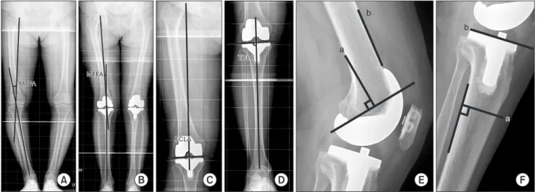
The radiographic assessment was performed at 2 years after surgery to measure five parameters: mechanical tibiofemoral angle (MTFA), coronal femoral prosthesis alignment, coronal tibial prosthesis alignment, sagittal femoral prosthesis align
ment, and sagittal tibial prosthesis alignment (Fig. 2). Two of the authors took all radiographic measurements using fulllength weight bearing anteroposterior radiographs, and lateral radio
graphs of the operated limb. To achieve the same foot rotation
MTFA MTFA
TCLA
TCLA a
b
A B C D E F
MTFA MTFA
b
a FCLA
FCLA
Fig. 2. (A) The radiograph shows the preoperative mechanical tibiofemoral angle (MTFA). (B) The radiograph shows the postoperative MTFA. (C) The radiograph shows the femoral component lateral angle (FCLA), defined as the lateral angle between the femoral mechanical axis and a line con
necting the most distal points of the medial and lateral condyles of the femoral component. (D) The radiograph shows the the tibial component lat
eral angle (TCLA), defined as the lateral angle between the tibial mechanical axis and a line parallel to the top surface of the tibial component. (E) The radiograph shows the femoral component flexion angle. To assess the sagittal alignment of the femoral component, we measured the angle between the line perpendicular to the tangent line of the femoral component box (a) and the line connecting the anterior cortical point of 10 cm proximal to the joint line and the anterior point of the metaepiphysis junction before metaphyseal diverging (b). (F) The radiograph shows the tibial component posterior slope. To assess the sagittal alignment of the tibial component, we measured the angle between the line parallel to the upper surface of the tibial tray (b) and the line perpendicular to the tangent line of the anatomical axis (the posterior cortical line of tibia at two points, 5 cm and 15 cm distal to the joint line) of the proximal tibia (a).
angle, a reference template was positioned on the platform of our plain radiographic system. All radiographic images were digitally acquired using a picture archiving and communication system (PACS; Infinitt, Seoul, Korea). Assessments were performed on a 20inch LCD monitor in portrait mode using the PACS software.
To determine intra and interobserver reliabilities of the radio
graphic assessments, the two investigators performed all radio
graphic assessments in 20 randomly selected radiographs twice, with an interval of 1 week. The intra and interobserver reliabili
ties were then evaluated using intraclass correlation coefficients, which were found to be >0.80 for all measurements. Since the measurements were judged as reliable, measurements taken by a single investigator (LSY) were used in the final analyses.
In order to gauge overall limb alignment, we measured the MTFA of the knee, which was defined as the angle formed by the intersection between the mechanical axis of the femur (the line from the femoral head center to the femoral intercondylar notch center) and the tibia (the line from the ankle talus center to the
center of tibial spine tips) (Fig. 2A and B). A negative value was given to the angle of the knees in varus alignment. To assess the coronal alignment of the femoral component, we measured the femoral component lateral angle (FCLA), defined as the lateral angle between the femoral mechanical axis and a line connecting the most distal points of the medial and lateral condyles of the femoral component (Fig. 2C). The coronal femoral prosthesis alignment was calculated by subtracting the FCLA from 90, and accordingly, a negative value was given to the angle of varus ori
entation of the femoral component. To assess the coronal align
ment of the tibial component, we measured the tibial component lateral angle, (TCLA) defined as the lateral angle between the tibial mechanical axis and the line parallel to the top surface of the tibial component (Fig. 2D). We calculated the coronal tibial prosthesis alignment by subtracting the TCLA from 90°, and accordingly, a negative value was assigned to the angle for varus orientation of the tibial component. To assess the sagittal align
ment of the femoral component, we measured the angle between
Table 1. Comparison of Demographic Characteristics, Preoperative Mechanical Axis (MA) and Outcome Scales among the VegaPS, E.motionPS, and Genesis II Groups
Variable VegaPS
(n=206) E.motionPS
(n=205) Genesis II
(n=216) pvalue pvaluea)
PV VG PG
Sex (female, %) 198 (96.1) 196 (95.6) 203 (98.5) 0.199
Age (yr) 70.7 (5.6) 68.3 (5.2) 69.7 (5.8) <0.001 <0.001 >0.05 0.033
Height (cm) 151.5 (6.0) 151.9 (6.0) 151.5 (5.8) 0.733
Weight (kg) 61.5 (8.7) 63.0 (8.2) 63.9 (9.9) 0.023 >0.05 0.020 >0.05
Body mass index (kg/m2) 26.8 (3.5) 27.3 (3.0) 27.8 (3.8) 0.013 >0.05 0.010 >0.05
Preoperative MA (°) –10.7 (4.8) –10.1 (5.1) –11.6 (6.7) 0.827 Motion arc (°)
Flexion contracture 11.2 (6.3) 9.9 (6.5) 12.0 (6.6) 0.006 (0.022) 0.152 (0.223) 0.005 (0.019) 0.685 (1.000) Maximum flexion 135.6 (13.8) 135.9 (13.5) 135.0 (14.2) 0.527 (0.782)
AKS score
Knee 46.6 (9.5) 45.9 (8.1) 45.7 (8.3) 0.586 (0.745)
Function 58.4 (9.8) 57.5 (10.0) 56.5 (13.2) 0.250 (0.221) WOMAC
Pain 9.8 (4.3) 12.2 (4.6) 11.0 (4.5) <0.001 (<0.001) <0.001 (<0.001) 0.032 (0.036) 0.016 (0.017) Stiffness 4.4 (2.1) 4.9 (2.0) 4.4 (1.9) 0.011 (0.013) 0.031 (0.050) 0.021 (0.019) 1.000 (1.000) Function 34.1 (13.9) 41.2 (14.5) 39.6 (12.2) <0.001(<0.001) <0.001 (<0.001) 0.813 (0.667) <0.001 (<0.001) Short Form 36
PCS 31.6 (8.0) 29.8 (7.1) 30.1 (8.1) 0.051 (0.053)
MCS 48.4 (11.5) 41.1 (12.3) 41.6 (12.3) <0.001 (<0.001) <0.001 (<0.001) 1.000 (1.000) <0.001 (<0.001) Values are presented as mean (standard deviation).
PV: E.motionPS vs. VegaPS, PG: E.motionPS vs. Genesis II, VG: VegaPS vs. Genesis II, AKS: American Knee Society, WOMAC: Western Ontario McMaster Universities Osteoarthritis Index, PCS: physical component summary, MCS: mental component summary.
a)pvalue is listed along with the adjusted pvalue (after taking the preoperative differences as covariates).
the line perpendicular to the tangent line of the femoral compo
nent box and the line connecting the anterior cortical point of 10 cm proximal to the joint line and the anterior point of the meta
epiphysis junction before metaphyseal diverging (Fig. 2E). A negative value was given to the angle for extension of the femoral component, while a positive value was given to the angle for flex
ion of the component. To assess the sagittal alignment of the tibi
al component, we measured the angle between the line parallel to the upper surface of the tibial tray and the line perpendicular to the tangent line of the anatomical axis (the posterior cortical line of the tibia at two points, 5 cm and 15 cm distal to the joint line) of the proximal tibia (Fig. 2F). A positive value was given to the posterior slope of the tibial component and a negative value was given to the anterior slope of the component.
Statistical analyses were carried out using SPSS ver. 21.0 (IBM Co., Armonk, NY, USA), and a pvalue <0.05 was considered significant. Knees with the VegaPS were compared to knees with the E.motionPS and knees with the Genesis II for functional out
come scales and incidence of adverse events. Patient satisfaction was also measured and compared. The KolmogorovSmirnov test was used to confirm that the clinical outcome scores, including maximal flexion, were normally distributed. As preoperative dif
ferences could have confounding effects on postoperative func
tional outcomes, all three groups were compared with respect to their demographic characteristics and preoperative clinical status (Table 1). There were a few parameters, which were different among the groups, and their confounding effects were adjusted using the analysis of covariance (ANCOVA) test, when compar
ing postoperative outcomes. To determine the significance of the differences in the functional outcome scores among the three implant systems, post hoc analysis was performed.
To determine adequacy of our sample size, we performed an a priori power analysis using the twosided hypothesis test at an alpha level of 0.05. Sixtyfour knees were required to detect a difference of 5° in a motion arc and a 6% difference in outcome scales. We considered these cutoff values to be clinically impor
tant because motion arc was measured to the nearest 5°, and a 6%
difference of maximum score has been suggested as the minimal clinically important difference for WOMAC and SF36 indices14). Thus, the sample sizes used were regarded as adequate.
Results
Knees replaced with the VegaPS had comparable func
tional outcomes with the Genesis II but better results than the E.motionPS. In the knees with the VegaPS, almost all of the
measured outcome scales improved at 2 years postoperatively (p<0.05) (Table 2). On comparisons among the three groups, the mean AKS knee score (VegaPS, 94.2; E.motionPS, 92.5; and Genesis II, 93.2) (p=0.046) and WOMAC stiffness score (Vega
PS, 1.8; E.motionPS, 2.3; and Genesis II, 2.0) (p=0.020) were found to be better for the knees replaced with the VegaPS than for the knees with the E.motionPS (Table 3). Although knees replaced with the VegaPS and Genesis II revealed similar results in terms of most of the outcome scores, the VegaPS knees had better WOMAC function scores compared with the Genesis II group (VegaPS, 11.8; E.motionPS, 16.8; and Genesis II, 18.5) (p<0.001). Conversely, knees replaced with the Genesis II had better AKS function scores than knees with the VegaPS and knees with the E.motionPS (Genesis II, 95.7; VegaPS, 93.0;
E.motionPS, 93.6) (p=0.022). Nevertheless, the three groups showed similar results in terms of flexion contracture, maximal flexion achieved, WOMAC pain score, SF36 mental component summary (MCS) scores, and physical component summary (PCS) scores (Table 3). Patient satisfaction was higher in the VegaPS and Genesis II groups than the E.motionPS group (p=0.001) (Table 4). No notable differences were found in limb and prosthe
sis alignment among the three groups postoperatively (Table 5).
Table 2. Comparison of Preoperative and 2Year Postoperative Out
comes of the VegaPS Prosthesis
Variable Preoperative Postoperative
2year pvalue Motion arc (°)
Flexion contracture 11.2 (6.3) 0.3 (1.5) <0.001 Maximum flexion 135.6 (13.8) 132.4 (10.4) <0.001 Range of motion 124.4 (17.3) 132.1 (10.6) <0.001 AKS score
Knee 46.6 (9.5) 94.2 (4.5) <0.001
Function 58.4 (9.8) 93.0 (9.2) <0.001 WOMAC
Pain 9.8 (4.3) 2.3 (2.7) <0.001
Stiffness 4.4 (2.1) 1.8 (1.5) <0.001
Function 34.1 (13.9) 11.8 (9.7) <0.001 Short Form 36
PCS 31.6 (8.0) 41.9 (8.0) <0.001
MCS 48.4 (11.5) 50.0 (11.6) <0.001
Values are presented as mean (standard deviation).
AKS: American Knee Society, WOMAC: Western Ontario McMaster Universities Osteoarthritis Index, PCS: physical component summary, MCS: mental component summary.
No noticeable differences were noted in the incidence of adverse events: VegaPS, 5 (2.4%; immediate postoperative infection 1, wound complication 2, and periprosthetic fracture 2); E.motion
PS, 8 (3.9%; immediate postoperative infection 1, wound compli
cation 4, periprosthetic fracture 2, and instability 1); and Genesis II, 2 (0.92%; immediate postoperative infection 1 and peripros
thetic fracture 1) (p>0.05). All the postoperative infection cases were treated with open debridement with prosthesis retention.
Wound complications were treated with prolonged antibiotic administration. All of the periprosthetic fracture cases, which were minimally displaced, were treated conservatively. Instability complications were treated by applying a knee brace for 6 weeks.
In addition, radiographic evaluation showed no radiolucent lines
or osteolysis in any of the cases in all three implant groups.
Discussion
The main finding of this study is that the VegaPS, a newly de
veloped PS fixed bearing prosthesis, had comparable or superior clinical performances in comparison with the two established fixed or mobile bearing PS prosthesis, without any added in
cidence of adverse events. PS knee design was developed with Table 3. Comparison of Functional Outcomes among Three Implant Groups at 2 Years after Surgery
Variable VegaPS
(n=206) E.motionPS
(n=205) Genesis II
(n=216) pvalue pvaluea)
PV VG PG
Motion arc (°)
Flexion contracture 0.3 (1.5) 0.1 (0.8) 0.2 (1.5) 0.329 (0.454) Maximum flexion 132.4 (10.4) 130.3 (11.1) 130 (13.8) 0.155 (0.067) AKS score
Knee 94.2 (4.5) 92.5 (6.2) 93.2 (6.4) 0.046 (0.046) 0.041 (0.046) 0.874 (1.000) 0.440 (0.319) Function 93.0 (9.2) 93.6 (9.1) 95.7 (8.2) 0.022 (0.003) 1.000 (1.000) 0.129 (0.011) 0.025 (0.007) WOMAC
Pain 2.3 (2.7) 3.0 (2.9) 3.0 (3.2) 0.034 (0.075) 0.077 (0.168) 1.000 (1.000) 0.082 (0.153) Stiffness 1.8 (1.5) 2.3 (1.5) 2.0 (1.6) 0.020 (0.004) 0.016 (0.003) 0.446 (0.449) 0.695 (0.238) Function 11.8 (9.7) 16.8 (10.4) 18.5 (11.8) <0.001 (<0.001) <0.001 (<0.001) 0.556 (1.000) <0.001 (<0.001) Short Form 36
PCS 41.9 (8.0) 39.3 (8.3) 41.6 (8.0) 0.017 (0.001) 0.022 (0.002) 0.082 (0.006) 1.000 (1.000) MCS 50.0 (11.6) 45.8 (10.3) 46.9 (10.6) 0.007 (0.065) 0.007 (0.063) 1.000 (1.000) 0.108 (0.509) Values are presented as mean (standard deviation).
PV: E.motionPS vs. VegaPS, PG: E.motionPS vs. Genesis II, VG: VegaPS vs. Genesis II, AKS: American Knee Society, WOMAC: Western Ontario McMaster Universities Osteoarthritis index, PCS: physical component summary, MCS: mental component summary.
a)pvalue is listed along with the adjusted pvalue (after taking the preoperative differences as covariates).
Table 4. Comparison of Patient Satisfaction among the Three Implant Groups at 2 Years after Surgery
Variable VegaPS
(n=206) E.motionPS
(n=205) Genesis II
(n=216) pvalue Satisfaction score
Enthusiastic 49 (23.8) 18 (8.8) 46 (21.2) Satisfied 157 (76.2) 173 (84.4) 158 (73.3) 0.001
Noncommittal 0 14 (6.8) 12 (5.5)
Disappointed 0 0 0
Values are presented as number (%).
Table 5. Comparison of Limb and Prosthesis Component Alignment between the Three Implant Groups at 2 Years after Surgery
Variable VegaPS
(n=206) E.motion
PS (n=205) Genesis II (n=216) pvalue Postoperative
mechanical axis (°) –0.6 (2.4) –0.6 (2.6) 0.2 (2.2) 0.110 Femoral coronal
alignment (°) –0.7 (1.6) –0.8 (1.6) –0.1 (1.7) 0.070 Tibial coronal
alignment (°) 0.3 (1.4) 0.4 (1.57) 0.6 (1.5) 0.850 Femoral sagittal
alignment (°) –0.4 (2.4) 1.4 (2.4) 0.8 (2.52) <0.001 Tibial sagittal
alignment (°) 1.4 (1.3) 0.7 (1.5) 2.0 (1.9) <0.001 Values are presented as mean (standard deviation).
the intent of achieving better stability in flexion and increased ROM21). Over the past two decades, various implants with en
hanced designs of PS knee system prostheses have been intro
duced, in hopes of improving clinical and functional outcomes and patient satisfaction. The VegaPS prosthesis was designed recently by Aesculap to achieve high flexion and shape optimiza
tion, to avoid overhang. However, whether or not these modifica
tions translate into better clinical performance is unknown. The present study was conducted to compare this new PS knee design with two wellestablished PS knee systems, the E.motionPS and the Genesis II, with respect to clinical performance in terms of early functional outcomes, patient satisfaction, and adverse events.
Several limitations of the study should be noted when interpret
ing our findings. First, our patient population is predominantly female, which could be a confounding factor when our findings are extrapolated to other study populations with a different sex composition. However, female sex dominance does reflect the true sex proportions of patients undergoing TKA in Korea22). Second, this study does not address longterm outcomes, and therefore, we were unable to investigate longevityrelated is
sues, such as wear and loosening, which are of real practical importance considering the design features of the three different prostheses. Lack of randomization can be considered as the third limitation. Although all surgeries were performed by the same surgeon and all clinical data were collected by the same clini
cal investigator, using predesigned data collection sheets, some confounding factors arising from the study design, particularly, the different periods involved, may have affected the study re
sults. However, we attempted to adjust for possible confounders arising from preoperative differences by conducting analysis of covariance. The fourth limitation is that we did not estimate the overhang value directly; therefore, it is inaccurate to estimate the specific result of the shape optimization for a lesser overhang design. However, we assume that the effect is reflected in the clinical outcomes. Nonetheless, our study had several strengths.
It was conducted at a tertiary care center specializing in TKA sur
geries and included a statistically adequate number of patients in each group, enhancing the generalizability of our results. All the surgeries were performed by a single high volume surgeon, and all the patients were treated according to the standard updated surgical and perioperative protocols of our center. Furthermore, comparison of the new prosthesis with the wellestablished fixed bearing and mobile bearing TKA designs helps to ensure more precise and valid comparisons. In addition, as the VegaPS and E.motionPS prostheses are manufactured by the same company,
any manufacturing related issues were addressed properly in the present study.
The study results partially support our first hypothesis in that they revealed reasonably good functional outcomes for the VegaPS knees, which were indistinguishable from the Genesis II knees, and better than the E.motionPS knees. In spite of the observed differences in the functional outcomes, there was no difference among the three groups concerning maximal flexion achieved 2 years after surgery. This lack of correlation between maximal flexion and functional outcomes is consistent with the results of previous studies, which demonstrated only a weak cor
relation between the postoperative maximum flexion and the clinical parameters for pain relief, function, and quality of life4,23). Additionally, it is asserted that a mobile bearing PS prosthesis provides greater maximum flexion, which is attributed to the femoral rollback and the rotation at the interface between the tibial tray and the bottom surface of the insert8,9).
Furthermore, these differences could be explained by the pre
operative differences among the three groups. In particular, patients’ weight and body mass index (BMI) were significantly lower in the VegaPS group than in the Genesis II group. In ad
dition, patients in the VegaPS group had significantly better WOMAC pain, stiffness, and function scores, and SF 36 MCS scores in the preoperative period. Although we attempted to ad
just for their confounding effects using analysis of covariance, it might have helped us to achieve only “satisfactory control” over their confounding effects rather than ensuring their complete neutralization. The controversy regarding the superiority of one over the other between fixed bearing and mobile bearing knees may exist in the present study. The realization of the intended design features of the VegaPS also seemed reflected in patient satisfaction. This finding is consistent with the results of previous studies20,24,25), which have proved that postoperative patient satis
faction is an important predictor of successful outcomes of TKA.
In this study, the VegaPS had a better clinical performance than the E.motionPS. The VegaPS was designed to achieve high flexion and avoid overhang. Its specific features are a low profile intercondylar box, reduced posterior condyle length, and nar
rowed mediolateral width of the femoral component, along with an anterior cutout and increased post inclination of the tibial insert. These characteristics seem to be reflected in the superior results of the knees with the VegaPS in the mean AKS knee score and WOMAC stiffness score than the knees with the E.motion
PS. Although the knees replaced with the VegaPS and Genesis II revealed similar results in the outcome scores, the VegaPS knees had better WOMAC function scores compared the Genesis II
group. Although not statistically significant, the clinical outcomes of the VegaPS were better than the Genesis II in most subscales, and patient satisfaction with the VegaPS was also better than that of the GenesisPS. The use of high flexion knee prostheses has become more prevalent recently. And it is controversial whether or not the high flexion TKA implants show improved ROM26). In this study, there was no difference in ROM among the three prostheses. However we suppose that such a design shape con
tributed indirectly to the functional outcome. Additional studies are required.
Our study also supports our second hypothesis in that knees replaced with the VegaPS did not have any added incidence of adverse events. No notable differences were seen among the three implant groups with regard to adverse events. An overall average incidence of adverse events of 2.4%, noted in the present study, is consistent with that of the several studies in the past2729), which have found good early clinical outcomes with few adverse com
plications, when using newly developed prostheses. However, some studies have reported unsatisfactory and adverse results in shortterm analyses57,23,30). For example, early results using high flex legacy PS knee prostheses found a high incidence of loosen
ing of the femoral component6,7). In our study, examination of serial radiographs at the 2year followup, showed no radiolucent lines, osteolysis, or other sign of loosening in any of the implant
ed knees. In that context, the VegaPS was found to be secure in the early postoperative period.
Conclusions
The present study demonstrates that the VegaPS prosthesis had improved functional outcomes at the 2year followup, and no notably high incidence of adverse events was observed. Knees implanted with the VegaPS seemed to have better functional outcomes and higher patient satisfaction than knees with the E.motionPS, but were comparable to the knees implanted with the Genesis II. Nevertheless, longterm followup evaluation is warranted to confirm the performance of this new prosthesis with regard to its overall function and endurance.
Conflict of Interest
No potential conflict of interest relevant to this article was re
ported.
References
1. Buechel FF Sr. Longterm followup after mobilebearing to
tal knee replacement. Clin Orthop Relat Res. 2002;(404):40
50.
2. Dixon MC, Brown RR, Parsch D, Scott RD. Modular fixed
bearing total knee arthroplasty with retention of the poste
rior cruciate ligament: a study of patients followed for a min
imum of fifteen years. J Bone Joint Surg Am. 2005;87:598
603.
3. Hoffmann C, Gosheger G, Gebert C, Jurgens H, Winkel
mann W. Functional results and quality of life after treat
ment of pelvic sarcomas involving the acetabulum. J Bone Joint Surg Am. 2006;88:57582.
4. Nicholls MA, Selby JB, Hartford JM. Athletic activity after total joint replacement. Orthopedics. 2002;25:12837.
5. Barrack RL, Nakamura SJ, Hopkins SG, Rosenzweig S. Win
ner of the 2003 James A: Rand Young Investigator’s Award:
early failure of cementless mobilebearing total knee arthro
plasty. J Arthroplasty. 2004;19(7 Suppl 2):1016.
6. Cho SD, Youm YS, Park KB. Three to sixyear followup results after highflexion total knee arthroplasty: can we al
low passive deep knee bending? Knee Surg Sports Traumatol Arthrosc. 2011;19:899903.
7. Han HS, Kang SB, Yoon KS. High incidence of loosening of the femoral component in legacy posterior stabilisedflex to
tal knee replacement. J Bone Joint Surg Br. 2007;89:145761.
8. Ranawat CS, Luessenhop CP, Rodriguez JA. The pressfit condylar modular total knee system. Fourtosixyear results with a posteriorcruciatesubstituting design. J Bone Joint Surg Am. 1997;79:3428.
9. Stern SH, Insall JN. Posterior stabilized prosthesis: results after followup of nine to twelve years. J Bone Joint Surg Am.
1992;74:9806.
10. Vince KG. Principles of condylar knee arthroplasty: issues evolving. Instr Course Lect. 1993;42:31524.
11. Kocmond JH, Delp SL, Stern SH. Stability and range of motion of InsallBurstein condylar prostheses: a computer simulation study. J Arthroplasty. 1995;10:3838.
12. Hozack WJ, Rothman RH, Booth RE Jr, Balderston RA. The patellar clunk syndrome: a complication of posterior stabi
lized total knee arthroplasty. Clin Orthop Relat Res. 1989;
(241):2038.
13. Haas BD. Tibial post impingement in posteriorstabilized to
tal knee arthroplasty. Orthopedics. 2006;29(9 Suppl):S835.
14. Lachiewicz PF, Soileau ES. The rates of osteolysis and loos
ening associated with a modular posterior stabilized knee replacement: results at five to fourteen years. J Bone Joint Surg Am. 2004;86:52530.
15. Tayot O, Aït Si Selmi T, Neyret P. Results at 11.5 years of a series of 376 posterior stabilized HLS1 total knee replace
ments: survivorship analysis, and risk factors for failure.
Knee. 2001;8:195205.
16. Thadani PJ, Vince KG, Ortaaslan SG, Blackburn DC, Cu
diamat CV. Ten to 12year followup of the InsallBurstein I total knee prosthesis. Clin Orthop Relat Res. 2000;(380):17
29.
17. Insall JN, Dorr LD, Scott RD, Scott WN. Rationale of the Knee Society clinical rating system. Clin Orthop Relat Res.
1989;(248):134.
18. Bellamy N, Buchanan WW, Goldsmith CH, Campbell J, Stitt LW. Validation study of WOMAC: a health status instrument for measuring clinically important patient relevant outcomes to antirheumatic drug therapy in patients with osteoarthritis of the hip or knee. J Rheumatol. 1988;15:183340.
19. Ware JE Jr, Sherbourne CD. The MOS 36item shortform health survey (SF36): I. conceptual framework and item se
lection. Med Care. 1992;30:47383.
20. Becker R, Döring C, Denecke A, Brosz M. Expectation, satisfaction and clinical outcome of patients after total knee arthroplasty. Knee Surg Sports Traumatol Arthrosc. 2011;
19:143341.
21. Insall JN, Lachiewicz PF, Burstein AH. The posterior stabi
lized condylar prosthesis: a modification of the total condy
lar design: two to fouryear clinical experience. J Bone Joint Surg Am. 1982;64:131723.
22. Kim HA, Kim S, Seo YI, Choi HJ, Seong SC, Song YW,
Hunter D, Zhang Y. The epidemiology of total knee replace
ment in South Korea: national registry data. Rheumatology (Oxford). 2008;47:8891.
23. Morrison TA, Nyce JD, Macaulay WB, Geller JA. Early adverse results with bicompartmental knee arthroplasty: a prospective cohort comparison to total knee arthroplasty. J Arthroplasty. 2011;26(6 Suppl):359.
24. Du H, Tang H, Gu JM, Zhou YX. Patient satisfaction after posteriorstabilized total knee arthroplasty: a functional spe
cific analysis. Knee. 2014;21:86670.
25. Kwon SK, Kang YG, Kim SJ, Chang CB, Seong SC, Kim TK. Correlations between commonly used clinical outcome scales and patient satisfaction after total knee arthroplasty. J Arthroplasty. 2010;25:112530.
26. Murphy M, Journeaux S, Russell T. Highflexion total knee arthroplasty: a systematic review. Int Orthop. 2009;33:887
93.
27. Kim TH, Lee DH, Bin SI. The NexGen LPSflex to the knee prosthesis at a minimum of three years. J Bone Joint Surg Br.
2008;90:130410.
28. Kim TK, Cho HJ, Kang YG, Kim SJ, Chang CB. Improved early clinical outcomes of RP/PS mobilebearing total knee arthroplasties. Clin Orthop Relat Res. 2009;467:290110.
29. Long WJ, Levi GS, Scuderi GR. Highly crosslinked poly
ethylene in posterior stabilized total knee arthroplasty: early results. Orthop Clin North Am. 2012;43:e358.
30. Kim YH, Yoon SH, Kim JS. Early outcome of TKA with a medial pivot fixedbearing prosthesis is worse than with a PFC mobilebearing prosthesis. Clin Orthop Relat Res.
2009;467:493503.