Clinical Outcomes of Arthroscopic Treatment of Calcific Tendinitis of the Shoulder
Jong-Won Kang, Sang-Yeop Shin, In-Soo Song , Chi-Hoon Ahn
Department of Orthopedic Surgery, Sun General Hospital, Daejeon, Korea
Background: Our study aimed to make a comparative analysis of clinical outcomes of arthroscopic decompression for rotator cuff cal- cific tendinitis by location of calcific deposits and by its size.
Methods: We enrolled a total of 38 patients, comprising 39 affected shoulders, who underwent arthroscopic decompression for calcific tendinitis. As our clinical scores, we evaluated the UCLA, the ASES, and the VAS scores and analyzed them by calcific location, by calcific deposit size, by the presence or absence of calcific remnants, and by whether concomitant cuff repair was performed.
Results: The clinical scores of those whose calcific deposit had an area greater than 77.0 mm2 and of those whose calcific deposit had an area smaller than 77.0 mm2 did not significantly differ (p=0.21 in ASES; p=0.19 in UCLA; p=0.17 in VAS). Nor did the clinical scores significantly differ with respect to the location of calcification (p=0.23). Further, the clinical scores did not significantly differ be- tween those who had calcific remnants and those who did not and between those who received additional cuff repair and those who did not.
Conclusions: We found that the clinical outcomes after arthroscopic decompression of calcific tendinitis were not significantly associ- ated with the cuff tendon in which the calcium deposits are found; the location of the calcium deposits in the supraspinatus tendon (if found in this tendon); the size of calcific deposits; the presence of calcific remnants; and concomitant cuff repairs.
(Clin Shoulder Elbow 2016;19(4):202-208)
Key Words: Calcification; Tendinopathy; Rotator cuff; Decompression
Copyright © 2016 Korean Shoulder and Elbow Society. All Rights Reserved. pISSN 2383-8337
Clinics in Shoulder and Elbow Vol. 19, No. 4, December, 2016 https://doi.org/10.5397/cise.2016.19.4.202
Received June 8, 2016. Revised July 22, 2016. Accepted August 1, 2016.
Correspondence to: In-Soo Song
Department of Orthopedic Surgery, Sun General Hospital, 29 Mokjung-ro, Jung-gu, Daejeon 34811, Korea Tel: +82-42-220-8220, Fax: +82-42-220-8220, E-mail: [email protected]
IRB approval (No. DSH-인-16-02).
Financial support: None. Conflict of interests: None.
Introduction
Calcific tendinitis can occur in any tendon of the body, but its occurrence in rotator cuff calcific tendinopathy has been shown to be most common.1) Varying incidence of calcific tendinitis has been reported in the literature; for instance, Bosworth2) reported an incidence of 2.7% in asymptomatic individuals, among whom 35% to 45% presented with a painful shoulder. Com- pared to Bosworth,2) Welfling et al.1) reported a higher incidence of 7.5%. Calcific tendinitis is commonly found in people over the age of 50 years.1,3,4) And it is generally treated through con- servative management, but surgical treatment may be indicated if in spite of conservative management calcific tendinitis that is
beyond the calcific stage is associated with recalcitrant shoul- der pain.5,6) Surgical treatment of calcific tendinits has involved arthroscopic decompression, but there has been much debate as to whether acromioplasty should be performed concomit- tantly.7,8) There also exist discrepancies among findings of studies that have compared the clinical outcomes of complete and of incomplete removal of calcium deposits.4,7,9) In this study, we performed arthroscopic decompression in patients with calcific tendinitis who were resistant to conservative treatment. Among the affected shoulders, we analyzed the distribution of the stages of calcification, of age, and the characteristics of the calcium deposits—its location within the rotator cuff and its size. We in- vestigated the effect of three factors on the clinical outcome after
surgical treatment of rotator cuff calcific tendinitis: the size of calcium deposits, the location of calcification within the tendons of the rotator cuff, and the presence and the absence of calcific remnants.
Methods
We enrolled a total of 38 patients who had received ar- throscopic removal for calcific tendinitis between March 2008 and April 2014. A total of 39 shoulder calcific tendinitis was diagnosed among the 38 patients. Those who had concomitant rotator cuff lesions independently of the calcific tendinitis or those who had received additional treatment for shoulder insta- bility were excluded from the study. As to the type of arthroscop- ic treatment, we performed arthroscopic decompression on 33 patients and arthroscopic decompression combined with rotator cuff repair on 6 patients. Our patient sample consisted of 34 women and 4 men, and calcific tendinitis was found on the right in 26 shoulders and on the left in 13 shoulders, which includes a single case of bilateral calcific tendinitis. The average age of the patients at the time of operation was 53.4 years (range, 34–77 years). And the average follow-up period was 43.4 months (range, 18–108 months). The average size of the calcium deposit was 77.0 mm2 (range, 14.4–476.2 mm2), which was calculated by multiplying the anteroposterior (AP) and the lateral lengths measured on coronal and on axial planes of the preoperative magnetic resonance imaging (MRI) radiographs.
Arthroscopic decompression was performed with the patients in a beach-chair position. The intra-articular cuff was viewed using a 30° posterior arthroscope. When impaired vascularity of the rotator cuff was seen through this portal, a spinal needle
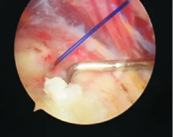
and a PDS No. 1 suture were used to first indicate the area of le- sion, and then the arthroscope was directed to the subacromial bursa to remove the inflammatory lesion, after which the rotator cuff calcific tendinitis was diagnosed. For calcium deposits with a toothpaste-like appearance, the deposit was excised using a probe as if squeezing out toothpaste. And for calcium deposits with a chalky appearance, it was excised after shaving off the su- perficial hypertrophic layer of the cuff tendon with a motorized shaver (Fig. 1). All calcific remnants were removed as much as possible using a curette, and sufficient postoperative lavage was carried out (Fig. 2). If we observed an overgrowth of osteophyte on the acromion, a section of it was excised using a flat burr.
Coracoacromial ligament release was not performed on any of the patients. If a concomitant cuff tear was present, we made a cuff repair at the cuff footprint using suture anchors if the tear occurred following the line of fibrosis of the cuff but did not make a repair if the tear was in parallel to the rotator cuff.
For our patient sample, we found that the distribution of age according to age group was as follows: one patient aged 30–39 years (2.6%); 12 patients aged 40–49 years (31.6%); 18 patients aged 50–59 years (47.4%); 4 patients aged 60–69 years (10.5%);
and 3 patients aged 70–79 years (7.9%). To compare the ef- fect of the location of calcific deposits within the rotator cuff on clinical outcome, we classified the patients with respect to the rotator cuff tendon in which the calcific tendinitis is found: the subscapularis tendon, the supraspinatus tendon, and the infra- spinatus tendon. For calcifications found in the supraspinatus tendon, we sub-classified them into more specified regions of the supraspinatus tendon: the anterior portion, the middle por- tion, and the posterior portion. To investigate the effect of the size of calcific deposits on the postoperative clinical outcome,
Fig. 1. We could make sure the No. 1 PDS which was marked in intra-articu- lar examination. The calcific deposit just nearby pre-marked PDS was identi- fied in subacromial space.
Fig. 2. Chalk-like calcific deposit in subacromial space was demonstrated.
we divided the patients around the average area of the calcific deposit (77.0 mm2): patients with calcific deposits of greater than 77.0 mm2 were allocated into group A, and those with calcific deposits of below 77.0 mm2 were allocated into group B (Fig. 3).
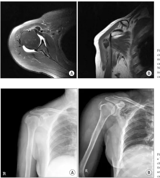
At the final follow-up, we confirmed using plain radiography that the calcification was completely excised—AP and axial views of the shoulders were taken in neutral position. The crite- rion for complete removal was an absence of any radiographic signs of calcification on at least two planes of radiography. We found that despite surgical treatment calcific deposits persisted in the supraspinatus tendons of two patients (the size of the cal- cific deposits decreased from 189.4 mm2 to 100.1 mm2 in one patient and from 200.5 mm2 to 137.4 mm2 in another) (Fig. 4).
We measured the following indicators of clinical outcome both preoperatively and postoperatively: the University of Califormia Los Angeles (UCLA) score; the American Shoulder and Elbow Society (ASES) score; and the visual analogue scale of pain (VAS) score. We investigated whether parameters such as cuff location,
calcific remnants, and concomitant cuff repair are significantly associated with the changes in these scores after surgery.
All statistical analyses were performed using the SPSS Win- dows ver. 13.0 (SPSS Inc., Chicago, IL, USA). Using the Student’s t-test, we tested for a significant difference in clinical outcomes between groups classified with respect to the size of calcium deposits and to whether or not a cuff repair was performed. We used one way-ANOVA to compare the clinical outcomes among three groups classified with respect to the location of calcium deposits in the rotator cuff. We used the chi-square test to as- sess whether the change in clinical scores between the pre- and postoperative measurements was significant. For all analyses, statistical significance was set to a p-value of less than 0.05.
Results
We found that the calcific deposits were mostly found in the middle supraspinatus tendon. Calcific tendinitis was found
A B
Fig. 3. (A) We measured gross mass of cal- cium deposit in axial and coronal image of magnetic resonance imaging (MRI), then was calculated the average of the two value. (B) We measured gross mass of calcium deposit in axial and coronal image of MRI, then was calculated the average of the two value.
A B
Fig. 4.(A) These preoperative radiograph of a 70-year-old female patient shows large cal- cific deposit on supraspinatus. (B) Postopera- tive radiograph shows small calcific remnant and this means incomplete removal of the calcium deposit.
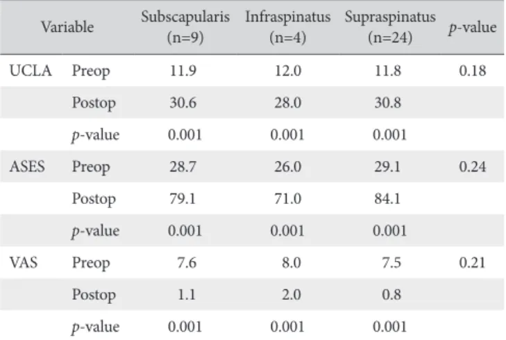
in the subscapularis tendon in 9 patients (23.7%); in the ante- rior supraspinatus tendon in 4 patients (10.5%); in the middle supraspinatus tendon in 17 patients (44.7%); in the posterior supraspinatus tendon in 5 patients (13.2%); and in the infraspi- natus tendon in 4 patients (10.5%). We found the average area of calcific deposit by location was as follows: 80.8 mm2 in the subscapularis tendon (range, 34.2–182.2 mm2); 68.9 mm2 in the anterior supraspinatus tendon (range, 17.1–122.1 mm2); 65.6 mm2 in the middle supraspinatus tendon (range, 14.4–200.4 mm2); 178.4 mm2 in the posterior supraspinatus tendon (range, 72.4–476.2 mm2); and 73.6 mm2 in the infraspinatus tendon (range, 32.1–131.8 mm2) (Table 1). We also evaluated the pre- operative and postoperative UCLA scores, ASES scores, and the VAS scores of the affected shoulder by rotator cuff tendon. The UCLA score improved from a preoperative average of 11.9 to a postoperative average of 30.6 in patients with calcific tendinitis affecting the subscapularis tendon; from 11.8 to 30.8 for the su- praspinatus tendon; and from 12.0 to 28.0 for the infraspinatus tendon. The ASES score improved from a preoperative aver- age of 28.7 to a postoperative average of 79.1 in patients with calcific tendinitis affecting the subscapularis tendon; from 29.1 to 84.1 for the supraspinatus tendon; and from 26.0 to 71.0 for infraspinatus tendon. The VAS score improved from a preopera- tive average of 7.6 to a postoperative average of 1.1 in patients with calcific tendinitis affecting the subscapularis tendon; from 7.5 to 0.8 for the supraspinatus tendon; and from 8.0 to 2.0 for the infraspinatus tendon. Thus, we found that the location of calcification within the rotator cuff does not have a significant influence on any of the clinical scores after arthroscopic surgery of calcific tendinitis (p=0.18 in UCLA; p=0.24 in ASES; p=0.21 in VAS) (Table 2).
We analyzed the distribution of calcification in the supraspi- natus tendon across the three regions: the anterior portion, the middle portion, and the posterior portion. We calculated the clinical scores by supraspinatus tendon location. We found that the UCLA score of patients with calcific deposits within the an- terior supraspinatus tendon improved from a preoperative aver- age of 11.3 to a postoperative average of 31.8; that of patients with calcific deposits within the middle supraspinatus tendon improved from a preoperative average of 11.4 to a postopera- tive average of 30.4; and for the posterior supraspinatus tendon, from 12.8 to 30.3. We found that the ASES score of patients with calcific deposits within the anterior supraspinatus tendon
improved from a preoperative average of 26.8 to a postopera- tive average of 88.8; that of patients with calcific deposits within the middle supraspinatus tendon improved from a preoperative average of 27.8 to a postoperative average of 85.2; and for the posterior supraspinatus tendon, from 32.9 to 78.2. We found that the respective scores for VAS were as follows: the anterior supraspinatus tendon, from 7.5 to 0.3; the middle supraspinatus tendon, from 7.7 to 1.0; and the posterior supraspinatus ten- don, from 7.3 to 1.0. Therefore, we found that the location of calcification within the supraspinatus tendon are not associated with postoperative clinical scores (p=0.28 in UCLA; p=0.44 in ASES; p=0.27 in VAS) (Table 3).
We also found that the clinical outcomes after arthroscopic treatment of calcific tendinitis were independent of calcifica- tion size. We divided the patients into two groups by taking the average calcification size 77.0 mm2 as the boundary to allocate patients into either group A (with larger than 77.0 mm2 calci- fication) or group B (with less than 77.0 mm2 of calcification).
For group A, we found that the UCLA score improved from a preoperative average of 11.3 to a postoperative average of 31.0;
the ASES score improved from a preoperative average of 22.5 to a postoperative average of 83.7; and the VAS score improved from a preoperative average of 7.7 to a postoperative average of 1.3. For group B, we found that the UCLA score improved from 12.0 to 29.6; the ASES score improved from 30.5 to 82.4; and
Table 1. Calcific Site and Size Distribution
Variable Subscap IST SST (ant. sup.) SST (central) SST (post. sup)
Patients (n=39), n (%) 9 (23.1) 4 (10.3) 4 (10.3) 17 (43.6) 5 (12.8)
Gross area (mm2) 80.8 73.6 68.9 65.6 178.4
Subscap: subscapularis, IST: infraspinatus, SST (ant. sup.): supraspinatus anterior superior part, SST (central): supraspinatus central part, SST (post. sup.): supra- spinatus posterior superior part.
Table 2. Clinical Results in according to Location of Calcific Deposit Variable Subscapularis
(n=9) Infraspinatus
(n=4) Supraspinatus (n=24) p-value
UCLA Preop 11.9 12.0 11.8 0.18
Postop 30.6 28.0 30.8
p-value 0.001 0.001 0.001
ASES Preop 28.7 26.0 29.1 0.24
Postop 79.1 71.0 84.1
p-value 0.001 0.001 0.001
VAS Preop 7.6 8.0 7.5 0.21
Postop 1.1 2.0 0.8
p-value 0.001 0.001 0.001
UCLA: University of California at Los Angeles shoulder rating scale, ASES:
American Shoulder and Elbow Surgeons evaluation form, VAS: visual analogue scale, Preop: preoperation, Postop: postoperation.
the VAS score improved from 7.5 to 1.3. Thus, we found that no significant differences were seen in the changes in clinical scores between the two groups (p=0.19 in UCLA; p=0.21 in ASES;
p=0.17 in VAS).
We analyzed the clinical scores according to the presence or the absence of calcific remnants to determine an association between this parameter and the improvements in clinical scores after arthroscopic treatment. In the 2 patients who had calcific remnants, we found that the UCLA score improved from a pre- operative average of 9.5 to a postoperative average of 32.0; the ASES score improved from a preoperative average of 21.8 to a postoperative average of 88.5; and the VAS score improved from a preoperative average of 8.0 to a postoperative average of 1.0. In the rest of patients in whom calcification was com- pletely removed, we found that the UCLA score improved to a postoperative average of 11.9; the ASES score improved to a postoperative average of 82.5; and the VAS score improved to a postoperative average of 1.1 (from the corresponding preopera- tive values mentioned above). As such, we found that whether or not the calcific remnants were completely removed, the post- operative clinical outcomes improved irrespectively (p=0.13 in UCLA; p=0.08 in ASES; p=0.16 in VAS).
We found that concomitant repairs of rotator cuff tears had no bearing on the clinical outcomes of arthroscopic treatment of calcific tendinitis. In the 6 patients who had received a concomi- tant rotator cuff repair, we found that the UCLA score improved from a preoperative average of 11.7 to a postoperative average of 31.3; the ASES score improved from a preoperative average of 25.6 to a postoperative average of 88.5; and the VAS score improved from a preoperative average of 7.8 to a postoperative average of 0.5. In the 33 patients who had received only simple decompression without cuff repair, we found that the UCLA
score improved to a postoperative average of 30.1; the ASES score improved to a postoperative average of 80.6; and the VAS score improved to a postoperative average of 1.2 (from the cor- responding preoperative values mentioned above). Altogether, these findings show that concomitant rotator cuff repairs do not have a statistically significant influence on clinical scores associ- ated with arthroscopic treatment of calcific tendinitis (p=0.18 in ASES; p=0.10 in UCLA; p=0.12 in VAS).
Discussion
The concept of calcific tendinitis was first established in 1970 by De Seze and Welfling.10) Studies have shown that calcific ten- dinitis is more prevalent in women than in men and in individu- als of the older age groups. According to findings by Bosworth,2) the incidence of calcific tendinitis is 2.7% among asymptomatic individuals. Bosworth2) reported the following proportion of each rotator cuff tendon affected by calcific tendinitis: 51% of calcific tendinitis involved the supraspinatus tendon; 44.5% in- volved the infraspinatus tendon; 23.3% involved the teres minor muscles; and 3% involved the subscapularis tendon. And De- Palma and Kruper3) reported that calcific tendinitis involving only the supraspinatus tendon composed 74% of the total incidence, and those involving the supraspinatus tendon in combination with another rotator cuff tendon composed 90%. In their study on 106 shoulders with calcific tendinitis, Rhee et al.4) found that 66% of calcific tendinitis affected the supraspinatus tendon;
17% affected the infraspinatus tendons; and 17% affected the subscapularis tendon. They also found that 92.9% of patients with calcific tendinitis affecting the supraspinatus tendon had satisfactory clinical outcomes (13 patients); 80% of patients with calcific tendinitis affecting the infraspinatus tendon had satisfac- tory clinical outcomes (4 patients); and none of the patients with calcific tendinitis affecting the subscapularis tendon had satisfac- tory clinical outcomes (2 patients).4)
In our study, we found that the distribution of cuff tendons af- fected by calcific tendinitis among our patients was as follows: 9 subscapularis tendons (23.1%); 4 anterior supraspinatus tendons (10.3%); 17 middle supraspinatus tendons (43.6%); 5 posterior supraspinatus tendons (12.8%); and 4 infraspinatus tendons (10.3%). Similar to the findings of other reports, we found that the prevalence of calcific tendinitis was highest in the middle supraspinatus tendon. Based on Painter11) report’s on the histo- pathological findings of calcific tendinitis using plain radiography in 1907, the classification system of calcific tendinitis devised by Bosworth) categorizes the lesions into large (>1.5 cm), in- termediate (0.5–1.5 cm), and small (<0.5 cm) sizes. Whereas, DePalma and Kruper3) and Patte and Goutallier12) subdivided the lesions into various classifications based on appearance. In this study, we did not apply the above criteria set by Bosworth2) but measured the size of calcific deposits using the AP and the Table 3. Clinical Results in according to Location Site of Calcific Deposit in
Supraspinatus Site in
supraspinatus Anterior
(n=4) Center
(n=16) Posterior
(n=4) p-value
UCLA Preop 11.3 11.4 12.8 0.28
Postop 31.8 30.4 30.3
p-value 0.001 0.001 0.001
ASES Preop 26.8 27.8 32.9 0.44
Postop 88.8 85.2 78.2
p-value 0.001 0.001 0.001
VAS Preop 7.5 7.7 7.3 0.27
Postop 0.3 1.0 1.0
p-value 0.001 0.001 0.001
UCLA: University of California at Los Angeles shoulder rating scale, ASES:
American Shoulder and Elbow Surgeons evaluation form, VAS: visual ana- logue scale, Preop: preoperation, Postop: postoperation.
lateral lengths derived from preoperative coronal and axial MRI radiographs. Through this method, we found that the average area of calcification was 77.0 mm2. This threshold was taken as the boundary to classify patients into two groups in terms of de- posit size; those with calcification of area greater than 77.0 mm2 and those with less than 77.0 mm2. Then, we made a compara- tive analysis of the clinical outcomes by deposit size. Although a very large calcific deposit may potentially cause pain that leads to an underestimation of the change in VAS score associated with the surgery, because we found that calcific deposit size did not influence the clinical outcomes after arthroscopic removal this was not an issue. Accordingly, we suggest that preoperative deposit size is not necessarily correlated with surgery outcome and therefore should not be used as an estimator or an indicator of clinical outcome.
Uhthoff13) described the process of calcific tendinitis in three stages based on histopathological findings: the pre-calcific phase in which there is fibrocartilaginous deposition secondary to his- tological changes to the tendon; the calcific phase comprising the formative and the resorptive phases; and the post-calcific phase during which tissue regeneration occurs after resorption.
During the resting phase, which in general is a painless stage, the calcium deposits have a chalky appearance. By the resorptive phase, the calcium deposits have a consistency of toothpaste.
The resorptive stage is associated with substantial pain and the stage at which macrophages and multinuclear giant cells remove the deposited calcium.14)
In general, more than 90% of patients with calcific tendi- nitis respond to conservative management. Examples of non- operative management of this condition includes non-steroidal anti-inflammatory drugs, steroid injections into the subacromial bursa, physiotherapy, extracorporeal shock-wave therapy, barbo- tage, and etc.15) Krasny et al.16) reported that ultrasound-guided needling enables an aspiration of up to 60% of calcium deposits, which is in comparison to the 32.5% of removal seen for ex- tracorporeal shock-wave therapy. For patients who are resistant to conservative management, a surgical excision of the calcific deposits is performed. The surgical excision was first suggested and carried out by Harrington and Codman et al. in 1902. The open excision via deltoid splitting with acromiplasty, which had since been established, is associated with satisfactory clinical out- comes in 71% to 82% of patients.15,17,18) Lately, the arthroscopic treatment has been employed, during which the intra-articular cherry red spot, caused by vascular proliferation, is marked and excision of the calcific deposit is made from the subacromion.
When removing the calcification, a mes is sometimes used to remove the deposit following the fibrosis of the rotator cuff, but generally a blunt tool like a probe is used to remove the calcific content in a squeezing motion, which results in a toothpaste- like content squeezed out as if from a tube. If the calcification has a hard chalky texture, then a curette and a rotating blade
are used to excise the calcific deposits; after, the area of concern is sufficiently irrigated to remove all remnants that if left would otherwise induce postoperative stiffuss.19,20)
Studies by Jerosch et al.7,21) suggest that the amount of rem- nant calcific deposits have an important effect on the clinical outcomes, yet recent conflicting reports show that completely removing the calcific deposits is not necessary and does not leave serious implications.4,6,9) In agreement with the latter statement, we found that calcific remnants did not have any significant effects on clinical outcomes. Thus we suggest that at- tempting to completely remove the calcification around chronic lesions or that of multiple lesions may be too time-consuming and may cause avoidable damage to surrounding tissues. As long as the calcific deposit that is implicated in the symptoms of cal- cific tendinitis is sufficiently removed, it may be that calcific rem- nants would not have a significant impact on clinical outcome.
Another issue in the treatment of calcific tendinitis that remains controversial is the issue of making a concomitant rotator cuff re- pair after removal of calcific deposits. There is clear and definite need to make either tendon-to-tendon or tendon-to-bone repair especially after removing large calcific deposits. But cuff repairs may negatively influence the postoperative rehabilitation be- cause the rotator cuff of a calcifying tendinitis shoulder tends to be thinner because of inflammation caused by calcification and by space-occupying lesions; consequently, the conditions for recovery are less favorable than in a standard rotator cuff repair.
Not only this, the long-term immobilization required after rota- tor cuff repair may induce postoperative shoulder stiffness. Tak- ing these points atogether, we attempted to minimize damage to the rotator cuff during the arthroscopic approach in various ways. For calcific deposits of toothpaste appearance, we made a small incision in the direction of the cuff tendons through which we carried out decompression by squeezing out the calcific de- posits. For chalky calcific deposits, we used a motorized shaver to carefully peel off the superficial subacromial layer and a cu- rette to remove the underlying calcification. For calcific deposits that encompass more than 50% of the width of the rotator cuff and has at least a medium-sized cuff tear, we considered per- forming a tendon-to-bone repair.
Because unnecessary tendon-to-tendon repair of the rota- tor cuff not only delays recovery but also promotes pain in- duced by compromised rotator cuff morphology, we did not perform tendon-to-tendon rotator cuff repairs. During removal of the calcific deposits, there is much debate as to whether ad- ditional subacromial decompression should be performed or not. Jerosch et al.7) reported that the presence of concomitant subacromial lesions of type 3 acromial morphology is an indica- tion for acromioplasty. Whereas Ellman et al.8) reported that the functional outcomes in those who received acromioplasty for calcific tendinitis and those who did not were comparable. They suggested that acromioplasty need not performed unless there
is a clear sign of impingement on radiographs or on arthroscopic findings. Although subacromial decompression, comprising both acromioplasty and coracoacromial ligament release, is neces- sary for the treatment of large calcific deposits and for instances of combined subacromical impingement syndrome, it must be indicated with caution because it necessitates unnecessary liga- ment release and acromioplasty, which leads to complications such as postoperative shoulder pain.22-24) In this study, we per- formed neither coracoacromial ligament release nor acromio- plasty on any of the patients.
Conclusion
Our findings revealed that the cuff tendon in which the cal- cific deposits are found; the location of the calcific deposits in the supraspinatus tendon; the size of calcific deposits; the pres- ence of calcific remnants; and concomitant cuff repairs did not have a statistically significant effect on the clinical outcomes after arthroscopic treatment of calcific tendinitis.
References
1. Welfling J, Kahn MF, Desroy M, Paolaggi JB, de Sèze S. Calci- fications of the shoulder. II. The disease of multiple tendinous calcifications. Rev Rhum Mal Osteoartic. 1965;32(6):325-34.
2. Bosworth BM. Calcium deposits in the shoulder and sub- acromial bursitis: a survey of 12, 122 shoulder. JAMA 1941;116:2477-82.
3. DePalma AF, Kruper JS. Long-term study of shoulder joints afflicted with and treated for calcific tendinitis. Clin Orthop.
1961;20:61-72.
4. Rhee YG, Kim YH, Park MS. Arthroscopic treatment in cal- cific tendinitis of the shoulder. J Korean Shoulder Elbow Soc.
2000;3(2):68-74.
5. Arrigoni P, Brady PC, Burkhart SS. Calcific tendonitis of the subscapularis tendon causing subcoracoid stenosis and cora- coid impingement. Arthroscopy. 2006;22(10):1139.e1-3.
6. Seil R, Litzenburger H, Kohn D, Rupp S. Arthroscopic treat- ment of chronically painful calcifying tendinitis of the supraspi- natus tendon. Arthroscopy. 2006;22(5):521-7.
7. Jerosch J, Strauss JM, Schmiel S. Arthroscopic treatment of calcific tendinitis of the shoulder. J Shoulder Elbow Surg.
1998;7(1):30-7.
8. Ellman H, Biglianini LU, Flatow E, et al. Arthroscopic treatment of calcific tendinitis. The American experience. Paper pre- sented at: 5th International Conference on Shoulder Surgery;
1992 Jul; Paris, France: p.1-15.
9. Ark JW, Flock TJ, Flatow EL, Bigliani LU. Arthroscopic treat- ment of calcific tendinitis of the shoulder. Arthroscopy.
1992;8(2):183-8.
10. De Seze S, Welfling J. Tendinities calcifications. Rhumatolocie.
1970;22:5-14.
11. Painter CF. Subdeltoid bursitis. Boston Med Surg J. 1907;156:
345-9.
12. Patte D, Goutallier D. Periarthritis of the shoulder. Cal- cifications. Rev Chir Orthop Reparatrice Appar Mot.
1988;74(4):277-8.
13. Uhthoff HK. Anatomopathology of calcifying tendinitis of the cuff. In: Gazielly DF, Gleyze P, Thomas T, eds. The cuff. Paris:
Elsevier; 1997. 144-7.
14. Uhthoff HK, Loehr JW. Calcific tendinopathy of the rotator cuff: pathogenesis, diagnosis, and management. J Am Acad Orthop Surg. 1997;5(4):183-91.
15. Lam F, Bhatia D, van Rooyen K, de Beer JF. Modern manage- ment of calcifying tendonitis of the shoulder. Curr Orthop.
2006;20(6):446-52.
16. Krasny C, Enenkel M, Aigner N, Wlk M, Landsiedl F. Ultra- sound-guided needling combined with shock-wave therapy for the treatment of calcifying tendonitis of the shoulder. J Bone Joint Surg Br. 2005;87(4):501-7.
17. Rochwerger A, Franceschi JP, Viton JM, Roux H, Mattei JP.
Surgical management of calcific tendinitis of the shoulder: an analysis of 26 cases. Clin Rheumatol. 1999;18(4):313-6.
18. Postel JM, Goutallier D, Lambotte JC, Duparc F. Treatment ofchronic calcifying or post calcifying shoulder tendinitis by acromioplasty without excision of the calcification. In: Gazielly DF, Gleyze P, Thomas T, eds. The cuff. Paris: Elsevier; 1997.
159-63.
19. Molé D, Kempf JF, Gleyze P, Rio B, Bonnomet F, Walch G. Re- sults of endoscopic treatment of non-broken tendinopathies of the rotator cuff. 2. Calcifications of the rotator cuff. Rev Chir Orthop Reparatrice Appar Mot. 1993;79(7):532-41.
20. Kempf JF, Bonnomet F, Nerisson D, Gastaud F, Lacaze F, Ger- aud H. Arthroscopic isolated excision of rotator cuff calcium deposits. In: Gazielly DF, Gleyze P, Thomas T, eds. The cuff.
Paris: Elsevier; 1997. 164-7.
21. Jerosch J, Strauss JM, Schmiel S. Arthroskopische therapie der tendinitis calcarea. Wie wichtig ist die Kalkentfernung? Ar- throskopie. 1996;9:241-5.
22. Tillander BM, Norlin RO. Change of calcifications after ar- throscopic subacromial decompression. J Shoulder Elbow Surg. 1998;7(3):213-7.
23. Loew M, Sabo D, Wehrle M, Mau H. Relationship between calcifying tendinitis and subacromial impingement: a prospec- tive radiography and magnetic resonance imaging study. J Shoulder Elbow Surg. 1996;5(4):314-9.
24. Resch H, Povacz P, Seykora P. Excision of calcium deposit and acromioplasty? In: Gazielly DF, Gleyze P, Thomas T, eds. The cuff. Paris: Elsevier; 1997. 169-71.