ISSN 2287-8130(Online) Particle and Aerosol Research
Part. Aerosol Res. Vol.14, No. 3: September 2018 pp. 49-63 http://dx.doi.org/10.11629/jpaar.2018.14.3.049
Electrospray technique for preparation of core-shell materials : A mini-review
Vinh Van Tran⋅Young-Chul Lee1)
1)Department of BioNano Technology, Gachon University, 1342 Seongnamdaero, Sujeong-gu, Seongnam-si 13120, Gyeonggi-do, Korea (Received 26 June 2018; Revised 3 July 2018; Accepted 12 July 2018)
Abstract
During the last decade, electrospray (ES) techniques have been used as potential methods for preparing of core-shell materials. Depending on the architecture of nozzle and design of devices, the ES techniques includes monoaxial, coaxial, multiple coaxial nozzle ES and microfluidic ES devices. ES operates based on a basic principle, in which a spray of monodisperse droplets is formed by dispensing an electrically conductive liquid through a capillary charged to a sufficiently high potential. In review of many recent research papers, we take a closer look at ES techniques and their applications for fabrication of core-shell materials. Several advantages of ES technique compared with other methods were emphasized and it may be regarded as a potential tool for fabrication of core-shell materials current and near future.
Keywords: Electrospray; Coaxial nozzle; Core-shell materials; Electrospraying mode
* Corresponding author.
Tel:+82-31-750-8751, Fax:+82-31-750-4748 E-mail:[email protected] (Y.-C. Lee)
1. Introduction
The synthesis of core-shell nanoparticles is a complex process. Although “top-down” approaches are possible to prepare the core-shell nanoparticles with different shapes and sizes, “bottom-up” techniques are mostly preferred (Gawande et al., 2015). Bottom-up synthetic methods have been used for the synthesis of core-shell materials in which there is a wide range of techniques have been developed such as precipitation, co-precipitation, hydrothermal degradation, deposition, sol-gel (Stöber method), reduction, template-assisted synthesis, polymerization, hydrolysis, ion-exchange process, low-temperature solid-state method (Almería et al., 2011; Chaudhuri and Paria, 2012; Gawande et al., 2015). However, most of them suffer from numerous shortcomings, including: modest encapsulation efficiency, batch-nature of the process, difficulty to scale-up, poor control of the particle size distribution, difficulty to generate sufficiently small particles, poor repeatability and limitations with respect to encapsulation of hydrophilic agents (Enlow et al., 2011; Hans and Lowman, 2002). Recently, electrospray (ES) technique has been used to produce and process micro-/
nanostructured materials (Chen et al., 2018; Xie et al., 2015). Compared with the mentioned traditional methods, ES techniques possess several advantages such as (1) producing lowest and uniform particle size as possible; (2) easy to control the operation parameters; (3) fast preparation and one step technique;
(4) simple set up; (5) extend for bulk production (Chen et al., 2005; Sridharab and Ramakrishna, 2013; Yeo et al., 2005). All these outstanding properties make ES as a fascinating method for fabricating a wide range of micro- and nanostructured core-shell materials applied in many fields like pharmaceutics, food, and healthcare to name a few. In this present review, we will provide an up-to-date appraisal of ES technique and their potential applications in preparation of core-shell materials.
2. Principle of electrospray technique
2.1 Basic setup and principle of electrospray technique
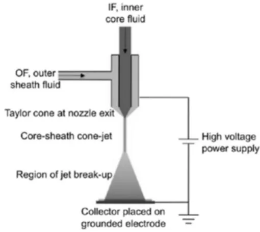
The basic setup for standard ES consists of several major components: a syringe pump, a syringe, a metal needle serving as nozzle, a high voltage power source and a grounded substrate serving as a collector (Fig. 1) (Smeets et al., 2017).
Fig. 1. Schematic representation of basic setup for standard electrospray (Reprinted (adapted) with permission from (Luo and Edirisinghe, 2014). Copyright (2018) American Chemical Society).
Although there are many different types of developed ES techniques, they all have operated based on a basic principle, in which a spray of monodisperse droplets is formed by dispensing an electrically conductive liquid through a capillary charged to a sufficiently high potential (Cloupeau and Prunet-Foch, 1994; Hartman et al., 1999; Zarrabi and Vossoughi, 2009). Briefly, an electrical source is installed between a capillary which liquid supply comes from a reservoir and a counter electrode placed some distance apart. In case of no voltage, a hemispherical droplet emerges from the capillary, while a voltage is applied, the air/liquid interface becomes polarized causing the meniscus to deform into a conical shape. The equilibrium then evolves toward the creation of a real cone called Taylor cone (Taylor, 1964, 1969). If the voltage is increased, it finally reaches a level where
the surface tension cannot maintain the liquid inside the droplet anymore resulting in the liquid ejection through a thin jet at the cone tip, where the electric field is the most important. This jet is directed toward the counter electrode. The jet breaks up further downstream into a spray of fine charged droplets (Bock et al., 2012; Lhernould and Lambert, 2011).
2.2 Electrospray modes
According to Wu et al. (2012a)’s review, the ES modes and resulting droplet sizes strongly depends on the process parameters such as the applied voltage, liquid flow rate, and liquid properties including electrical conductivity, surface tension, and liquid density. Liquid jet breaks up into droplet due to flow instability such as varicose instability. Electrical forces make very thin liquid jet, and, because of the surface tension, thin liquid jet undergoes flow instability. The obtainment of spraying modes will depend on the strength of the electric stresses relative to the surface tension stress on the liquid surface and on the kinetic energy of liquid leaving the nozzle (Smeets et al., 2017; Yurteri et al., 2010), it was demonstrated that various electrospray modes including dripping, micro-dripping, spindle, Taylor cone-jet and multi-jet mode are formed when increasing the applied voltage (Wu et al., 2012a; Zarrabi and Vossoughi, 2009). The balance between the surface tension and the electric stress will identify the eventually obtained mode. To determine the operating mode for ES system, it is necessary to consider and evaluate several important factors like the electric field strength, flow rate, the physical properties of the solution (electric conductivity, viscosity and surface tension) and their interplay.
Despite the existence of the diverse modes, Taylor cone-jet mode is the most common used ES mode due to producing highly monodisperse particles in a stable manner and a reproducible way (Smeets et al., 2017).
ES in the cone-jet mode may describe by three different processes: (1) The first process is the acceleration of the liquid in the liquid cone as a result
of the force balance of surface tension, gravity, electric stresses in the liquid surface, inertia and viscous stresses; (2) The second process is the breakup of the jet into droplets; (3) The third process is the development of the spray after droplet production (Gañán-Calvo et al., 1994; Hartman et al., 1999). In the cone-jet mode, a size segregation effect could be produced by different inertia which is formed by electrical interaction between highly charged with various sizes. Hence, small sizes will present at the edge of the spray, while the larger droplets will present in the spray centre. In addition, the droplet size in cone-jet mode is directly proportional with the liquid flow rate, but in inverse proportion to the liquid conductivity. Generally, the droplet size is difficult to be determined. However, many studies supported that it can be calculated by the following scaling law (Jaworek, 2007) with the confirmation of many experiment results (equation 1) (Ganan-Calvo, 1999).
dD
Q
(with α = 2.9) (1) where droplet sizes dD (m), Q is the liquid flow rate (m3· s-1), γ is the liquid conductivity (S/m), ε0 is permittivity of a vacuum (C V-1 m-1), ρ is the liquid density (g/mL), σ is the surface tension (N · m-1).
When decreasing flow rate and increasing electrical conductivity of the liquid, droplet size will decrease.
Wu et al. (2012a) indicated that the droplets with the suitable size may be produced through changing the flow rate and the liquid properties in a well-controlled manner. Therefore, the final particle size (dp) is related to the initial droplet size (dp) by the equation 2:
dp dDp
C (2)
Where: C is the concentration of solute and p is the density of the particle.
2.3 Formation mechanisms of core-shell particle in electrospray technique
The formation of core-shell materials by ES techniques may be explained through three different mechanisms depending on the surface charge density and the droplet surface tension (Zarrabi and Vossoughi, 2009). First mechanism, in case of the low surface charge density, no coulomb fission appears because the Rayleigh limit will be not attained even when the diameter of the droplets keeps shrinking due to solvent evaporation. Second mechanism, the high surface charge density will cause the particles to disintegrate rapidly and form nanoparticles. Especially, in the third mechanism, it was suggested that although the surface charge density is not very high, the Rayleigh limit will still reach after solvent evaporation due to shrinking of the droplets and coulomb fission occurs. When coulomb fission occurs, the particle size distribution will change and it causes the disintegration of particles to form nanoparticles.
The formation of a core-shell structure relating the ratio of charge relaxation lengths and inertial breakup lengths was explained in some studies of Mei and Chen (2007, 2008). They demonstrated that to form the core-shell structured droplets, several specific requirements must be satisfied by the ratio of the length scales. In order to evaluate the formation of the droplets, Xu et al. (2013) simplified the surface tension model and simulated the tip of the nozzle. The studies about effects of nozzle electric potentials on the structure of core-shell droplets and droplet size prepared by ES process indicated that the cone-jet formation near the nozzle tip could lead to the core-shell droplet formation due to force balance among gravity, surface tension, interfacial tension, electrical stresses, inertial and viscous stresses (Yan et al., 2016). It was demonstrated that the crucial parameters (electrical conductivity, viscosity, dielectric constant, flow rate, and voltage) and additional parameters (interfacial tension, flow rates of liquids, and the nozzle configuration) will affected the outcome of the core-shell particle formation in the ES process.
3. Development of types of electrospray technique in preparation of core- shell materials
Generally, there are three types of ES commonly used to synthesize core-shell materials including monoaxial ES, coaxial ES, multiple nozzle ES. Recent applications in the synthesis of core-shell particle relating to these types of ES will be discussed in the following section and summarized in table 1.
3.1 Monoaxial electrospray
Monoaxial ES is a traditional ES technique developed to synthesize micro/nanomaterials due to cheap cost and simple operation. For principle of monoaxial ES, a polymer solution is slowly injected into a capillary nozzle through a needle by a syringe pump and it is charged by a strong external electric field. An electric stress condition generated from the free charge will cause the acceleration of the liquid away from the needle. In case of increasing the electrical potential to several kilovolts, the liquid meniscus at the needle opening develops into a conical shape or the Taylor cone. Soon, a fine polymer jet will be formed as a result of the electrical conduction of liquid. The jet eventually breaks up into charged droplets that fly towards the grounded collector. To allow the formation of particles with smooth surface morphology, a closed chamber with continuous air or nitrogen flow often used to reduce the evaporation rate of solvent (Cloupeau and Prunet-Foch, 1994; Davoodi et al., 2015; Jaworek, 2007; Loscertales et al., 2002;
Salata, 2005).
According to Bock et al. (2012)’s review, core-shell particulate may be fabricated by monoaxial ES where the active agent (drug) is mixed with the polymer solution before ES commences. Core-shell particulate systems will be formed through aqueous nanoprecipitation, emulsification and solid dispersion (Nikolaou and Krasia-Christoforou, 2018). In other study, Yu et al.
(2014) was successfully prepared CeO2 microspheres by using the monoaxial ES technique with morphology in
Nozzle/ needle geometry
Nozzle size (μm)
Particle size (μm)
Core materials Shell materials
Application Refs
Monoaxial 150 1.34 Doxorubicin
hydrochloride
PLGA Drug delivery (Almería et al., 2011)
19 coaxial nozzles 70 1.32 Doxorubicin hydrochloride
PLGA Drug delivery (Almería et al., 2011)
Single coaxial nozzle
- 30 PLGA poly-DL-
lactic acid Drug encapsulation
(Xu et al., 2013)
Monoaxial 880 5.0 Fe3O4 Eudragit
RS PO
Enhancing gastric antimicrobial delivery
(Hao et al., 2014)
Monoaxial 5000 2.8 Peptides, proteins poly-DL-
lactic acid Drug encapsulation
(Yeo et al., 2005).
Double-nozzle coaxial
500 - Poly-(lactic acid) (PLA)
PLGA Drug delivery (Yan et al., 2016)
Single coaxial nozzle
- 300 Living cells Alginate Medicine (Zhao et al., 2014)
Single coaxial nozzle
75 ~100 Living cells Alginate Medicine (Zhang and He,
2009) Single coaxial
nozzle
508 1.2 Budesonide PLGA Drug
encapsulation
(Lee et al., 2010)
Single coaxial nozzle
720 400 Poly-L-lactide PLGA Drug delivery (Nie et al., 2010)
Monoaxial - 6-9 Congo red/
albumin
PLGA Drug delivery (Yao et al., 2016)
Tetraaxial nozzle 2800 2900 Insulin B. longum Medicine (Paz-Samaniego et
al., 2018) Tri-needle coaxial 28500 13.5 Acridine yellow Nile blue Pharmaceutic (Zhang et al.,
2017)
25 coaxial emitters 7000 17-39 Ethylene glycol Sesame oil Drug delivery (Olvera-Trejoab and Velásquez-García, 2016)
Single coaxial nozzle
1000 - Sodium
carboxymethylcellu lose
Na-Alg Biocatalysis and Biomimetic
(Wang et al., 2018) Table 1. The recent application of electrospray technique in fabrication of core-shell materials
solid particles. The particle sizes of these structured CeO2
microspheres with different morphologies are 1–2 µm, which suggested that monoaxial ES may be a tool that has much significance in the production of many other photocatalysts. In addition, monoaxial ES also showed a high efficiency in the fabrication of core-shell materials for active agent encapsulation in drug delivery. For example, a novel stomach-specific sinking magnetic microparticles enhancing gastric antimicrobial delivery were formed via the monoaxial electrospray method practice (Hao et al., 2014). The size of this material was approximately 5 μm and it showed a high density when increasing the Fe3O4 concentration in the electrospray inlet flow. This study stated that monoaxial ES is an effective technique to prepare the high density gastroretentive dosage forms. Especially, a new monoaxial ES using high frequency AC electric fields above 10 kHz was developed for encapsulation of DNA, peptides, proteins and other therapeutic molecules (Yeo et al., 2005). It was demonstrated the AC monoaxial ES may be regarded as a potential technique with a greater flexibility to encapsulate organic phase soluble therapeutic molecules and drug compounds, compared
with conventional methods in the nanoparticle fabrication for drug encapsulation.
It was demonstrated that monoaxial ES exhibits a monotonic dependence of droplet size on liquid flow rates (Davoodi et al., 2016; Xu et al., 2013). The size of obtained core-shell particles increases when increasing flow rate, therefore the optimal condition for operating monoaxial ES is the extremely low flow rate.
In other words, the productivity will be limited by the low flow rate. In addition, for large scale production, monoaxial ES also exists restriction due to the low throughput (Yan et al., 2016).
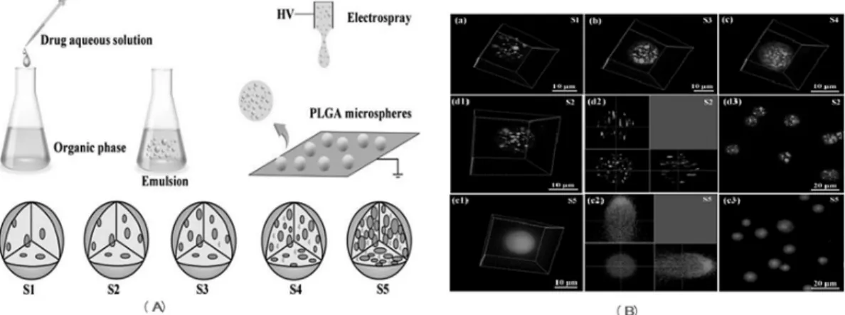
For the emulsion ES method, electric field is added to the drug emulsion solution to spray particles (Bock et al., 2012; Malik et al., 2016). Based on this technique, a novel microsphere of PLGA polymer was successfully prepared to encapsulate two kind of aqueous model drugs, Congo red and albumin from bovine serum (BSA) (Yao et al., 2016). In the preparation process (Fig. 2A), the emulsion solution formed by adding the aqueous phase of two drugs into the organic phase of PLGA polymer was passed an ES system to fabricate drug entrapped PLGA microspheres. It was shown that the morphology
Fig. 2. (A) Schematic representation of the emulsion ES process (S1 to S5, the Vw/Vo were 5, 10, 20, 50 and 100 µL/mL, respectively) (B) The BSA-FITC distribution in the PLGA microspheres with different Vw/Vo
of 5 (a, S1), 10 (d1–d3, S2), 20 (b, S3), 50 (c, S4) and 100 µL/mL (e1–e3, S5). (a–c), (d1) and (e1) are the 3D reconstruction of z-stack confocal images. (d2) and (e2) are the cross-section of PLGA microspheres;
(d3) and (e3) are the drug phase (BSAFITC was green) distribution in the PLGA microspheres (red was rhodamine). (Reprinted (adapted) with permission from (Yao et al., 2016). Copyright (2018) Oxford University Press).
of these PLGA microspheres was affected by the volume ratio of aqueous drug phase and organic PLGA phase (Vw/Vo) and the molecule weight of model drugs. With increasing the volume ratio of aqueous drug phase Vw/Vo
from 5 to100 µL/mL (S1-S5), the distribution of aqueous nanodroplets in the PLGA microspheres was different and the number of nanodroplets also increased gradually. Yao et al. (2016) showed the drug distributions within the different formulation of PLGA microspheres prepared by the emulsion monoaixal ES method (Fig. 2B). The 3D reconstructions of laser scanning confocal microscope (LSCM) images for fluorescein conjugated BSA (BSA-FITC) (Fig. 2B(a–c), (d1–d3) and (e1–e3)) confirmed that more drugs were encapsulated with more aqueous solution added. And the formation of nanodroplets of aqueous phase solution (the green dots) dispersed over the PLGA emulsion homogenously. Also, it is notable that the formed nanodroplets in the emulsification process was likely to be maintained in the subsequent electrospray and the density of the drug nanodroplets increased with the Vw/Vo ratio. In summary, the authors stated that emulsion monoaxial ES will provide an efficient and simple system to prepare the PLGA microspheres with the great drug release control at a desired rate satisfying the need of the practices.
3.2 Coaxial electrospray
The coaxial ES technique has gained considerable attention in the synthesis of biodegradable polymeric particles for encapsulating therapeutic agents in controlled and sustained drug release applications (Xie et al., 2006).
It can produce core-shell particles with exceptional properties such as uniform drug distribution within the particles and high loading capacity (Bock et al., 2012;
Chakraborty et al., 2009). Recently, ES technique has often been employed to synthesize polymeric composite microspheres, well-known core-shell particles, widely used for drug delivery applications due to its mentioned advantages. Xu’s group applied this technique to successfully fabricate a polymeric composite microspheres consisting of a poly (D,L-lactic-co-glycolic acid) (PLGA) core and a poly (D,L-lactic acid) (PDLLA) shell (Xu et
al., 2013). The authors used a computational fluid dynamics (CFD) model to simulate coaxial ES process with conditions including nozzle voltage and polymer solution flow rates. It was suggested that CFD model may be used to predict the production of consistent compound droplets and the expected core–shell structured microspheres. The simulation results indicated that the operating conditions (nozzle voltage and flow rates) of ES system influenced on the particle size of microspheres.
When increasing nozzle voltages, the particle size decreased and above 6kV, the polymer jet became unstable. For various flow rates, the particle size increased almost linearly with increasing shell flow rate.
Also, Nie et al. (2010) demonstrated that coaxial ES can be used for the fabrication of microspheres with distinct core-shell structure. These core-shell particles have ability to encapsulate both hydrophobic and hydrophilic drugs in one single step. Especially, hydrophobic drug is located in the shell, while hydrophilic drug can be encapsulated in the core. In addition, different drug distributions are reached in the microspheres by altering drug solutions between the inner and outer needles, which allow different temporal release profiles of each drug (Nie et al., 2010).
Also, coaxial ES has been extensively used for cell microencapsulation using alginate probably given that simple operation, reusability, and high production rate (Chakraborty et al., 2009). However, current ES techniques have still existed two main limitations including producing homogeneous, solid-like microbeads that lack an aqueous liquid core or use highly evaporative organic solvents in the fabricating process of a core–shell structure (Kim et al., 2013; Lee et al., 2010; Zhang and He, 2009). The latter indicated that the applicability of ES techniques in the encapsulation of living cells is impossible because of the high cytotoxicity of organic solvents. To overcome this problem, a novel coaxial ES technology was developed to synthesize a core-shell microcapsule based on a hydrogel shell of alginate and an aqueous liquid core of living cells, embryonic stem cells (Fig. 3a) (Zhao et al., 2014). This coaxial ES technique produced a great core-shell material for ES encapsulation. This was demonstrated by generation
of microcapsules with high viability (>90%), but using two aqueous fluids without any cytotoxic organic solvent usually needed in ES technology to fabricate core–shell microcapsules. Moreover, it was suggested that compared with the 3D solid-like hydrogel and conventional 2D culture methods, coaxial ES could provide a better biomimetic, miniaturized 3D liquid culture microenvironment for culturing stem cells (Fig. 3b).
3.3. Complex multiple coaxial nozzle systems Similar to mono-axial ES, the single coaxial ES process exhibits a limitation of the productivity due to the low flow rate. (Davoodi et al., 2016; Xu et al., 2013;
Yan et al., 2016). For mass production of core-shell particles, to overcome the restriction of the low throughput with single nozzle while maintaining the particle size, multi coaxial nozzle ES system has been regarded as an indispensable solution (Deng et al., 2009;
Oh et al., 2008). According to this approach, multi coaxial nozzle ES has gained considerable attention in the synthesis of core-shell particle.
Based on the basic coaxial ES technique, Yan et al.
(2017) designed a double-nozzle coaxial ES system to fabricate core-shell microspheres toward large-scale production. The nozzle spacing is one of the most
important factors for designing multi-nozzle ES array for large scale production (Regele et al., 2002). This study results revealed that the formed particle quality (morphology and core-shell structure) is strongly influenced by the interference between neighboring nozzles and suggested that optimal spacing should be
~3000 nozzle/m2 packing density which may be achieved by adjusting the nozzle-to-nozzle distance greater than 0.018 m. In addition, the authors conducted a comparison between single-nozzle system and double nozzle system and calculated the electrical field distribution and simulated ES process for both single and double-nozzle system to explain the interactions between adjacent nozzles. It has been found that using cone shaped nozzle tip can enhance the productivity (Yan et al., 2016). The distributions of electric filed line released from the nozzle tips displayed (Fig. 4a, b). While the electric lines display an axisymmetric distribution along the axis of nozzle single-nozzle system, the electric field interference between two neighboring nozzles causes a bend of electric lines in double-nozzle system. The simulation of droplets distribution for single-nozzle system and double-nozzle system is shown in (Fig. 4c, d). For single-nozzle system, the ES displays an axisymmetric distribution, while it shifts from nozzle Fig. 3. (a) schematic illustration of the coaxial electrospray system for generating microcapsules with a liquid core and alginate hydrogel shell; (b) Expression of pluripotency protein markers of ES cells obtained under three different culture conditions: quantitative analysis of mean intensity showed that cells obtained from the miniaturized 3D liquid core have significantly higher expression of Nanog and Sox-2 on average than 3D hydrogel and 2D culture (Reprinted (adapted) with permission from (Zhao et al., 2014). Copyright (2018) Royal Society of Chemistry)
axis along X direction in double-nozzle system. In conclusion, to design a double-nozzle coaxial ES system, it is very necessary to avoid and minimize the interference of electric field from neighboring nozzle (Yan et al., 2017).
These day, a single-dosage form platform encapsulating and delivering multiple drugs has been considered as one of the most advanced techniques for the development of advanced therapies. In addition, co-encapsulation of multiple active agents can deliver synergistic therapeutic effects and reduce side effects (Parhi et al., 2012).
Moreover, the coencapsulation of both hydrophilic and hydrophobic drugs in a single-dosage form like core-shell particle is an ideal method to enhance the drug
dispersibility, active release, and therapeutic impact (Nii and Ishii, 2005). Therefore, the development of unique techniques applied for the synthesis of core-shell particles encapsulating two or more active drug will be a potential approach. Hence, new ES systems with a tri-needle coaxial, tetra-axial or even multiple coaxial nozzles has been considered for fabrication of core-shell particle.
Zhang et al. (2017) developed novel core-shell particles, namely magnetic polymer yolk-shell particles (YSPs), using a tri-needle coaxial ES technique to encapsulate multidrug in the compartments (Figure 5a).
The architecture of YSPs showed that magnetic Fe3O4
nanoparticles (MNPs) and Nile blue (NB) entrapped in Fig. 4. Distributions of electric filed line released from the nozzle tips: (a) single-nozzle system; (b) double-nozzle system; Spatial distributions of charged droplets for (c) single-nozzle system and (d) double-nozzle system (Reprinted (adapted) with permission from (Yan et al., 2017). Copyright (2018) Wiley Materials).
Fig. 5. (a) Tri-needle coaxial electrospray (Reprinted (adapted) with permission from (Zhang et al., 2017).
Copyright (2018) American Chemical Society); (b) Tetraaxial nozzle electrospray (Reprinted (adapted) with permission from (Paz-Samaniego et al., 2018). Copyright (2018) Wiley Materials)
the outer shell, Sudan red G (SRG) incorporated in the central layer and acridine yellow (AY) contained in the inner core. Also, the study stated that three solutions were dispensed into the tri-needle coaxial nozzle and the jetting stabilities observed is similar to single and conventional two needle coaxial ESs. Tetraaxial nozzle ES is next version of multiple coaxial nozzles ES developed for preparing the core-shell materials.
Paz-Samaniego et al. (2018) employed tetraaxial nozzle ES to fabricate core-shell particles composed of maize bran arabinoxylans MBAX) with insulin in the core, and maize wastewater arabinoxylans (MWAX) with Bifidobacterium in the shell (Figure 5b). For encapsulation efficiency, the study stated that tetraaxial nozzle ES produced a core-shell particle with higher encapsulation efficiency of Bifidobacterium compared with other methods (Zou et al., 2011).
As mentioned, standard coaxial ES technologies usually have a great limitation of productivity because of only possessing one emitter and operating in the cone-jet emission mode. Also, a coaxial ES emitter is limited by low flow rates. Moreover, the adverse effects in a given application can be caused by increasing the particle size as a result of increasing the flow rates of the emitter. An approach to improve the throughput of a coaxial ES source without affecting the
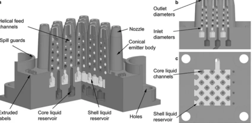
size variation has gained a great interest (Denga et al., 2006; Velásquez-García et al., 2006; Wang et al., 2004). With an attempt to find an upgraded version of coaxial ES for large-scale production of core-shell particles, Olvera-Trejo and Velasquez-Garcia (2016) reported the first MEMS multiplexed coaxial ES sources in the literature. The device design of the MEMS multiplexed coaxial ES sources is shown (Fig.
6). The device consists two separate liquid reservoirs:
the inner liquid reservoir and the outer liquid reservoir provide the feedstock for the core layer and the shell layer of the particles, respectively (Fig. 6a). The structure of the liquid reservoirs includes inlet channels opening to the bottom of the device. An array of rounded square columns will support the ceilings of the liquid reservoirs to avoid the collapse of the liquid reservoirs. Each reservoir feeds a microchannel embedded within each conical emitter that ends in a coaxial nozzle (Fig. 6b). In this study, the miniaturized coaxial ES emitters (up to 25 emitters) were designed as monolithic planar arrays to work uniformly in parallel, which significantly increased the throughput of the compound micro-droplet source and made this technology compatible with low-cost commercial applications. Contrary to the predicted theory, the study results demonstrated that the per-emitter current is
Fig. 6. (a) Intersected cross-section of a 3D schematic of a planar array of 25 coaxial electrospray emitters.
The hydraulics for the inner and outer liquids are coloured in red and yellow, respectively. (b) Lateral cross-section of the central emitter row (Reprinted (adapted) with permission from (Olvera-Trejoab and Velásquez-García, 2016). Copyright (2018) Royal Society of Chemistry)
proportional to the flow rate of the liquid and it did not depend on the flow rate of the driven liquid. In addition, the diameters and the size distribution of the formed core-shell particles were likely to be modulated by controlling the flow rates (Olvera-Trejoab and Velásquez-García, 2016). With similar approach, Almeria et al. (2011) prepared a biodegradable polymer particle using a multiplexed ES system. The system comprises an array of 19 microfabricated nozzles in silicon. The study indicated that multiplexed ES technique have many advantages: (i) allow coating agents for drug delivery in a single-step process; (ii) control the particle size and monodispersity of formed particles, (ii) avoid particle aggregation, (iv) increases the throughput by orders of magnitude, (v) suitable for a semi-batch process.
4. Electrospray incorporation with others technique for fabrication of core- shell material
4.1 Microfluidic electrospray devices
To construct the capillary microfluidic device, two ground capillaries was coaxially assembled on a glass slide. It is notable that the combination of microsphere particles prepared by microfluidic ES and the enzymatic inverse opal particles a unique biocatalyst system with specific functions of the enzyme cascade reaction (Wang et al., 2018). To conduct the encapsulation, a coaxial capillary microfluidic chip was integrated with three-bore capillary injection channels using the ES collection device. This study demonstrated that ES can be incorporated with microfluidic to fabricate the core-shell system with great properties for encapsulation application. Similarly, Wu and coworkers introduced a novel static micromixer coaxial ES (MES) system, which has been designed from the combination of the advantages between static micromixer and coaxial ES to produce uniform theranostic lipolexes in a single step with high reproducibility (Fig 7) (Wu et al., 2012b). The synthetic process was described as follows: The static
micromixer was utilized to bring together the imaging reagent stream and the therapeutic reagent stream in the range of microscale and further reduce the stream width to the nanoscale through the repeated stretching folding function of the static mixing units. The flows of the multilayered mixture of imaging reagent and therapeutic reagent were formed the inner needle of the coaxial ES system. The lipids were dissolved in ethanol and flow through the outer needle of the coaxial electrospray system. After the quick evaporation of ethanol, theranostic lipoplexes were formed. As a result, compared with traditional methods, lipoplexes produced by MES were distributed uniformly with a diameter of ~194 nm and a polydispersity of ~0.024 and also inhibited good therapeutic and imaging ability with little toxicity.
Fig. 7. Schematic diagram of the static micromixercoaxial electrospray device used for theranostic lipoplexes synthesis (Reprinted (adapted) with permission from (Wu et al., 2012b). Copyright (2018) American Chemical Society)
4.2 Combination of electrospinning and electrospray techniques
Electrospinning is usually used to synthesize continuous nano/microfibers deriving from natural and synthetic polymers, ceramics and composites (Chronakis, 2005).
Because of the same in the experimental setup, both electrospinning and ES also have the same influencing factors. Besides their independent use for the fabrication of various materials, the combination of electrospinning and ES have been also considered to create hybrid particle composite materials (Ramasundarama et al., 2015). Some reviews indicated that the combination of the two
techniques can be conducted by following 3 different routes: (1) the simultaneous occurrence of electrospinning and ES; (2) the ES and electrospinning process is performed onto the same collector, but electrospinning complete first; (3) the ES process takes place onto a preformed electrospun mat (Jaworek et al., 2009; Nikolaou and Krasia-Christoforou, 2018). In Luo and Edirisinghe (2014)’s study about combination of these two techniques, it was demonstrated for the first time that the sheath polymer solution (low viscosity) to electrospin instead of electrospray in a coaxial electrified jet may be caused by simply infusing a liquid in the core nozzle without increasing the polymer concentration. It is the presence of core liquids that contributes to the suppression of Rayleigh instability, which lead to the transition from ES to electrospinning of beads-on-strings. This process has some crucial parameters such as high surface tension of the core solvent, high interfacial tension at the core-sheath interface and electrohydrodynamic operating parameters.
Moreover, the solvents in this process facilitate the effective fabrication of core-shell polymeric structures by coaxial electrohydrodynamic processes.
5. Conclusion
In this review, we have summarized the recent research into the development of electrospray techniques for fabrication of core-shell materials, and we have also emphasized several important types of commonly used ES techniques. Recent applications in the synthesis of core-shell particle relating to monoaxial, coaxial, multiple coaxial, microfluidic ES was discussed. Several advantages of ES technique compared with other methods was pointed. ES technique has been attracting a great interest and it could be regarded as a potential tool for fabrication of core-shell materials current and near future.
Acknowledgement
This work was supported by the Basic Science Research Program through the National Research Foundation of Korea funded by the Ministry of Education (NRF-2017 R1D1A1A09000642).
References
Almería, B., Fahmy, T.M., and Gomez, A. (2011). A multi- plexed electrospray process for single-step syn- thesis of stabilized polymer particles for drug delivery, Journal of Controlled Release, 154, 203-210.
Bock, N., Dargaville, T.R., and Woodruff, M.A. (2012).
Electrospraying of polymers with therapeutic molecules: State of the art, Progress in Polymer Science, 37, 1510-1551.
Chakraborty, S., Liao, I.-C., Adler, A., and Leong, K.W.
(2009). Electrohydrodynamics: A facile techni- que to fabricate drug delivery systems, Advanced Drug Delivery Reviews, 61, 1043-1054.
Chaudhuri, R.G., and Paria, S. (2012). Core/shell nano- particles: Classes, properties, synthesis mecha- nisms, characterization, and applications, Chemical Reviews, 112, 2373-2433.
Chen, G.-C., Kuo, C.-Y., and Lu, S.-Y. (2005). A general process for preparation of core-shell particles of complete and smooth shells, Journal of the American Ceramic Society, 88, 277-283.
Chen, J., Cui, Y., Xu, X., and Wang, L.-Q. (2018). Direct and effective preparation of core-shell PCL/PEG nanoparticles based on shell insertion strategy by using coaxial electrospray, Colloids and Surfaces A, 547, 1-7.
Chronakis, I. (2005). Novel nanocomposites and nano- ceramics based on polymer nanofibers using elec- trospinning process-A review, Journal of Materials Processing Technology, 167, 283-293.
Cloupeau, M., and Prunet-Foch, B. (1994).
Electrohydrodynamic spraying functioning
modes: A critical review, Journal of Aerosol Science, 25, 1021-1036.
Davoodi, P., Feng, F., Xu, Q., Yan, W.-C., Tong, Y.W., Srinivasan, M.P., Sharma, V.K., and Wang, C.-H.
(2015). Coaxial electrohydrodynamic atom- ization: Microparticles for drug delivery applica- tions, Journal of Controlled Release, 205, 70-82.
Davoodi, P., Ng, W.C., Yan, W.C., Srinivasan, M.P., and Wang, C.-H. (2016). Double-walled micro- particles-embedded self-cross-linked, injectable, and antibacterial hydrogel for controlled and sus- tained release of chemotherapeutic agents, ACS Applied Materials & Interfaces, 8, 22785-22800.
Deng, W., Waits, C.M., Morgan, B., and Gomez, A. (2009).
Compact multiplexing of monodisperse electro- sprays, Journal of Aerosol Science, 40, 907-918.
Denga, W., Klemic, J.F., Li, X., Reed, M.A., and Gomez, A. (2006). Increase of electrospray throughput using multiplexed microfabricated sources for the scalable generation of monodisperse droplets, Journal of Aerosol Science, 37, 696-714.
Enlow, E.M., Luft, J.C., Napier, M.E., and Desimone, J.M.
(2011). Potent engineered PLGA nanoparticles by virtue of exceptionally high chemotherapeutic loadings, Nano Letters, 11, 808-813.
Ganan-Calvo, A.M. (1999). The surface charge in electro- spraying: Its nature and its universal scaling laws, Journal of Aerosol Science, 30, 863-872.
Gañán-Calvo, A.M., Lasheras, J.C., Dávila, J., and Barrero, A. (1994). The electrostatic spray emitted from an electrified conical meniscus, Journal of Aerosol Science, 25, 1121-1142.
Gawande, M.B., Goswami, A., Asefa, T., Guo, H., Biradar, A.V., Peng, D.-L., Zboril, R., and Varma, R.S.
(2015). Core–shell nanoparticles: synthesis and applications in catalysis and electrocatalysis, Chemical Society Reviews, 44, 7540-7590.
Hans, M.L., and Lowman, A.M. (2002). Biodegradable nanoparticles for drug delivery and targeting, Current Opinion in Solid State & Materials Science, 6, 319-327.
Hao, S., Wang, Y., and Wang, B. (2014). Sinking-magnetic
microparticles prepared by the electrospray meth- od for enhanced gastric antimicrobial delivery, Molecular Pharmaceutics, 11, 1640-1650.
Hartman, R.P.A., Brunner, D.J., Camelot, D.M.A., Marijnissen, J.C.M., and Scarlett, B. (1999).
Electrohydrodynamic atomization in the cone-jet mode physical modeling of the liquid cone and jet, Journal of Aerosol Science, 30, 823-849.
Jaworek, A., (2007). Micro- and nanoparticle production by electrospraying, Powder Technology, 176, 18-35.
Jaworek, A., Krupa, A., Lackowski, M., Sobczyk, A.T., Czech, T., Ramakrishna, S., Sundarrajan, S., and Pliszka, D. (2009). Nanocomposite fabric for- mation by electrospinning and electrospraying technologies, Journal of Electrostatics, 67, 435-438.
Kim, J., Sachdev, P., and Sidhu, K. (2013). Alginate micro- capsule as a 3D platform for theefficient differ- entiation of human embryonicstem cells to dop- amine neurons, Stem Cell Research, 11, 978-989.
Lee, Y.-H., Mei, F., Bai, M.-Y., Zhao, S., and Chen, D.-R.
(2010). Release profile characteristics of bio- degradable-polymer-coated drug particles fab- ricated by dual-capillary electrospray, Journal of Controlled Release, 145, 58-65.
Lhernould, M.S., and Lambert, P. (2011). Compact polymer multi-nozzles electrospray device with integrated microfluidic feeding system, Journal of Electrostatics, 69, 313-319.
Loscertales, I.G., A. Barrero, Guerrero, I., Cortijo, R., Marquez, M., and Ganan-Calvo, A.M. (2002).
Micro/nano encapsulation via electrified coaxial liquid jets, Science, 295, 1695-1698.
Luo, C.J., and Edirisinghe, M. (2014). Core-liquid-induced transition from coaxial electrospray to electro- spinning of low-viscosity poly(lactide-co- glyco- lide) sheath solution, Macromolecules, 47, 7930-7938.
Malik, S.A., Ng, W.H., Bowen, J., Tang, J., Gomez, A., Kenyon, A.J., and Day, R.M. (2016).
Electrospray synthesis and properties of hier-
archically structured PLGA TIPS microspheres for use as controlled release technologies, Journal of Colloid and Interface Science, 467, 220–229.
Mei, F., and Chen, D.-R. (2007). Investigation of compound jet electrospray: Particle encapsulation, Physics of Fluids, 19, 103303.
Mei, F., and Chen, D.-R. (2008). Operational modes of dual-capillary electrospraying and the formation of the stable compound cone-jet mode, Aerosol and Air Quality Research, 8, 218-232.
Nie, H., Dong, Z., Arifin, D.Y., Hu, Y., and Wang, C.-H.
(2010). Core/shell microspheres via coaxial elec- trohydrodynamic atomization for sequential and parallel release of drugs, Journal of Biomedical Materials Research A, 95A, 709-716.
Nii, T., and Ishii, F. (2005). Encapsulation efficiency of water-soluble and insoluble drugs in liposomes prepared by the microencapsulation vesicle meth- od, International Journal of Pharmaceutics, 298, 198-205.
Nikolaou, M., and Krasia-Christoforou, T. (2018).
Electrohydrodynamic methods for the develop- ment of pulmonary drug delivery systems, European Journal of Pharmaceutical Sciences, 113, 29-40.
Oh, H., Kim, K., and Kim, S. (2008). Characterization of deposition patterns produced by twin-nozzle electrospray, Journal of Aerosol Science, 39, 801-813.
Olvera-Trejoab, D., and Velásquez-García, L.F. (2016).
Additively manufactured MEMS multiplexed co- axial electrospray sources for high-throughput, uniform generation of core–shell microparticles, Lab Chip, 16, 4121-4132.
Parhi, P., Mohanty, C., and Sahoo, S.K. (2012).
Nanotechnology-based combinational drug de- livery: an emerging approach for cancer therapy, Drug Discovery Today, 17, 1044-1052.
Paz-Samaniego, R., Rascon-Chu, A., Brown-Bojorquez, F., Carvajal-Millan, E., Pedroza-Montero, M., Silva-Campa, E., Sotelo-Cruz, N., Lopez-Franco, Y.L., and Lizardi-Mendoza, J. (2018).
Electrospray-assisted fabrication of core-shell arabinoxylan gel particles for insulin and pro- biotics entrapment, Journal of Applied Polymer Science, 135, 46411.
Ramasundarama, S., Son, A., Seid, M.G., Shim, S., Lee, S.H., Chung, Y.C., Lee, C., Lee, J., and Hong, S.W. (2015). Photocatalytic applications of pa- per-like poly(vinylidene fluoride)–titanium diox- ide hybrids fabricated using a combination of electrospinning and electrospraying, Journal of Hazardous Materials, 285, 267-276.
Regele, J.D., Papac, M.J., Rickard, M.J.A., and Dunn-Rankin, D. (2002). Effects of capillary spacing on EHD spraying from an array of cone jets, Journal of Aerosol Science, 33, 1471-1479.
Salata, O.V. (2005). Tools of nanotechnology: Electrospray, Current Nanoscience, 1, 25-33.
Smeets, A., Clasen, C., and Mooter, G.V.d. (2017).
Electrospraying of polymer solutions: Study of formulation and process parameters, European Journal of Pharmaceutics and Biopharmaceutics, 119, 114-124.
Sridharab, R., and Ramakrishna, S. (2013). Electrosprayed nanoparticles for drug delivery and pharmaceut- ical applications, Biomatter, 3, e24281.
Taylor, G. (1964). Disintegration of water drops in an electric field, Proceedings of the Royal Society of London A, 280, 383-397.
Taylor, G. (1969). Electrically driven jets, Proceedings of the Royal Society of London A, 313, 453-475.
Velásquez-García, L.F., Akinwande, A.I., and Martínez–
Sánchez, M. (2006). A planar array of micro-fab- ricated electrospray emitters for thruster applica- tions, Journal of Microelectromechanical Systems, 15, 1272-1280.
Wang, H., Zhao, Z., Liu, Y., Shao, C., Bian, F., and Zhao, Y. (2018). Biomimetic enzyme cascade reaction system in microfluidic electrospray micro- capsules, Science Advances, 4, 2816.
Wang, Y.-X., Cooper, J.W., Leec, C.S., and DeVoe, D.L.
(2004). Efficient electrospray ionization from polymer microchannels using integrated hydro-
phobic membranes, Lab Chip, 4, 363-367.
Wu, Y., Duong, A., Lee, L.J., and Wyslouzi, B.E. (2012a).
Electrospray production of nanoparticles for drug/nucleic acid delivery, The Delivery of Nanoparticles, InTech.
Wu, Y., Li, L., Mao, Y., and Lee, L.J. (2012b). Static micromixercoaxial electrospray synthesis of theranostic lipoplexes, ACS Nano, 6, 2245-2252.
Xie, J., Jiang, J., Davoodi, P., Srinivasan, M.P., and Wang, C.-H. (2015). Electrohydrodynamic atomization:
A two-decade effort to produce and process mi- cro-/nanoparticulate materials, Chemical Engineering Science, 125, 32-57.
Xie, J., Marijnissen, J.C.M., and Wang, C.-H. (2006).
Microparticles developed by electro- hydrodynamic atomization for the local delivery of anticancer drug to treat C6 glioma in vitro, Biomaterials, 27, 3321-3332.
Xu, Q., Qin, H., Yin, Z., Hua, J., Pack, D.W., and Wang, C.-H. (2013). Coaxial electrohydrodynamic atomization process for production of polymeric composite microspheres, Chemical Engineering Science, 104, 330-346.
Yan, W.-C., Davoodi, P., Tong, Y.W., and Wang, C.-H.
(2016). Computational study of core-shell droplet formation in coaxial electrohydrodynamic atom- ization process, AIChE Journal, 62, 4259-4276.
Yan, W.-C., Tong, Y.W., and Wang, C.-H. (2017). Coaxial electrohydrodynamic atomization toward large scale production of core-shell structured micro- particles, AIChE Journal, 63, 5303-5319.
Yao, S., Liu, H., Yu, S., Li, Y., Wang, X., and Wang, L. (2016). Drug-nanoencapsulated PLGA micro- spheres prepared by emulsion electrospray with controlled release behavior, Regenerative Biomaterials, 309-317.
Yeo, L.Y., Gagnon, Z., and Chang, H.-C. (2005). AC elec- trospray biomaterials synthesis, Biomaterials, 26, 6122-6128.
Yu, W., Ma, Q., Wang, C., Dong, X., Wang, J., and Liu, G. (2014). Electrospray ionization preparation and photodegradation properties of CeO2 micro- spheres with tunable morphologies, Materials Express, 4, 435-440.
Yurteri, C.U., Hartman, R.P.A., and Marijnissen, J.C.M.
(2010). Producing pharmaceutical particles via electrospraying with an emphasis on nano and nano structured particles – a review, Kona Powder and Particle Journal, 28, 91-115.
Zarrabi, A., and Vossoughi, M. (2009). Electrospray: Novel fabrication method for biodegradable polymeric nanoparticles for further applications in drug de- livery systems, Nanocon 2009, Conference Proceedings, 2009, 324–331.
Zhang, C., Yao, Z.-C., Ding, Q., Choi, J.J., Ahmad, Z., Chang, M.-W., and Li, J.-S. (2017). Tri-needle coaxial electrospray engineering of magnetic pol- ymer yolk-shell particles possessing dual-imag- ing modality, multiagent compartments, and trig- ger release potential, ACS Applied Materials &
Interfaces, 9, 21485-21495.
Zhang, W., and He, X. (2009). Encapsulation of living cells in small (approximately 100 microm) algi- nate microcapsules by electrostatic spraying: a parametric study, Journal of Biomechanical Engineering, 131, 074515.
Zhao, S., Agarwal, P., Rao, W., Huang, H., Zhang, R., Liu, Z., Yu, J., Weisleder, N., Zhangi, W., and He, X. (2014). Coaxial electrospray of liquid core–hydrogel shell microcapsules for encapsula- tion and miniaturized 3D culture of pluripotent stem cells, Integrative Biology, 6, 874-884.
Zou, Q., Zhao, J., Liu, X., Tian, F., Zhang, H.p., Zhang, H., and Chen, W. (2011). Microencapsulation of Bifidobacterium bifidum F‐35 in reinforced alginate microspheres prepared by emulsifica- tion/internal gelation, International Journal of Food Science & Technology, 46, 1672-1678.