Volume 13, Number 2, December, 2010
※ 통신저자: 송 석 환
서울특별시 영등포구 여의도동 62 여의도성모병원 정형외과
TEL: 02) 3779-1192 FAX: 02) 783-0252 E-mail: [email protected] 접수일: 2010년 9월 2일, 게재확정일: 2010년 12월 8일
가토에서 졸레드론산이 다공성 삽입물의 초기 골결합에 미치는 영향
가톨릭대학교 의과대학 정형외과학교실1), 병리학교실2)
서유준1)・김형민1)・송석환1)・유남진2)
= Abstract =
Effect of Zoledronic Acid on Early Osseointegration of Porous Implant in Rabbit
Yoo-Joon Sur, M.D.1), Hyoung-Min Kim, M.D.1), Seok-Whan Song, M.D.1), Nam-Jin Yoo, M.D.2)
Department of Orthopaedic Surgery1), Department of Pathology2), College of Medicine, The Catholic University of Korea, Seoul, Korea
Purpose:The purpose of this study was to determine whether intravenous injection of the zoledronic acid could promote osseointegration of the porous implant inserted into the rabbit medullary cavity.
Materials and Methods: A rabbit intramedullary osseointegration model was used. A specially designed porous nitinol implant (Bio-Smart, Sungnam, Gyeonggi-do, Korea) was inserted in the right distal femur of twenty six rabbits. They were randomized into the control or the experimental groups. In the control group, an intravenous injection of normal saline 2 ml/kg (Daihan Pharm, Seoul, Korea) was given at the end of the opera- tion. In the experimental group, an intravenous injection of zoledronic acid 0.1 mg/kg (AclastaGⓇ2 ml/kg, Nor- vatis, Stein, Switzerland) was given at the end of the operation. Six weeks later, all animals were sacrificed and undecalcified histologic sections were prepared. Then, histomorphometric measurement of bone affinity index (%) and bone ingrowth area rate (%) was carried out.
Results:Six rabbits were excluded due to death and wound infection. Nine rabbits in the control group and eleven rabbits in the experimental group were included for the analysis. The bone affinity indices were 19.9±
7.9% in the control group, and 28.4±7.2% in the experimental group. Although there was no statistical signifi- cance (p=0.056), the bone affinity index of the experimental group was higher than that of the control group.
The bone ingrowth area rates were 8.7±3.7% in the control group, and 12.1±4.1% in the experimental group (p=0.046), indicating zoledronic acid had an positive effect on the promotion of bone ingrowth into the porous implant.
Conclusion:In our rabbit intramedullary osseointegration model, intravenous injection of the zoledronic acid promoted early osseointegration of the porous implant. Zoledronic acid might be useful to promote the early osseointegration of the joint replacement implants.
Key Words:Zoledronic acid, Osseointegration, Bone ingrowth, Porous implant, Nitinol
서 론
인공 관절 치환술은 정형외과 영역에서 보편화 된 치료 방법 중 하나이며 질병이나 외상으로 관 절이 비가역적으로 손상된 경우에 통증 완화와 기 능 회복을 도모하는 좋은 치료 방법이다. 그러나 인공 관절은 수술 후 수년에서 십여 년이 지나면 재수술이 불가피한 문제점이 있기 때문에, 인공 관절의 수명을 연장시키기 위한 연구가 계속되고 있다. 인공 관절 재치환술의 흔한 원인은 폴리에 틸렌의 마모와 삽입물의 해리인데, 삽입물의 해리 가 발생하지 않으려면 주변 골 조직의 골 내성장 에 의한 삽입물의 생물학적 고정(biologic fixa- tion)이 이루어져야 한다. 골결합(osseointegra- tion)이란 정상적인 골 조직과 삽입물이 직접 접 촉되어서 단단하게 결합되어 있는 상태를 일컫는 용어로서 가장 이상적인 생물학적 고정 상태라고 할 수 있다. 인공 관절 삽입물의 골결합 능력을 향상시키기 위한 연구는 활발히 진행되고 있는데, 다공성(porous) 물질은 비다공성 물질보다 골결 합 능력이 우수해서 인공 관절 재료로의 사용이 시도되고 있으며15), 새로운 다공성 생물질(bio- material)의 개발4)이나 인공 관절 삽입물의 표면 처리 방법17), 여러 가지 골결합 촉진 물질에 대한
연구3,12,18)등도 많이 시행되고 있다.
비스포스포네이트(bisphosphonate)는 골다공 증 치료제로 사용되고 있으며 주작용은 파골세포 (osteoclast)에 의한 골 흡수(bone resorption) 를 억제하는 것이지만, 골모세포(osteoblast)에 의한 골 형성(bone formation)도 촉진하는 것으 로 보고된 바 있다8,11,23). 신연 골형성술(distrac- tion osteogenesis)이나 골절의 치유에 비스포스 포네이트의 투여가 골 형성을 촉진하는 것이 확인
되었으며1,21), 인공 관절과 관련하여 비스포스포네
이트의 투여가 응력 차단 효과(stress shielding effect)에 의한 골 조직의 약화와 마모 입자 (wear debris)에 의한 골용해(osteolysis)를 억 제하는 것으로 보고되었다9,14). 또한, 알렌드로네 이트(alendronate)의 투여가 삽입물의 초기 골 결합을 촉진한다는 결과도 보고된 바 있다12).
졸레드론산(zoledronic acid)은 3세대 비스포 스포네이트로서 알렌드로네이트보다 약효가 20배
이상 뛰어나고 무엇보다도 1회의 정맥 주사로 1 년간 효과가 지속되는 장점이 있다13). 이러한 장 점 때문에 졸레드론산은 현재 널리 사용되고 있는 경구용 비스포스포네이트 제제들을 대체할 것으로 기대된다. 이에 본 실험은 가토 골수내 골결합 모 델18)을 이용하여 졸레드론산의 투여가 다공성 삽 입물의 초기 골결합에 어떠한 영향을 미치는지 알 아보고자 하였다.
대상 및 방법 1. 실험 재료
실험에 사용된 삽입물(바이오스마트, 성남, 경 기도, 한국)은 다공성 니티놀(nitinol)로 제작되 었으며 윗면과 밑면의 직경이 각각 3 mm와 4 mm, 길이가 10 mm인 원추형 모양으로 기공도 (porosity)는 60%, 기공의 크기(pore size)는 340 μm이다(Fig. 1). 니티놀은 니켈과 티타늄의 합금으로서 구성 성분인 니켈과 티타늄의 원자량 비는 50대 50, 중량비는 55대 45이며, 인장 강도 (tensile strength)와 압축 강도(compression strength)는 각각 35 Mpa, 50~80 Mpa로 다 른 금속에 비해서 기계적 물성이 골 조직과 유사 하고, 초탄성(super-elasticity)이 있어서 치과
Fig. 1. Porous nitinol implant used in this study. It is 10 mm long and has a truncated conical shape with the topside diameter of 3 mm and the bot- tom side diameter of 4 mm. It is fabricated using a self-propagating high-temperature syn- thesis technique.
및 정형외과 영역에서 많은 사용이 기대되고 있는 재료이다4).
실험에 사용된 약물은 3세대 비스포스포네이트 제재인 졸레드론산(AclastaⓇ, Norvatis, Stein, Switzerland)이다.
2. 실험 동물 및 시술 방법
체중 3.0~3.5 kg의 뉴질랜드 백색 토끼 26마 리를 실험 시작 일주일 전부터 동일한 사료, 동일 한 조건의 사육장에서 사육하였으며, 무작위적으 로 13마리씩 대조군과 실험군으로 분류하였다.
토끼를 tiletamine과 zolazepam 혼합액 (Zoletil 50Ⓡ, Virbac, Carros, France) 15 mg/kg 및 xylazine (Xylazine 20Ⓡ, Kepro, Deventer, Netherlands) 5 mg/kg 근육 주사 로 마취 시킨 후에 우측 슬부의 털을 제거하고 알 코올과 포비딘 용액으로 소독하였다. 무균 처리된 수술 도구를 사용하여 정중앙 피부 절개 및 내측 슬개 주위 관절 도달법(medial para-patellar approach)을 이용해서 슬관절을 노출시켰다. 대 퇴골 관절 면의 중앙부에 대퇴골의 골수 강에 평 행하도록 전동 연마기(burr)와 확공기(reamer) 를 이용하여 직경 3 mm, 깊이 10 mm의 구멍을 뚫고서 삽입물을 삽입하되, 삽입물이 관절 면에 돌출되지 않도록 압박 고정(press fit) 하였다. 삽 입물은 직경 3 mm인 윗면이 대퇴골의 근위부에 위치하고 직경 4 mm인 밑면이 원위부, 즉 관절 면에 위치하도록 삽입하였다. 생리 식염수로 세척 한 후에 Vicryl 3-0 봉합사(VicrylⓇ, Ethicon, Livingston, UK)를 이용하여 관절낭과 근막을 봉합하고, Prolene 3-0 봉합사(ProleneⓇ, Ethicon, Livingston, UK)로 피부를 봉합하였 다. 피부 봉합 후에 대조군 13마리에게는 생리 식 염수(대한멸균생리식염수 100ML, 대한약품 공 업, 서울, 한국) 2 ml/kg, 실험군 13마리에게는 졸레드론산 0.1 mg/kg(AclastaⓇ 2 ml/kg)을 자동수액주입기(STC-503, TerumoⓇ, Tokyo, Japan)를 이용하여 15분간에 걸쳐서 천천히 정 맥 주사하였다. 술 후 단순 방사선 검사를 시행하 여 삽입물의 삽입 위치가 적절한지 확인하였으며, 감염 방지 및 통증 조절을 위하여 1주일 동안 1일
1회씩 겐타마이신(국제겐타마이신주, 국제약품, 성남, 경기도, 한국) 4 mg/kg과 케토프로펜(부 광케토프로펜주, 부광약품, 서울, 한국) 3 mg/
kg을 근육 주사하였다.
3. 추시 관찰
실험 동물은 일반적인 실험 동물 사육 환경에서 사육하였고, 술 후에 즉시 정상적인 체중 부하 보 행을 허용하였다. 술 후 06주간 사육하였으며, 창 상 감염이나 수술 부위의 관절 강직이 발생한 개 체는 실험 대상에서 제외하였다.
4. 슬라이드 제작
술 후 06주에 토끼를 Zoletil 50Ⓡ 30 mg/kg 및 Xylazine 20Ⓡ 10 mg/kg 근육 주사로 마취 시키고 염화칼륨(염화칼륨주사액, 제일제약, 서 울, 한국)을 과량 정맥 주사하여 희생시킨 후에 우측 대퇴골 원위부를 약 2 cm 길이로 채취하였 다. 채취한 검체는 PBS (phosphate buffered saline)로 세척한 후에 10% 포르말린 용액으로 1주일간 고정하였다. 고정된 검체를 소형 절단기 (Proxxon, Niersbach, Germany)를 이용하여 삽입물의 장축에 평행하게 삽입물의 정중앙 부위 에서 절단하고 흐르는 물에 1일간 세척하였다. 다 음 과정으로 알코올을 이용한 탈수 과정을 진행하 였는데, 80% 알코올 1회, 90% 알코올 1회, 95% 알코올 2회, 100% 알코올 3회를 각각 4시 간씩 순차적으로 시행하였다. 포매(embedding) 는 Osteo-Bed Bone Embedding KitⓇ (Poly- sciences, Warrington, PA, USA)를 이용하여 시행하였다. 조직에 Osteo-Bed resin을 침투시 키기 위해서 Osteo-Bed resin 용액에 조직을 2 일간 담그는 과정을 2회 반복하고, Osteo-Bed resin 용액에 과산화벤조일(benzoyl peroxide) 을 각각 1.4 g, 3.5 g 용해한 혼합액에 동일한 과정을 2회씩 순차적으로 반복하였다. 최종적으로 Osteo-Bed resin 용액에 과산화벤조일 3.5 g을 용해한 혼합액을 사용하여 포매하였다. 포매된 검 체로부터 EXAKT grinding & cutting sys- tem (EXAKT, Norderstedt, Germany)과
TechnovitⓇ resin (TechnovitⓇ4000, Tech- novitⓇ7210, Heraeus Kulzer, Wehrheim, Germany)을 이용하여 두께 약 35~40 μm의 시 편을 제작하고 헤마톡실린-에오신 염색을 시행하 여 슬라이드를 완성하였다(Fig. 2A).
5. 조직형태학적 계측
완성된 슬라이드는 Photoshop 7.0(Adobe Systems, San Jose, CA, USA)과 영상 분석 프로그램인 ImageJ (NIH, Bethesda, MD, USA)를 이용하여 조직형태학적 계측을 시행하였 다. 각 슬라이드마다 bone affinity index (%)19)와 bone ingrowth area rate (%)19)를 측 정하여 대조군과 실험군을 비교하였다. Bone affinity index는 삽입물의 네 면 중에서 대퇴골 관절 면에 인접한 밑면을 제외한 세 면의 길이에 대한 골 조직과 삽입물이 직접 접촉되어 있는 부분 의 길이(bone to implant contact length)의 백 분율로 정의하였으며(Fig. 2B), bone ingrowth area rate는 삽입물의 전체 단면적(total area) 중에서 금속이 차지하는 면적(strut area)(Fig.
2C)을 제외한 기공 면적(pore area)을 기준으로
골 조직이 차지하고 있는 면적의 백분율로 정의하 였다(Fig. 2D).
bone affinity index (%)=
bone to implant contact length (mm) ×100 total length of 3 surfaces of implant (mm)
bone ingrowth area rate (%)=
bone ingrowth area (mm2) ×100 pore area (total area-strut area) (mm2)
6. 통계학적 분석
대조군과 실험군의 결과 비교를 위한 통계 처 리는 SPSS 12.0 통계 프로그램 (SPSS for Windows Release 12.0, Chicago, IL, USA) 으로 시행하였으며 Mann-Whitney U-test를 이용하였고 p<0.05를 유의한 것으로 간주하였다.
결 과
추시 기간 중 대조군과 실험군에서 각각 2마리 의 토끼가 죽고, 대조군에서 2마리의 창상 감염이 발생하여 실험 대상에서 제외하였으며, 최종적으
Fig. 2. Undecalcified histologic section of porous nitinol implant after implantation in the right femur for six weeks. From the original histologic section (A), the direct bone to implant contact area on the implant surface was marked in yellow line (solid arrow) (B). The bone to implant con- tact length is the total summation of white lines. (C) The strut area means the area occupied by th implant itself. The strut area from the outer dimension of implant (35 mm2) leaves the pore area. (D) The blue area indicates the bone ingrowth area (hematoxylin-eosin stain, origi- nal magnification×4).
A
C D
B
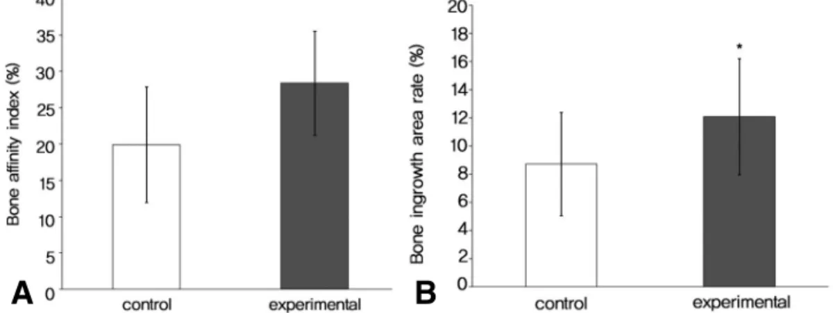
로 대조군 9마리와 실험군 11마리를 대상으로 실 험 결과를 도출하였다. Bone affinity index는 대조군이 19.9±7.9%, 실험군이 28.4±7.2%로 실험군이 더 높게 측정 되었으나(Fig. 3A), 통계 학적으로 유의하지는 않았다(p=0.056). Bone ingrowth area rate는 대조군이 8.7±3.7%, 실험군이 12.1±4.1%로 측정되었으며(Fig.
3B), 실험군이 대조군보다 통계학적으로 유의하 게 높았다(p=0.046).
고 찰
비스포스포네이트는 골 조직의 수산화인회석 (hydroxyapatite)에 높은 친화도를 갖고 있는 데, 골 흡수 억제 능력의 정도는 곁 사슬(side chain) 화학 조성에 의하여 결정된다. 에티드로네 이트(etidronate)나 클로드로네이트(clodronate) 와 같은 1세대 비스포스포네이트는 알킬기(alkyl group)나 할로젠화물(halide)을 갖고 있으며, 파 미드로네이트(pamidronate)나 알렌드로네이트 등의 2세대 비스포스포네이트는 아미노기(amino group)를, 졸레드론산이나 이반드로네이트(iban- dronate) 등의 3세대 비스포스포네이트는 링 사 슬(ring chain) 작용기를 갖고 있다.
비스포스포네이트는 질소 원자의 유무에 따라서 이분할 수 있는데, 질소 원자가 포함되어 있지 않 은 1세대 비스포스포네이트는 class Ⅱ aminoa- cyl-transfer RNA synthetase에 의하여 ATP 유사체(analogue)를 형성해서 파골세포의 자멸
사를 유도한다7). 반면에 질소 원자를 포함하는 2, 3세대 비스포스포네이트(아미노비스포스포네이 트)는 콜레스테롤과 지질 형성에 중요한 meval- onic acid pathway에서 중요한 역할을 하는 farnesyl pyrophosphate synthase (FPP synthase)의 작용을 억제하여 파골세포의 자멸사 를 유도한다6).
비스포스포네이트의 주된 역할은 파골세포에 의 한 골 흡수를 억제하는 것이지만, 골모세포에 의 한 골 형성 촉진 효과도 있는 것으로 보고되고 있
다8,11,23). 콜레스테롤 합성을 억제하는 statin계열
약물은 아미노비스포스포네이트처럼 mevalonic acid pathway를 억제하는데, statin 계열 약물 들이 골모세포에 의한 골 형성 촉진 효과가 있는 것이 보고된 바 있으며3), 이는 아미노비스포스포 네이트도 골 형성 촉진 효과가 있을 것으로 추정 할 수 있는 근거라 하겠다22). 실제로 아미노비스 포스포네이트가 골모세포의 증식과 성숙을 촉진하 고, 골모세포의 자멸사를 억제하며, 기원세포의 골모세포로의 분화를 유도한다는 실험 결과도 보 고된 바 있다8,11,16,23)
.
정형외과 영역에서는 비스포스포네이트의 골 흡 수 방지 및 골 형성 촉진 효과를 임상적으로 이용 하기 위한 연구가 많이 시행되고 있으며, 특히, 비스포스포네이트의 동화 작용 효과(anabolic effect)를 이용하여 인공 관절의 내구성을 향상시 키기 위한 연구가 활발하다. 비스포스포네이트의 투여가 마모 입자에 의한 골용해를 억제하는 것은 동물 실험을 통해서 보고된 바 있으며14), 응력 차
Fig. 3. Histomorphometric results for the bone affinity index (A) and the bone ingrowth area rate (B). Data are presented as mean (%) ± standard deviation (*p<0.05).
A B
단 효과에 의한 인공 관절 주변 골 조직의 약화 방지 효과는 실제 임상적으로도 적용되어서 우수 한 결과가 보고되었다2,24). 또한, 인공 관절의 해 리를 방지하기 위해서는 초기 골결합이 매우 중요 한데, 동물 실험을 통해서 비스포스포네이트의 투 여가 초기 골결합에도 우수한 효과가 있는 것으로 보고 되었고12), Hilding 등10)은 임상 실험을 통해 서 클로드로네이트의 투여가 인공 슬관절의 초기 이동을 방지하는 효과가 있다고 보고하였다.
널리 사용되고 있는 2세대 비스포스포네이트 제 재들은 경구 복용 약제로서 식도염 등 소화기계의 부작용 발생 우려로 인해서 복용법이 까다롭기 때 문에 환자의 순응도가 떨어지는 문제점이 있다.
이 점을 고려할 때 1년에 1회의 정맥 주사로 약효 가 유지되는 졸레드론산은 향후 경구 복용 약제들 을 대체할 것으로 예상된다. Bobyn 등5)은 다공 성 tantalum을 개의 척골에 6주간 삽입한 뒤에 졸레드론산의 투여 여부에 따른 골결합 정도를 비 교한 실험에서 졸레드론산을 투여한 군의 bone ingrowth area rate가 12.2%로 투여하지 않은 군의 6.6%보다 더 우수하다고 보고하였고, Tanzer 등20)은 수산화인회석이 피복된 다공성 tantalum의 표면에 일정량의 졸레드론산을 도포 하고 개의 척골에 12주간 삽입한 뒤 골결합 정도 를 비교한 실험에 졸레드론산을 도포한 군의 bone ingrowth area rate가 19.8%로 도포하 지 않은 군의 12.5%보다 더 우수하다고 보고한 바 있다. 본 실험에서 bone affinity index는 대조군과 실험군이 각각 19.9±7.9%, 28.4±
7.2%로 통계학적으로 유의한 차이는 없었지만 (p=0.056), 대체적으로 졸레드론산을 투여한 실 험군의 수치가 생리 식염수를 투여한 대조군의 수 치보다 높은 경향을 보였다. Bone ingrowth area rate는 대조군과 실험군이 각각 8.7±
3.7%, 12.1±4.1%로 실험군의 수치가 통계학적 으로 유의하게 높았다(p=0.046). 이는 앞서 설명 한 Bobyn 등5)과 Tanzer 등20)의 실험 결과와 일 치하는 것으로 졸레드론산의 투여가 다공성 삽입 물의 골결합을 촉진한다는 것을 의미한다.
Bobyn 등5)과 Tanzer 등20)은 개의 척골에 다공 성 tantalum을 삽입하고 backscattered scan- ning electron microscopy를 이용하여 골 내성
장 정도를 분석하는 동일한 실험 방법을 사용하였 다. Bobyn 등5)은 졸레드론산에 의한 다공성 삽 입물의 골결합 촉진 효과를 입증하기 위해서 다양 한 실험 방법으로 동일한 결과가 도출되어야 함을 지적한 바 있다. 본 실험은 가토와 다공성 니티놀 을 사용하여 기존의 방법과는 다른 조직형태학적 계측 방법을 사용하여 졸레드론산에 의한 다공성 삽입물의 골결합 촉진 효과를 입증했다는 점에서 의미가 있다고 판단된다.
결 론
졸레드론산의 약효가 기존의 2세대 비스포스포 네이트보다 우수하며, 경구 복용 약제에 비해서 약물의 투여가 훨씬 간단한 점을 감안할 때 임상 적으로 인공 관절의 내구성을 향상시키기 위한 방 법으로 졸레드론산의 투여가 좋은 방법의 하나가 될 수 있을 것으로 생각된다.
REFERENCES
1) Amanat N, Brown R, Bilston LE, Little DG: A single systemic dose of pamidronate improves bone mineral content and accelerates restoration of strength in a rat model of fracture repair. J Orthop Res, 23: 1029-1034, 2005.
2) Arabmotlagh M, Pilz M, Warzecha J, Rausc- hmann M:Changes of femoral periprosthetic bone mineral density 6 years after treatment with alendronate following total hip arthroplasty. J Orthop Res, 27: 183-188, 2009.
3) Ayukawa Y, Okamura A, Koyano K: Simvas- tatin promotes osteogenesis around titanium implants. Clin Oral Implants Res, 15: 346-350, 2004.
4) Bansiddhi A, Sargeant TD, Stupp SI, Dunand DC:Porous NiTi for bone implants: a review.
Acta Biomater, 4: 773-782, 2008.
5) Bobyn JD, Hacking SA, Krygier JJ, Harvey EJ, Little DG, Tanzer M:Zoledronic acid caus- es enhancement of bone growth into porous implants. J Bone Joint Surg, 87-B: 416-420,
2005.
6) Coxon FP, Thompson K, Rogers MJ: Recent advances in understanding the mechanism of action of bisphosphonates. Curr Opin Pharmacol, 6: 307-312, 2006.
7) Drake MT, Clarke BL, Khosla S: Bisphospho- nates: mechanism of action and role in clinical practice. Mayo Clin Proc, 83: 1032-1045, 2008.
8) Fromigue′′O, Body JJ: Bisphosphonates influ- ence the proliferation and the maturation of nor- mal human osteoblasts. J Endocrinol Invest, 25:
539-546, 2002.
9) Goodship AE, Blunn GW, Green J, Coathup MJ: Prevention of strain-related osteopenia in aseptic loosening of hip prostheses using periop- erative bisphosphonate. J Orthop Res, 26: 693- 703, 2008.
10) Hilding M, Ryd L, Toksvig-Larsen S, Aspen- berg P:Clodronate prevents prosthetic migra- tion: a randomized radiostereometric study of 50 total knee patients. Acta Orthop Scand, 71: 553- 557, 2000.
11) Im GI, Qureshi SA, Kenney J, Rubash HE, Shanbhag AS:Osteoblast proliferation and mat- uration by bisphosphonates. Biomaterials, 25:
4105-4115, 2004.
12) Jensen TB, Bechtold JE, Chen X, Søøballe K:
Systemic alendronate treatment improves fixa- tion of press-fit implants: a canine study using nonloaded implants. J Orthop Res, 25: 772-778, 2007.
13) Lewiecki EM: Intravenous zoledronic acid for the treatment of osteoporosis. Curr Osteoporos Rep, 6: 17-23, 2008.
14) Millett PJ, Allen MJ, Bostrom MP: Effects of alendronate on particle-induced osteolysis in a rat model. J Bone Joint Surg, 84-A: 236-249, 2002.
15) Patil N, Lee K, Goodman SB: Porous tantalum in hip and knee reconstructive surgery. J Biomed Mater Res B Appl Biomater, 89: 242-251, 2009.
16) Plotkin LI, Weinstein RS, Parfitt AM, Rober-
son PK, Manolagas SC, Bellido T:Prevention of osteocyte and osteoblast apoptosis by bisphos- phonates and calcitonin. J Clin Invest, 104:1363- 1374, 1999.
17) Simmons CA, Valiquette N, Pilliar RM:
Osseointegration of sintered porous-surfaced and plasma spray-coated implants: An animal model study of early postimplantation healing response and mechanical stability. J Biomed Mater Res, 47: 127-138, 1999.
18) Stewart M, Welter JF, Goldberg VM: Effect of hydroxyapatite/tricalcium- phosphate coating on osseointegration of plasma-sprayed titanium alloy implants. J Biomed Mater Res A, 69: 1-10, 2004.
19) Takemoto M, Fujibayashi S, Neo M, Suzuki J, Kokubo T, Nakamura T:Mechanical proper- ties and osteoconductivity of porous bioactive titanium. Biomaterials, 26: 6014-6023, 2005.
20) Tanzer M, Karabasz D, Krygier JJ, Cohen R, Bobyn JD:The Otto Aufranc Award: bone aug- mentation around and within porous implants by local bisphosphonate elution. Clin Orthop Relat Res, 441: 30-39, 2005.
21) Tekin U, Tu¨ z HH, Onder E, Ozkaynak O, Korkusuz P: Effects of alendronate on rate of distraction in rabbit mandibles. J Oral Maxillofac Surg, 66: 2042-2049, 2008.
22) Von Knoch F, Eckhardt C, Alabre CI, Schnei- der E, Rubash HE, Shanbhag AS:Anabolic effects of bisphosphonates on peri-implant bone stock. Biomaterials, 28: 3549-3559, 2007.
23) von Knoch F, Jaquiery C, Kowalsky M et al:
Effects of bisphosphonates on proliferation and osteoblast differentiation of human bone marrow stromal cells. Biomaterials, 26: 6941-3649, 2005.
24) Yamasaki S, Masuhara K, Yamaguchi K, Nakai T, Fuji T, Seino Y:Risedronate reduces postoperative bone resorption after cementless total hip arthroplasty. Osteoporos Int, 18: 1009- 1015, 2007.