115
서 론
원위부 요골의 골절은 전체골절의 1/6을 차지하는 매우 흔한
골절로서7,15), 대부분이 저 에너지 손상에 해당되며 비 수술적인
치료가 가능한 경우가 많다. 그러나 고 에너지 손상으로 인하여 요골 원위부 배측의 분쇄가 있는 관절 내 골절의 경우에는 비 관혈적 정복 및 석고고정만으로는 정복의 유지가 어려우며, 수 술적 처치가 필요하다4). 원위부 요골 골절에 대하여 여러 가지 수술적 치료법이 알려져 있지만, 골절의 정복 및 유지에 있어서 더 나은 치료방법에 대해서는 많은 논란이 있다6-8). 저자들은 시행되고 있는 수술법 중 외고정장치 단독치료 및 외고정장치 에 골 이식을 보강한 치료, 그리고 수장측 잠김 압박 금속판을 이용한 관혈적 정복술을 받은 환자들의 수술 후 기능적, 및 방 사선학적 결과를 후향적으로 분석하여 보고하고자 한다.
대상 및 방법
2006년 4월부터 2009년 5월까지 본원에 원위 요골 관절 내
골절로 입원한 217명의 환자 중, 배측 분쇄가 있고 수술 시행 후 1년 이상 추시가 가능했던 49예의 환자를 대상으로 후향적 연구 를 하였다. 수술적 치료의 적응증으로는 배측 분쇄가 있는 원위 부 요골의 관절 내 골절 중 불안정성을 동반한 경우로서, 불안정 성의 정의는 Lafontaine 등9)에 근거하여, (1) 20도 이상의 배측 굴곡이 있는 경우, (2) 배측 분쇄골절이 동반된 경우, (3) 관절 내 골절이 있는 경우, (4) 동반된 척골 골절이 있는 경우, (5) 60 세 이상 중 3가지 이상이 포함된 것으로 정의하였다. 전체 49예 중 골 이식 없이 외고정장치 혹은 외고정장치에 보조적 K-강선 고정술을 시행한 환자군이 19예 였고, 외고정 장치 혹은 외고정 장치에 보조적 K-강선 고정술을 시행하고 여기에 DBM (Orthoblast�3 cc)으로 골 이식을 시행한 환자군이 16예, 수장 측 잠김 압박 금속판을 이용한 환자군이 14예였다. 외고정장치 단독 치료군의 평균 나이는 68.7세(60세~78세)이었으며, 골이 식을 보강한 군은 67.8세(60세~78세), 금속판 이용군은 67.6 세(60세~82세)였고, 각각 남녀의 비율은 외고정장치 단독 치료 군에서는 7:12, 골이식을 보강한 군에서는 8:8, 수장측 잠김 압 박 금속판 이용군에서는 7:7이었다(Table 1). 치료 결과의 판정 을 위해 Mayo Wrist Score를 이용한 임상적 평가 및 2, 4, 6주 및 6, 12개월 추시의 방사선학적 평가를 시행하였다.
결 과
방사선학적으로 2, 4, 6주 및 6, 12개월 추시하였으며 통계
배측 분쇄가 있는 원위부 요골 관절내 골절의 3가지 수술적 치료 결과 비교: 외고정 기구 단독, 외고정 기구와
동종골이식 및 수장측 잠김 압박 금속판
최준철∙나화엽∙이영상∙김우성∙오경수∙송우석∙김세준 분당제생병원 정형외과
목적: 배측 분쇄가 있는 원위부 요골 골절에서 세가지 수술법에 따른 결과를 알아보고자 하였다.
대상 및 방법: 2006년 4월부터 2009년 5월사이의 60세 이상의 배측 분쇄가 있는 원위부 요골 골절이 있으면서 불안정성을 보 인 환자 49명을 대상으로 하였다. 수술 1년 후까지의 방사선학적 추시관찰을 통하여 골절 정복의 유지 여부 및 Mayo Wrist Score를 통한 임상적 결과를 비교하였으며, 일원배치 분산분석(One-Way ANOVA)을 통하여 연관성을 검정하였다.
결과: 세군 모두에서 수술 직후에는 만족스러운 정복을 얻을 수 있었으나, 추시 소견상 외고정장치 단독으로 고정시에는 외고 정장치에 동종골 이식을 추가한 군 및 수장측 잠김 압박 금속판을 사용한 군에 비해서 유의하게 정복 소실이 있음을 확인하였 다.(p<0.05) Mayo Wrist Score는 세군 모두에서 의미있는 차이는 없었다.
결론: 배측 분쇄가 심한 원위부 요골의 관절내 골절에서 외고정 장치만 사용한 치료시에는 해면골 결손으로 인한 정복유지의 어려움이 있어 불충분한 치료로 판단된다.
색인 단어: 요골 원위부 골절, 외고정장치, 동종 골 이식, 수장측 압박 금속판
통신저자: 김 우 성
경기도 성남시 분당구 서현동 255-2 대진의료재단 분당제생병원 정형외과 TEL: 031) 779-0175∙FAX: 031) 779-0176 E-mail: w00wa@dmc.or.kr
학적 검정은 일원배치 분산분석(One-Way ANOVA)을 사용 하였다(Table 2). 방사선학적으로 전 예에서 골유합 소견을
보였으며, 외고정장치 단독 치료군에서는 수술 전과 마지막 추 시상에서 요골길이는 1.63 mm에서 8.85 mm로, 요골 경사는
Table 1. Epidemiologic values and Mayo Wrist Score in 3 groups
External Fixation External Fixation with allo bone graft Volar anatomical locking Plate p-value
Age(year) 68.7 (60-78) 67.8 (60-78) 67.6 (60-82) 0.856
Sex (M:F) 7:12 8:8 7:7 0.680
Mayo wrist score 77.63 79.68 82.5 0.096
Table 2. Mean radiological measurements in 3 groups
External Fixation External Fixation Volar anatomical
p-value with allo bone graft locking Plate
Pre-OP Volar tilt
-21.18 -22.67 -22.90- 0.6330
(degrees from neutral) Radial inclination
-14.11 -13.42 13.80 0.1850
(degrees)
Radial length (mm) -01.63 -01.75
01.52 0.755
Post-OP Volar tilt 0
-11.94 -11.48 11.42 0.6040
(degrees from neutral) Radial inclination
-23.71 -23.24 24.86 0.3740
(degrees)
Radial length (mm)
-11.34
-11.57 11.7
0 0.819
2 weeks Volar tilt 0
-12.14 -11.29 11.27 0.4710
post-operatively (degrees from neutral) Radial inclination
-23.06 -22.98 24.80 0.3100
(degrees)
Radial length (mm)
-10.86
-11.34 11.59 0.652
4 weeks Volar tilt 0
-11.61 -11.32 11.09 0.7590
post-operatively (degrees from neutral) Radial inclination
-22.27 -21.95 24.45 0.2490
(degrees)
Radial length (mm)
-10.60 134 11.24 0.609
6 weeks Volar tilt 0
-10.88 -11.15 11.09 0.7850
post-operatively (degrees from neutral) Radial inclination
-21.77 -22.78 24.62 0.1020
(degrees)
Radial length (mm)
-10.14
-11.25 11.48 0.135
6 months Volar tilt -0 0
9.33 -10.93 11.00 0.0030
post-operatively (degrees from neutral) Radial inclination
-20.14 -22.70 24.64 0.0100
(degrees)
Radial length (mm)
-10.88
-10.97 11.45 0.024
12 months Volar tilt 0
-08.20 -10.84 10.85 0.0001
post-operatively (degrees from neutral) Radial inclination
-18.83 -22.30 24.37 0.0001
(degrees)
Radial length (mm) 8.85
-10.70 11.41 0.0040
14.11도에서 18.83도로, 수장측 경사는 -21.18도에서 8.20 도로 측정되었으며, 골 이식을 보강한 군에서는 요골길이는 1.75 mm에서 10.70 mm로, 요골 경사는 13.42도에서 22.30 도로, 수장측 경사는 -22.67도에서 10.84도로 측정되었고 수 장측 잠김 압박 금속판을 이용한 군에서는 요골길이는 1.52 m 에서 11.41 mm로, 요골 경사는 13.8도에서 24.37도로, 수장 측 경사는 -22.9도에서 10.85도로 측정되었다. 세군 모두에 서 수술 전후에는 만족스러운 정복을 얻을 수 있었으며, 수술 후 6,12개월째의 추시에서는 외고정장치 단독 치료군에서 다 른 두 군에 비해 유의한 정도로 정복소실이 발생하였고 (P<0.05), 그 중 3예는 수술 후 4주에 추시한 방사선 소견상에
서 수술 전과 큰 차이를 보이지 않을 정도의 정복소실이 있었 다(Fig 1, 2, 3). 임상적 결과는 Mayo Wrist Score를 통한 기 능적 평가에 따라 외고정장치 단독 치료군 에서는 평균 77.63 점이었으며, 골 이식을 보강한 군에서는 평균 79.68점, 수장측 잠김 압박 금속판 이용군에서는 평균 82.5점으로 세군 전례에 서 양호 이상의 결과를 보였다 (P>0.05)(Table 1).
고 찰
원위부 요골의 치료 목표가 해부학적 정복, 안정된 고정을 통한 정복 유지 그리고 수근관절 운동 회복을 통한 일상 생활
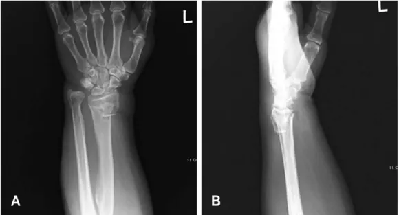
Fig. 1. Preoperative radiographs of 74-year-old female who had fallen down. Distal radius intraarticular fracture with dorsal com- minution and loss of radial shortening, radial inclination, volar tilting was seen. (A: anteroposterior view, B: lateral view)
A B
Fig. 2. Immediately postoperative radiographs tried external fixation without allogenous bone graft. We get a satisfactory restoration in radial length, radial inclination and volar tilting. (A: anteroposterior view, B: lateral view)
A B
이나 직업으로 조기 복귀라는 점에서 골절 정복 후 정복 유지 는 매우 중요한 부분이다. 그러나 특히 불안정성 원위부 요골 골절은 골 간단부에 위치하며, 피질골의 분쇄가 있어 정복 및 정복의 유지가 쉽지 않다2). 외고정장치는 심한 분쇄양상과 관 절면을 침범하는 요골 원위부 골절에서 인대 정복술(liga- mentotaxis)을 이용하여 해부학적 정복을 얻을 수 있고, 핀이 고정되는 위치를 자유롭게 조정할 수 있는 장점이 있어7)널리 사용되어져 왔다. 그러나 수근부 및 수지의 구축이 올 수 있으 며4)저자들은 구축을 최소화하기 위하여 술 후 4주부터 조기운 동을 시행하였다. 또한 분쇄가 심한 골절에서는 해면골의 소실 로 인한 정복 소실이 발생할 수 있으며1), 저자들의 경우도 요골 단축 및 요골 경사각의 소실을 경험하였다. 술 후 이러한 문제 점들을 최소화하기 위해 골 간단부 결손에 대해 구조적인 지지 대의 필요성이 보고되었으며11), 현재 임상적으로 자가 골 이식 및 동종 골 이식이 가장 많이 이용되는 골 결손부 대체물이다.
자가 골 이식은 생체적합성이 우수하면서 적당한 생역학적 강 도를 제공한다는 장점을 가지지만14), 공여부의 혈종과 감염, 동 통, 장골 능의 골절, 신경손상, 수술시간 지연, 출혈량 증가 등 의 합병증이 보고되었다3). Herrera 등은 동종 골 이식이 HIV (human Immunodeficiency virus) 같은 질병의 전염 위험성 이 있으나12), 전염의 가능성은 200만분의 1에서 800만 분의 1 정도로 낮으며, 현대적인 살균 기술로 처리할 때 HIV가 비활성 화되는 것을 확인하여, 그 안전성을 언급하였고, 골전도능을 가질 뿐 아니라 제한적이지만 골 유도능도 있어 동종골을 대신 할 수 있는 골 결손부 대치물로 보고하였다3). 금속판을 이용한 내고정술은 외고정 장치에 비해 보다 정확한 해부학적 정복을 얻을 수 있고 견고한 고정을 통해 조기 관절운동이 가능하다는 장점을 가진다15). 그 중에서도 잠김 압박 금속판은 전통적인 수 장측 금속판 (conventional volar plate)에 비해 나사못과 금
속판 사이의 움직임이 없어서 골편의 단단한 고정이 없이 버팀 목 작용만으로도 정복의 유지가 가능하여 심한 분쇄 골절시 정 복의 유지에 있어 수장측에 사용시에도 우수한 결과를 기대할 수 있다5,10,13).
본 연구에서는 세 군간 비교에서 외고정장치 단독 치료군에 서 유의하게 정복의 소실이 있는것으로 나타났으며(P<0.05), 외고정 장치를 단독으로 사용하였을 시 정복 소실의 가능성이 높으므로, 배측 분쇄가 심한 원위부 요골의 관절 내 골절에서 는 골 이식을 추가하거나 수장측 잠김 압박 금속판을 이용하는 방법이 정복유지에 유용한 수술법이라 생각된다.
결 론
배측 분쇄가 심한 원위부 요골의 관절 내 골절에서 외고정장 치만 사용한 치료시에는 해면골 결손으로 인한 정복유지의 어 려움이 있어 불충분한 치료로 판단되며 외고정장치에 DBM 골 이식을 보강한 치료 및 수장측 잠김 압박 금속판을 이용한 치 료에서는 임상적, 그리고 방사선학적으로 양호한 결과를 얻을 수 있었다.
참고문헌
01. Arora J, and Malik AC.:External fixation in comminut- ed, displaced intra-articular fractures of the distal radius:
is it sufficient? Arch Orthop Trauma Surg, 125(8): 536-40, 2005.
02. Arota R, Lutz M, Fritz D, Zimmerman R, Oberladstatter J, and Gabl M: Palmar locking plate for treatment of unstable dorsal dislocated distal radius fractures. Arch Fig. 3. postoperative 4-weeks radiographs shows loss of reduction. (A: anteroposterior view, B: lateral view)
A B
Orthop Trauma Surg, 125:399-404, 2005.
03. Arrington ED, Smith WJ, Chambers HG, Bucknell AL and Davido NA: Complications of iliac crest bone graft harvesting. Clin Orthop, 329: 300-309, 1996.
04. Capo JT, Swan KG Jr, and Tan V.: External fixation techniques for distal radius fractures. Clin Orthop Relat Res, 445:30-41, 2006.
05. Chung KC, Watt AJ, Kotsis SV, Margaliot Z, Haase SC, and Kim HM: Treatment of unstable distal radial fractures with the volar locking plating system. J Bone Joint Surg Am, 88: 2687-2694, 2006.
06. Egol K, Walsh M, Tejwani N, McLaurin T, Wynn C, and Paksima N.: Bridging external fixation and supple- mentary Kirschner-wire fixation versus volar locked plat- ing for unstable fractures of the distal radius: a random- ized, prospective trial. J Bone Joint Surg Br, 90(9):1214- 21. 2008.
07. Herrera M, Chapman CB, Roh M, Strauch RJ and Rosenwasser MP: Treatment of unstable distal radius fractures with cancellous allograft and external fixation. J Hand Surg, 24-A: 1269-1278, 1999.
08. Jung HG, Choi JB, Seo SY and Choi YS: Radiologic reduction loss after surgical treatment of distal radial fracture. J Korean Fractures Soc, 19: 454-459, 2006.
09. Lafontaine M, Hardy D, and Delince P.: Stability assessment of distal radius fractures. Acta Orthorp Scand
S, 108:208-210, 1989.
10. Leung F, Tu YK, Chew WY, and Chow SP.: Comparison of external and percutaneous pin fixation with plate fixation for intra-articular distal radial fractures. A randomized study. J Bone Joint Surg Am, 90(1): 16-22, 2008.
11. Leung KS, Shen WY, Leung PC, Kinninmouth AW, Chang JC, and Chan GP: Ligamentotaxis and bone grafting for comminuted fractures of the distal radius. J Bone Joint Surg Br, 71-B: 838-842, 1989.
12. Mellonig JT, Prewett AB and Moyer MP: HIV inactiva- tion in a bone allograft. J Periodontol, 63: 979-983, 1992.
13. Orbay JL, and Touhami A.: Current Concepts in Volar Fixed-angle Fixation of Unstable Distal Radius Fractures.
Clin Orthop Relat Res, 445:58-67, 2006.
14. Rogachefsky RA, Lipson SR, Applegate B, Ouellette EA, Savenor AM, and McAuliffe JA.: Treatment of severely comminuted intra-articular fractures of the distal end of the radius by open reduction and combined internal and external fixation. J Bone Joint Surg Am, 83-A(4);509- 19, 2001.
15. Willis AA, Kutsumi K, Zobitz ME, and Cooney WP 3rd: Internal fixation of dorsally displaced fractures of the distal part of the radius. A biomechanical analysis of volar plate fracture stability. J Bone Joint Surg Am, 88: 2411- 2417, 2006.
Comparison of Three Surgical Method Outcomes for Dorsal Communition Distal Radial Fractures:
External Fixation, External Fixation with Allogenous Bone Graft (DBM) and Volar Locking Plate Fixation
Joon-Cheol Choi, M.D., Hwa-Yeop Na, M.D.,Young-Sang Lee, M.D., Woo-Sung Kim, M.D., Kyung-Soo Oh, M.D., Woo-Suk Song, M.D., Se-Jun Kim, M.D.
Department of Orthopedic Surgery, Bundang Jesaeng General Hospital, Daejin Medical Center, Seonggnam, Gyounggi, Korea
Purpose: To evaluate the outcomes of three different surgical approaches in treating a distal radius fracture with dorsal communition.
Materials and Methods: From April, 2006 to May, 2009, 49 patients over age of 60years old with unstable dorsal communition distal radius fracture were analyzed. We compared maintainability of reduction by radiological follow-up observations and clinical outcomes using Mayo wrist scores.
One-Way ANOVA test was performed to examine the statistical correlation between the methods.
Results: All three approaches provided satisfactory post-op reduction results. However, follow-up observations showed significant loss of reduction in the external fixation only group compared to the added bone graft group and the volar anatomical locking plate group (P<0.05). Mayo wrist score was indifferent between the three groups.
Conclusion: Treatment of a distal radius fracture with severe dorsal communition using an external fixation only appears to be insufficient in maintaining reduction of a cancellous bone defect.
Key Words: Distal radius fracture, External fixation, Allogenous bone graft, Volar locking compres- sion plate
Address reprint requests to Woo-Sung Kim, M.D.
Department of Orthopaedic Surgery, Bundang Jesaeng General Hospital, Daejin Medical Center, 255-2, Seohyun-dong, Bundang-gu, Seongnam-si, Gyeonggi-do, Korea
TEL: 82-31-779-0175, FAX: 82-31-779-0176, E-mail: w00wa@dmc.or.kr
= ABSTRACT =