Introduction
Secundum atrial septal defect (ASD) is a common congeni- tal heart disease identified in adulthood. Patients with ASD and left to right shunt are at risk for developing pulmonary arterial hypertension (PAH).1) However, a minority of patients (< 1%) develop precocious, severe pulmonary hypertension with shunt reversal.2) It is a current practice to assess the re- versibility of pulmonary hypertension by means of pulmonary vasodilator testing during right heart catheterization in pa- tients with shunt-related pulmonary hypertension.3) Patients with irreversible PAH are considered ineligible for shunt clo- sure because of the risk of right ventricular (RV) decompensa- tion after the intervention.4)
There are several reports about successful surgical or percu- taneous ASD closure in patients with severe and seemingly ir- reversible pulmonary hypertension.4-6) Here, we report a pa- tient who showed remarkable recovery of severe PAH after percutaneous closure following 1 year use of the endothelin re- ceptor antagonist, bosentan (Tracleer, Actelion, Allschwil, Switzerland).
Case
A 20-year-old woman presented with progressively worsen- ing dyspnea for 6 months. She had no history of evaluation or treatment for her symptoms, although the symptoms started at the age of high school. Her symptoms became worse with time, and she presented with NYHA class III exertional dys- pnea and orthopnea. Her height, weight, and body surface area were 156 cm, 49 kg, and 1.45 m2 respectively. Her vital signs were blood pressure 104/68 mmHg, heart rate 104/min, and respiratory rate 22/min. A regular heart beat was detected with wide fixed splitting of S2 at the pulmonary valve area, and no clubbing of the fingers or nails was observed. A simple chest X-ray showed cardiomegaly and a dilated pulmonary trunk with cephalization of pulmonary vascular marking.
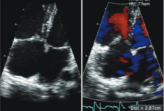
Electrocardiography revealed normal sinus rhythm with com- patible findings of RV hypertrophy. A transthoracic echocar- diogram revealed a large tissue defect of 29 mm with a bi-di- rectional shunt through the interatrial septum (Fig. 1). The RV was prominently dilated and revealed significantly de- creased contractility with a D-shaped left ventricle (LV) show- Joo Young Lee, RDCS1, Nam Jin Park, RDCS1, Dea Sung Ahn, MD1, Jae Hoon Jung, MD1, Dong Hee Shin, MD1 and Dal Soo Lim, MD1
Departments of 1Internal Medicine, 2Pediatrics, Sejong General Hospital, Bucheon, Korea
The presence of severe pulmonary arterial hypertension (PAH) in patients with atrial septal defect (ASD) is still thought to preclude shunt closure, although there are several reports of good clinical outcomes after vasodilator therapy. We report the case of a young woman with ASD and severe PAH who was able to successfully undergo percutaneous shunt closure following 1 year use of the oral endothelin receptor antagonist, bosentan.
KEY WORDS: Atrial septal defect · Pulmonary hypertension · Septal occlude device.
• Received: April 1, 2013 • Revised: May 29, 2013 • Accepted: August 12, 2013
• Address for Correspondence: In Hyun Jung, Department of Internal Medicine, Sejong General Hospital, 28 Hohyeon-ro 489beon-gil, Sosa-gu, Bucheon 422-711, Korea Tel: +82-32-340-1447, Fax: +82-32-340-1236, E-mail: [email protected]
• This is an Open Access article distributed under the terms of the Creative Commons Attribution Non-Commercial License (http://creativecommons.org/licenses/by-nc/3.0) which permits unrestricted non-commercial use, distribution, and reproduction in any medium, provided the original work is properly cited.
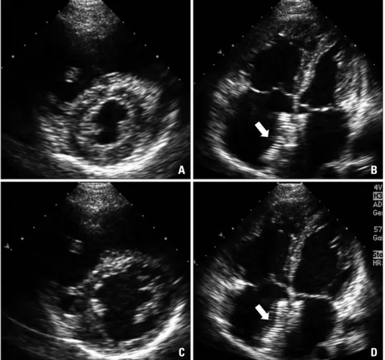
Fig. 2. Initial echocardiography reveals D-shaped left ventricle compressed by dilated right ventricle. A and B:
End-systole. C and D: End-diastole.
A
C
B
D
Fig. 1. Transthoracic echocardiogram shows a large tissue defect of 29 mm with a bi-directional shunt through the interatrial septum.
tension with a mean pulmonary arterial pressure of 48 mmHg (76/36 mmHg absolute) and pulmonary vascular resistance (Rp) of 9.6 Wood units (WU) on room air. The mean system- ic arterial pressure was 84 mmHg (97/73 mmHg absolute).
After administering oxygen (10 L/min for 10 minutes via a nasal prong) and inhaling iloprost, her mean pulmonary arte- rial pressure was 50 and 46 mmHg respectively, and had not changed significantly. We failed to identify the pulmonary ar- terial responsibility after pulmonary vasodilator administra- tion. Based on these findings, closure of the ASD was not per- formed. Instead, medical treatment including oral bosentan was started to improve patient’s symptom.
Her symptoms and exercise tolerance improved from NYHA class III to NYHA class I to II after 1 year of using bosentan. She was able to walk 426 m during the 6-min walk test without desaturation. A follow-up echocardiogram showed no significant interval change except slight improve- ment in RV contractility compared to the initial echocardio- gram, even though her symptoms were much improved. TR Vmax was 4.3 m/sec by continuous wave Doppler test, but there was no significant improvement in comparison with the initial test. Although the follow-up echocardiogram result was very disappointing, we decided to perform a cardiac cath- eterization and balloon occlusion test to confirm the change in
minutes. As a result, mean pulmonary arterial pressure de- creased to 36 mmHg (48/25 mmHg absolute), and mean aortic pressure increased to 85 mmHg (118/70 mmHg absolute).
We decided to close the ASD with a transcatheter occluder device considering her young age and the high perioperative risk. We performed percutaneous closure of the defect with a 34 mm Amplatzer® septal occluder (AGA Medical Corp., Minneapolis, MN, USA).
No residual leak was observed on the follow-up transthorac- ic echocardiography. The patient was given antiplatelet thera- py and was maintained on 62.5 mg bosentan bid with diuret- ics and an angiotensin receptor blocker for 1 year. Finally, her physical activity and symptoms were much improved as she could walk 554 m on the walk test 1 year after the device was deployed. A chest X-ray showed remarkably decreased pul- monary vascularity, but cardiomegaly remained. Follow-up transthoracic echocardiography showed increased left atrial size from 37 to 42 mm and LV chamber size of end-diastole/
end-systole from 40/23 to 46/28 mm. RV size had decreased significantly, and RV contractility was much improved in comparison with the initial study, but a compressed LV still remained on the parasternal short axis view (Fig. 4). TR Vmax also decreased markedly from 4.6 m/sec to 3.0 m/sec (Fig. 2).
Fig. 3. The maximal tricuspid regurgitation velocity (TR Vmax) was measured by continuous wave Doppler to estimate pulmonary arterial systolic pressure. Initial TR Vmax was 4.6 m/sec (A), which was suggestive of severe resting pulmonary arterial hypertension (estimated pulmonary arterial systolic pressure was 95 mmHg). The follow-up TR Vmax decreased remarkably to 3.0 m/sec (B) 1 year after percutaneous device closure.
A B
Discussion
The presence of irreversible PAH in patients with ASD is still thought to preclude shunt closure. Closure of such shunts is as- sociated with decrease of cardiac output and increase of right- sided heart failure and death. Thus, defect closure in these pa- tients should be performed only if the benefits of abolishing the shunt outweigh the risks of surgical or percutaneous closure.7) An additional reason for concern when contemplating surgery in ASD patients with PAH is the high perioperative risk.
Perioperative risk can be reduced in patients with the de- fects amendable by percutaneous closure. Balint et al.8) from Canada reported the outcomes of percutaneous closure in pa- tients with ASD and PAH. They concluded that transcatheter closure in patients with secundum ASD and PAH can be suc- cessfully performed in selected patient with good outcomes.
However, they diagnosed pulmonary hypertension based on echocardiographic data and not cardiac catheterization data.
Thus, they did not show the value of Rp and did not perform lung biopsies. Additionally, their patients were not so serious- ly compromised. Therefore, caution should be used when in- terpreting their results.
Several reports have shown good results of defect closure even in irreversible severe PAH with ASD after advanced therapy.4-6)9) Previously published articles suggested that pul- monary vasodilator therapy may offer patients with irrevers- ible anatomical changes of the pulmonary vascular bed a change for further improvement of pulmonary pressures.7)10) In recent years, potent oral vasodilators aimed at the pulmo- nary circulation have been available, with promising results.
The vasodilators appear to be effective in reducing Rp and symptoms in patients with near-systemic pulmonary pressure, previously thought to have irreversible pulmonary vascular disease.11)12) We thought that our patient was one of those who demonstrate a significant response to vasodilator therapy, be- cause her physical activities and symptoms were much im- proved and mean pulmonary arterial pressure reduced from 45 mmHg to 36 mmHg with temporary balloon occlusion.
The exact mechanism of how preoperative pulmonary vaso- dilator therapy works on a pathologically irreversibly changed pulmonary artery is poorly understood. One of the possible hypotheses is that there is possible reverse remodeling of pul- monary vascular changes with endothelin receptor antagonists
Fig. 4. The follow-up transthoracic echocardiogram shows the markedly reduced size of the right ventricle and increased left ventricular dimensions with slight compression after percutaneous closure. A and B: End-systole. C and D: End-diastole. Arrows indicate the device located in the interatrial septum (B and D).
A
C
B
D
In summary, we experienced a case of a young woman who had ASD with irreversible severe pulmonary hypertension, but the pulmonary hypertension improved remarkably after successful percutaneous device closure following 1 year of bosentan.
References
1. Konstantinides S, Geibel A, Olschewski M, Görnandt L, Roskamm H, Spillner G, Just H, Kasper W. A comparison of surgical and medical therapy for atrial septal defect in adults. N Engl J Med 1995;333:469-73.
2. Besterman E. Atrial septal defect with pulmonary hypertension. Br Heart J 1961;23:587-98.
3. Therrien J, Gatzoulis M, Graham T, Bink-Boelkens M, Connelly M, Niwa K, Mulder B, Pyeritz R, Perloff J, Somerville J, Webb GD.
Canadian Cardiovascular Society Consensus Conference 2001 update: Rec- ommendations for the Management of Adults with Congenital Heart Dis- ease--Part II. Can J Cardiol 2001;17:1029-50.
4. Schwerzmann M, Zafar M, McLaughlin PR, Chamberlain DW,
geted pulmonary arterial hypertension therapy. Int J Cardiol 2008;129:
163-71.
8. Balint OH, Samman A, Haberer K, Tobe L, McLaughlin P, Siu SC, Horlick E, Granton J, Silversides CK. Outcomes in patients with pul- monary hypertension undergoing percutaneous atrial septal defect closure.
Heart 2008;94:1189-93.
9. Yamauchi H, Yamaki S, Fujii M, Iwaki H, Tanaka S. Reduction in re- calcitrant pulmonary hypertension after operation for atrial septal defect.
Ann Thorac Surg 2001;72:905-6; discussion 906-7.
10. Jung JW. Pulmonary arterial hypertension of congenital heart diseases:
from reversible pulmonary hypertension to eisenmenger syndrome. Korean Circ J 2007;37:287-97.
11. Diller GP, Gatzoulis MA. Pulmonary vascular disease in adults with congenital heart disease. Circulation 2007;115:1039-50.
12. Galiè N, Beghetti M, Gatzoulis MA, Granton J, Berger RM, Lauer A, Chiossi E, Landzberg M; Bosentan Randomized Trial of Endo- thelin Antagonist Therapy-5 (BREATHE-5) Investigators. Bosentan therapy in patients with Eisenmenger syndrome: a multicenter, double-blind, randomized, placebo-controlled study. Circulation 2006;114:48-54.