Journal of Bacteriology and Virology 2008. Vol. 38, No. 3 p.161 – 166
Effect of Culture Conditions on the Protein Fibril Expression of Candida albicans
Kyoung-Ho Lee
1, Young Chul Yang
2, Sun Ju Choi
1, Su Jung Park
1, Joo Young Park
1, Yoon Sun Park
3and Woon-Seob Shin
1*1
Department of Microbiology,
2Department of Anatomy, Yonsei University Wonju College of Medicine and Institute of Basic Medicine, Yonsei University, Wonju, Republic of Korea
3
Department of Microbiology, Kwandong University College of Medicine, Gangneung, Republic of Korea
Received : July 14, 2008 Revised : September 3, 2008 Accepted : September 12, 2008
Candida albicans is an important human pathogen that causes systemic infections, predominantly among population with weakened immune system. Cell wall structures of C. albicans are important to adhere to host tissue and evade to host immune system. Among cell wall structure, the outermost fibrillar layer of C. albicans is of interest since it may play important roles in antigenicity, phagocytosis, and adherence. The expression of virulent factors could be affected by the growth conditions. The dynamic nature of the cell surface alters the physical properties of the fungal interface with host cells and thereby influences adhesion to the host and recognition by components of the host immune system.
In this study, we investigated the effects of culture conditions on cell surface fibril expression of C. albicans by a transmitting electron microscopy and SDS-PAGE. The protein fibril of C. albicans was expressed in the presence of whole serum, however, the fibril expression was decreased in 25% serum and serum containing 1% glucose. Also, germ tube can be induced by serum, RMPI medium, N-acetyl glucosamine, and 39℃ culture condition, hence, the fibrillar structure of C. albicans was detected only in serum-induced germ tube. The expression of fibril layer and the major fibril proteins of 66, 47, 30 kDa were reduced as increasing cell concentration of intial inoculum from 2×10
7cells/ml to 8×10
7cells/ml. The fibrillar layer of C. albicans was expressed in serum early within 10 min, and the thickness of fibril layer was increased according to the increase of culture time. When the fibrillar proteins were analysed by SDS-PAGE, major protein of 30 kDa was maintained continuously during over night culture although expression of the other proteins were various. These results suggest that expression of serum induced protein fibril is influenced by culture conditions and is not related to hyphal transition of C. albicans.
Key Words: Candida albicans, Fibril, Culture condition
서 론
Candida albicans는 정상균총으로 존재하고 일반적으로 질병을 유발하지 않으나, 면역저하된 사람에게는 치명적
인 전신증상을 일으킬 수 있다 (22). 이러한 칸디다증은 AIDS, 장기이식, 면역저하자, 노년인구의 증가 등으로 지속적으로 문제가 되고 있다 (9,25). 칸디다균의 주요 병독인자로 adhesin, 가수분해효소, 발아관 형성, 세포소 수성, 균막형성능 등이 알려져 있으며 (1,6,8,20,21), 이들 병독인자들은 복합적으로 작용하여 질병을 유발한다. 칸 디다균이 질병을 유발하기 위해서는 첫째로 숙주세포에 부착해야 하고, 이어서 가수분해효소에 의한 조직침습 을 통하여 숙주 내로 들어가야 하고 숙주 내에서 생존 161
*Corresponding author: Woon-Seob Shin. Department of Microbiology, Kwandong University College of Medicine, Gangneung, 210-701, Republic of Korea.
Phone: +82-33-649-7470, Fax: +82-33-641-1074, e-mail: [email protected]
해야 한다 (1).
칸디다균의 구조물 중 세포표면 물질은 숙주세포, 카 테터에의 부착뿐만 아니라 숙주 내에서의 생존에도 중 요한 역할을 한다. C. albicans의 세포표면 물질로 laminin receptor (28), compliment receptor (7), fibrolectin receptor (26), mannoprotein (14), chitin (19), hydrophobic protein (11) 등이 보고되었으며 이들은 칸디다증 병인과정 중에서 숙주 조 직에 부착을 촉진하는 것으로 알려졌다. 이들 세포표면 물질들은 칸디다균의 외부환경에 의해 발현이 조절될 수 있으며 시험관 내에서 보다 숙주의 생체 내에서 높게 발 현된다고 알려져 있다 (10).
칸디다균의 최외각 fibril 구조물은 항원성, 항탐식성, 부착인자로서 중요한 의미가 있으며, 이 fibril 구조물 성 분이나 형태학적 구조도 배양 조건에 의하여 영향을 받 는다 (2,10). 본 연구자들은 숙주 내 환경과 유사한 혈청 에서 C. albicans를 배양하여 fibril 구조물의 발현 및 그 성분의 변화를 보고한 바 있으나 (17), 혈청에 의해서는 발아관도 유도된다. 따라서 본 연구에서는 다양한 조건 으로 유도된 발아관에서 단백 fibril이 발현되는 것을 조 사하였으며, 또한 다양한 배양 조건이 단백 fibril 발현에 미치는 영향들을 조사하였다.
재료 및 방법
1. 균주 및 배양
C. albicans는 원주의과대학 미생물학교실에 보관중 인 C. albicans ATCC 10231을 발아관 실험, fibril 발현실 험에 사용하였으며, 실험에 사용하기 전에 균을 실온에 서 YEPD 배지 (1% yeast extract, 2% glucose, 2% peptone) 에 하룻밤 미리 배양한 후 원심 분리 (2000×g, 15분)로 균을 수거하고 멸균된 인산완충액 (100 mM phosphate buffer, saline, pH 7.4)로 3회 세척하여 사용하였다.
2. 세포농도의 측정
C. albicans의 세포농도는 YEPD 배지에서 하룻밤 배양 한 것을 인산완충액으로 세척하여 혈구계수기를 이용하 여 균수를 측정한 후 시험배지에 세포농도가 1×10⁷ ~ 16
×107 cells/ml가 되도록 접종하여 각 실험에 사용하였다.
3. 발아관 형성실험
C. albicans의 발아관 형성을 위하여 C. albicans ATCC
10231을 25℃ YEPD에서 하루 배양한 후 인산완충액으 로 세척하고 세포농도를 2×10⁷ cells/ml 이하로 맞추어 토끼혈청, YEPD, RPMI 배지, N-acetyl glucosamine 배지 에 접종하여 2시간 동안 배양하여 발아관 형성을 조사하 였다.
4. Fibril 형성
Fibril 구조물 발현을 위해 균을 토끼혈청에서 2시간 동안 진탕 배양하였다. 배양 후 균을 4℃에서 원심 분리 (2000×g, 15분)로 수거하고 냉각된 인산완충액으로 두 번 세척한 후 fibril 구조 관찰과 fibril 단백발현을 위한 시료 로 사용하였다.
5. 투과 전자현미경
전자현미경의 표본제작은 Methods for general and molecular bacteriology 방법 (3)에 따라 제작하였다. 칸디 다균을 4℃ 인산완충액으로 세척한 후 pH 7.4의 인산완 충액으로 조정된 2.5% glutaraldehyde 용액으로 4℃에서 24시간 전 고정시키고, 동일한 glutaraldehyde 용액으로 세척 후 2% OsO4 용액으로 90분간 후 고정하였다. 이후 에탄올 농도를 60% 에탄올에서 무수 에탄올까지 순차적 으로 각각 10분씩 처리하여 탈수시킨 후 propylene oxide 로 치환하고 epon 혼합액에 포매하였다. 다이아몬드 나 이프를 사용하여 epon block을 ultramicrotome (Sorvall MT- 5000; DuPont, Boston, MA, USA)으로 50~60 nm 두께로 초박절하고 uranyl acetate와 lead citrate로 30분간 이중 염 색한 후, 투과 전자현미경 (JEM-1200EXII, JEOL, Japan) 으로 세포표면의 구조를 관찰하였다.
6. 세포벽 처리
원침된 칸디다균 100 μl을 인산완충액 440 μl로 희석하 고 여기에 lyticase (1250 unit/ml)를 60 μl 넣어 25℃에서 2 시간 동안 반응시킨 후 5000×g에서 10분간 원심 분리 하여 침전물과 상등액을 얻었다. 침전물은 형태 분석에 사용하였고, 상층액은 에탄올을 90%되게 첨가하여 단백 질을 회수하여 단백 분석에 사용하였다.
7. 세포벽 단백 분석
위에서 분리된 단백질을 Bradford 방법 (4)으로 정량하 였으며, 전기영동하려는 시료와 sample buffer (1% SDS, 5% β-mercaptoethanol, 0.02% bromophenol blue, 10% glycerol,
1.25 M Tris-HCl, pH 6.8)를 동량의 비율로 섞은 후 100℃
에서 2분간 전 처리하여 시료로 사용하였다. SDS-PAGE 는 Laemmli의 방법 (18)으로 slab gel을 사용하는 vertical electophoresis unit (SE 600, Hoefer Scientific Instruments, San Francisco, CA, USA)를 사용하였다. 10~15%의 polyacryla- mide gel을 사용하여 전류가 닿는 slab gel의 상위 표면적 1 cm2 당 3 mA의 전류를 흘려 전기영동하고, 완충 용 액으로 tris-glycine buffer (pH 8.3)를 사용하였다. 염색은 Coomassie briliant blue G-250 (Bio-Rad, Richmond, CA, USA).
으로 염색한 후 메탄올과 acetic acid 혼합 용액으로 탈색 하여 단백 밴드를 확인하였다.
결 과
1. Fibril 발현에 미치는 혈청 및 포도당의 영향
C. albicans의 최외각 구조를 관찰하기 위하여 C.
albicans를 YEPD와 토끼혈청에서 2시간 배양한 후 전자 현미경으로 관찰한 결과, YEPD에서 배양한 세포에서는 fibril 층을 관찰할 수 없었으나, 토끼혈청에 배양한 세포 의 최외각에서는 100~150 nm 두께의 fibril 발현을 볼 수 있었다 (Fig. 1). 또한 C. albicans의 단백 fibril 발현에 미 치는 혈청 및 포도당의 영향을 조사하기 위하여 토끼혈 청에 포도당이 1%가 되도록 첨가하고 C. albicans를 2×
107 cells/ml의 농도로 맞추어 37℃에서 2시간 배양하였을 때 세포표면을 전자현미경으로 관찰하였다. 토끼혈청에 의한 단백 fibril 구조의 발현이 혈청을 증류수로 4배 희 석하여 배양한 C. albicans에서는 발현이 억제되었으며, 혈청에 포도당을 1% 첨가한 C. albicans에서도 단백 fibril 발현이 억제되었다 (Fig. 2).
2. 다양한 조건으로 유도한 발아관에서의 fibril 발현
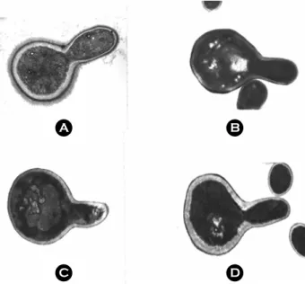
발아관는 혈청에 의하여 유도되며 혈청 이외에도 39℃
온도, RMPI 배지, N-acetyl glucosamine에 의해서도 유도될 수 있다 (12). 본 실험에서는 혈청에서 유도되는 단백 fibril이 발아관 형성시 발현되는 것인지 또는 발아관과 상관없이 혈청에 의해 발현되는 것을 조사하기 위하여 다양한 조건에서 발아관을 형성시켜 전자현미경으로 표 면구조를 관찰하였다. 혈청에 의해 유도된 발아관에서 만 단백 fibril이 발현되었으며, RPMI 배지 및 N-acetyl
A B
Figure 1. Electron micrographs of the fibrillar layer of C.
albicans. A, C. albicans was grown in YEPD at 37℃ for 2h; B, C. albicans was grown in rabbit serum at 37℃ for 2h. Initial cell concentration was 2×10⁷ cells/ml.
Figure 2. Effect of glucose and serum on the expression of fibrillar layer in C. albicans. C. albicans was grown at 37℃ for 2h in whole serum, 25% serum, and serum containing 1% glucose.
Initial cell concentration was 2×10⁷ cells/ml.
A B
C D
Figure 3. Expression of fibrillar layer of C. albicans in various germ tubes. Germ tube was induced for 2h by serum (A), 39℃
(B), RPMI medium (C), and N-acetyl glucosamine (D). Initial cell concentration was 2×10⁷ cells/ml.
glucosamine 첨가에 의해 유도되거나 39℃ 배양으로 얻어 진 발아관에서는 단백 fibril이 관찰되지 않았다 (Fig. 3).
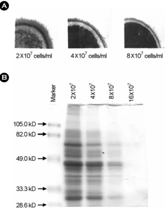
3. 세포농도에 따른 단백 fibril 발현
세포의 농도는 발아관 형성뿐만 아니라 세포표면 소 수성 및 최외각의 fibril 구조에도 영향을 미친다 (16,17).
C. albicans의 혈청배양시 세포농도에 따른 fibril 단백의 발현을 알아보기 위하여 C. albicans를 2×10⁷ ~ 16×107 cells/ml의 농도로 맞추어 혈청배양한 후 C. albicans의 세 포표면 단백질을 lyticase로 처리하여 SDS-PAGE로 분석 하였다. Fibril 구조물을 전자현미경으로 관찰한 결과, 혈 청에서 유도되는 fibril 구조물은 2×10⁷ cells/ml 이하의 세포농도에서 잘 발현되었으나, 4×10⁷ cells/ml 이상의 농 도에서 억제되기 시작하여 8×10⁷ 이상의 농도에서는 거 의 억제되었다. C. albicans 표면의 fibril 단백발현을 조사 하기 위하여 세포를 lyticase를 처리하여 SDS-PAGE로 분 석한 결과 66, 47, 30 kDa이 주요 fibril 단백이었으며 혈 청에서 유도되는 세포표면 단백들은 세포가 증가할수록 단백발현이 감소되었으며 16×107 cells/ml의 농도에서는
완전히 억제되었다 (Fig. 4).
4. 배양시간에 따른 fibril 발현
혈청으로 유도되는 C. albicans의 fibril을 배양시간에 따 른 변화를 관찰하기 위하여 C. albicans를 2×10⁷ cells/ml 세포농도로 조정하여 37℃에서 배양하면서 시간별로 세 포를 수거하여 전자현미경과 SDS-PAGE로 fibril 발현을 분석하였다. 전자현미경 관찰 결과 fibril 발현은 배양 10 분부터 발현되기 시작하였으며, C. albicans를 lyticase로 처리하여 fibril 층의 단백발현 양상 및 배양시간에 따른 fibril 단백의 변화 15% SDS-PAGE로 분석한 결과, 혈청에 의해 발현되는 66, 47, 30 kDa 주요 단백들 중 66 kDa 단 백은 배양 10분부터 발현되어 점차 감소되었으며, 47 kDa 단백은 배양 1시간부터 발현되기 시작하여 계속 유지되 었다. 반면에 30 kDa 단백은 배양 10분부터 2시간까지 B
A
Figure 4. Effect of cell concentration on the expression of fibril of C. albicans. A, Electron micrographs of the fibrillar layer of C.
albicans cultured at each cell concentration for 2h. B, SDS-PAGE analysis of fibril protein obtained by lyticase treatment.
B A
Figure 5. Effect of culture time on the expression of fibril of C.
albicans. A, Electron micrographs of the fibril layer of C. albicans cultured at each culture time; B, SDS-PAGE analysis of fibril protein obtained by lyticase treatment.
B A
발현 양상에 변화가 없었다 (Fig. 5).
고 찰
C. albicans의 세포표면 구조물은 숙주세포에의 부착 및 숙주 내에서의 생존에 영향을 미친다 (27). 칸디다균의 fibril 층은 주로 당 성분으로 되어 있어 일반적인 전자현 미경 고정과 염색법으로는 잘 관찰되지 않는다. 따라서 당 성분에 변형을 주지 않거나 염색이 가능한 ultrarapid- cryofixation (24), 혹은 망간 염색법 (13) 등 특수 염색으로 fibril 구조를 관찰할 수 있다. 그러나 혈청에 의해 발현 되는 단백 fibril은 일반적인 전자현미경 고정 및 염색법 으로 관찰된다 (17). 당 구조를 가진 fibril 구조는 배양 하는 배지 성분으로 발현 유도가 가능한데, galactose나 sucrose 등을 첨가한 배지나 세포배양배지인 M-199에서 배양하면 fibril 층이 생성되고 상피세포 부착능력도 증가 한다 (5,15). 본 연구자들은 혈청에서 C. albicans를 배양 하여 일반적인 시료 고정과 염색법으로도 fibril 구조물을 관찰할 수 있었으며 이들 fibril의 주성분은 단백질이라고 보고하였다 (17).
혈청배양 조건은 발아관을 유도하는 조건이다. 즉 혈 청배양하면 발아관과 일반 전자현미경 염색법으로 관찰 가능한 단백 fibril이 같이 유도된다. 그러므로 혈청에 의 해 유도되는 단백 fibril이 발아관과 관련 있는지를 조 사하기 위해 다양한 조건으로 발아관을 유도한 후 단백 fibril을 조사하였다. 본 연구에서 C. albicans 세포벽의 fibril 층은 혈청에서 배양할 때에만 관찰되었으며 (Fig. 1), 혈청을 희석하였을 때나 혈청에 포도당을 첨가하였을 때 단백 fibril의 발현이 억제되었다 (Fig. 2). 또한 혈청 이외 의 N-acetyl glucosamine 첨가나 RPMI 배지로 배양한 경 우와 39℃에서 균을 배양하여 유도한 발아관에서는 fibril 층이 관찰되지 않았다 (Fig. 3). 발아관 형성은 혈청, N- acetyl glucosamine, 세포농도, 포도당 제한 등, 여러 가지 조건에 영향을 받는데 (23), 본 실험의 결과로 칸디다균 의 단백 fibril은 발아관 구조와는 관계없이 혈청에 의해 서만 발현됨을 보여준다.
단백 fibril의 생성은 초기 세포농도가 4×10⁷ cells/ml 이 상의 농도에서 억제되었으며 (Fig. 4) 세포벽을 lyticase 처 리하여 SDS-PAGE로 분석한 결과, 세포농도에 따라 단백 질 band의 강도가 점차 감소하였다. 혈청에 의해 유도되 는 주요fibril 단백 66, 47, 30 kDa은 발현시간 및 발현유
지 시간이 각각 달랐다 (Fig. 5). 이러한 결과는 혈청에 의해 유도되는 단백 fibril이 C. albicans 감염 초기에 중요 한 역할을 할 것으로 생각할 수 있으며, 앞으로 생체 내 에서 발현되는 병독인자에 대한 진전된 연구가 필요할 것으로 사료된다. 더욱이 세포표면 물질은 부착 및 숙주 내에서의 생존에도 중요한 기능을 보임으로서 숙주 내 감염환경과 유사한 혈청 내 배양으로 유도된 fibril 단백 의 역할에 대한 연구는 칸디다균의 병인 연구에 반드시 필요할 것으로 생각된다.
참 고 문 헌
1) Agabian N, Odds FC, Poulain D, Soll DR, White TC:
Pathogenesis of invasive candidiasis. J Med Vet Mycol 32:
S229-237, 1994.
2) Antley PP, Hazen KC: Role of yeast cell growth temperature on Candida albicans virulence in mice. Infect Immun 56:
2884-2890, 1988.
3) Beveridge TJ, Popkin TJ, Cole RM: Electron microscopy.
pp 42-71. In Methods for general and molecular bacteriology, Gerhardt P, Murray RGE, Wood WA, Krieg NR (Ed), American society for microbiology, Washington DC, 1994.
4) Bradford MM: A rapid, sensitive method for the quantitation of micrograme quantities of protein utilizing the principle of protein-dye binding. Anal Biochem 72: 248-254, 1976.
5) Brawner DL, Cutler JE: Ultrastructural and biochemical studies of two dynamically expressed cell surgace determinants on Candida albicans. Infect Immun 51: 327-336, 1986.
6) Calderone RA, Fonzi WA: Virulence factors of Candida albicans. Trends Microbiol 9: 327-335, 2001.
7) Calderone RA, Linehan L, Wadsworth E, Sandberg AL:
Identification of C3d receptors on Candida albicans. Infect Immun 56: 252-258, 1988.
8) Cutler JE: Putative virulence factors of Candida albicans.
Annu Rev Microbiol 45: 187-218, 1991.
9)Enoch DA, Ludlam HA, Brown NM: Invasive fungal infections: a review of epidemiology and management options.
J Med Microbiol 55: 809-818, 2006.
10) Glee PM, Sundstrom P, Hazen KC: Expression of surface hydrophobic proteins by Candida albicans in vivo. Infect Immun 63: 1373-1379, 1995.
11) Hazen KC, Hazen BW: Hydrophobic surface protein masking by the opportunistic fungal pathogen Candida albicans. Infect
Immun 60: 1499-1508, 1992.
12) Hilmioglu S, Ilkit M, Badak Z: Comparison of 12 liquid media for germ tube production of Candida albicans and C.
tropicalis. Mycoses 50: 282-285, 2007.
13) Hubbard MJ, Sullivan PA, Shepherd MG: Morphological studies of N-acetylglucosamine induced germ tube formation by Candida albicans. Can J Microbiol 31: 696-701, 1985.
14) Kanbe T, Han Y, Redgrave B, Riesselman MH, Cutler JE:
Evidence that mannans of Candida albicans are responsible for adherence of yeast forms to spleen and lymph nodes tissue.
Infect Immun 61: 2578-2584, 1993.
15) Kennedy MJ, Sandin RL: Influence of growth conditions on Candida albicans adhesion, hydrophobicity and cell wall ultrastructure. J Med Vet Mycol 26: 79-92, 1988.
16) Kim D, Shin WS, Lee KH, Kim K, Park JY, Koh CM:
Rapid differentiation of Candida albicans from other Candida species using its unique germ tube formation at 39 degrees C.
Yeast 19: 957-962, 2002.
17)Koh CM, Lee KH, Shin WS, Kim DH: Serum induces protein fibrils expression in Candida albicans. J Korean Soc Microbiol 34: 277-283, 1999.
18) Laemmli UK: Cleavage of structural proteins during the assembly of the head of bacteriophage T4. Nature 227: 680 -685, 1970.
19) Lehrer N, Segal E, Lis H, Gov Y: Effect of Candida abicans cell wall components on the adhesion of the fungus to human and murine vaginal mucosa. Mycopathologia 102: 115-121, 1988.
20) Monod M, Borg-von Zepelin M: Secreted proteinases and
other virulence mechanisms of Candida albicans. Chem Immunol 81: 114-128, 2002.
21) Odds FC: Candida species and virulence. ASM News 60: 313 -318, 1994.
22) Odds FC: Disseminated candidosis (Candida septicemia). pp 206-230. In Candida and candidiosis, 2nd ed, Odds FC (Ed), WB Saunders, London, 1988.
23)Odds FC: Morphogenesis in Candida albicans. Crit Rev Microbiol 12: 45-93, 1985.
24) Osumi M: The ultrastructure of yeast: cell wall structure and formation. Micron 29: 207-233, 1998.
25) Pagano L, Caira M, Candoni A, Offidani M, Fianchi L, Martino B, Pastore D, Picardi M, Bonini A, Chierichini A, Fanci R, Caramatti C, Invernizzi R, Mattei D, Mitra ME, Melillo L, Aversa F, Van Lint MT, Falcucci P, Valentini CG, Girmenia C, Nosari A: The epidemiology of fungal infections in patients with hematologic malignancies: the SEIFEM-2004 study. Haematologica 91: 1068-1075, 2006.
26) Pendrak ML, Krutzsch HC, Roberts DD: Structural require- ments for hemoglobin to induce fibronectin receptor expression in Candida albicans. Biochemistry 39: 16110-16118, 2000.
27) Ruiz-Herrera J, Elorza MV, Valentin E, Sentandreu R:
Molecular organization of the cell wall of Candida albicans and its relation to pathogenecity. FEMS Yeast Res 6: 14-29, 2006.
28) Sakata N, Yamazaki K, Koqure T: Identification of a 21 kDa laminin-binding component of Candida albicans. Zentralbl Bakteriol 289: 217-25, 1999.