ORIGINAL ARTICLE
J Bacteriol Virol. Vol 48. No 4. December 2018; 48(4): 130-136 http://dx.doi.org/10.4167/jbv.2018.48.4.130
eISSN 2093-0249
JBV
Gene Expression Profiles of Th1-type Chemokines in Whole Blood of Mycobacterium avium subsp. paratuberculosis -Infected Cattle
Min-Kyoung Shin1,2, Hyun-Eui Park2, Hong-Tae Park2, Myunghwan Jung1, Hyung-Lyun Kang1, Seung Cheol Baik1, Woo-Kon Lee1, Young Hoon Jung3 and Han Sang Yoo2,4*
1
Department of Microbiology, Research Institute of Life Science, College of Medicine, Gyeongsang National University, Jinju, Korea
2
Department of Infectious Diseases, College of Veterinary Medicine, Seoul National University, Seoul, Korea
3
National Institute of Animal Science, Rural Development Administration, Wanju, Korea
4
Institute of Green Bio Science and Technology, Seoul National University, Pyeongchang, Korea
Corresponding Han Sang Yoo
Department of Infectious Diseases, College of Veterinary Medicine, Seoul National University, Seoul 08826, Korea.
Phone : +82-2-880-1263 Fax : +82-2-874-2738 E-mail : yoohs@snu.ac.kr
Received : October 08, 2018 Revised : October 30, 2018 Accepted : November 01, 2018
No potential conflict of interest relevant to this article was reported.
Copyright © 2018 Journal of Bacteriology and Virology
©This is an Open Access article distributed under the terms of the Creative Commons Attribution Non-Commercial License (http://creativecommons.org/
license/by-nc/3.0/).
Johne's disease (JD) is a chronic, debilitating disease of ruminants including cows, and is caused by Mycobacterium avium subsp. paratuberculosis (MAP). MAP is not only important in animal husbandry, but also in public health as it is associated with the onset of Crohn's disease, a chronic inflammatory bowel disease in humans.
JD, like other mycobacterial diseases including tuberculosis, is classified into different stages based on the progression of infection. In addition, development of diagnostic assays that can distinguish between subclinical and clinical stages of JD is essential to control mycobacterial infection by providing an effective treatment. For the development of novel diagnostic methods of JD, it is important to investigate and understand the mRNA expression of the various immune markers in individuals at each stage of infection. In this study, we measured the levels of Th1-type chemokines, CXCR3, CCL4, CCL5, CXCL9, CXCL10, and CXCL11 in MAP-infected bovine blood by interferon (IFN)-γ release assay (IGRA) using IFN-γ as an alternative biomarker.
The association of mRNA expression patterns of these chemokines with the MAP infection stages was analyzed and IFN-γ, CCL5, and CXCL10 were found to be significantly upregulated compared to IFN-γ, the biomarker used in IGRA. Our results further indicate that IFN-γ levels significantly increased in individuals with MAP- specific antibody, and CCL5 and CXCL10 levels significantly increased in those with MAP DNA. In particular, CCL5 was significantly upregulated in individuals, in which both MAP-specific antibody and MAP DNA were detected, but the expression of CXCL10 was specifically elevated in MAP DNA-detected individuals without MAP- specific antibody.
Key Words: Th-1 type chemokines, Paratuberculosis, Biomarkers, Cattle
INTRODUCTION
Mycobacterium avium subsp. paratuberculosis (MAP) is the causative agent of Johne's disease (JD), which is characterized by chronic granulomatous enteropathy in ruminants (1). When MAP is infected with calves, it takes a long latency period to induce severe chronic diarrhea and consumption, leading to death (2). In addition, although JD causes massive economic losses to the dairy industry, MAP is also
disease in humans (3, 4). MAP is a causative agent that is difficult to eradicate once it is inside the farm because it spreads through feces and milk of the MAP-infected individuals at the asymptomatic stage. Therefore, in order to control the disease, it is important to quickly diagnose and eliminate the infected individual.
Although fecal culture is the gold standard for the diagnosis of MAP, it is not widely used for diagnosis because of the long cultivation periods of MAP and its contamination with other bacteria (5). To date, diagnosis of JD is focused at detecting MAP-specific antibodies and MAP DNA in the sera and feces, respectively. Although polymerase chain reaction (PCR) can readily detect MAP in feces and milk, PCR-based methods have low sensitivity and reproducibility because of PCR inhibitors present in these samples (6). Enzyme-linked immunosorbent assay (ELISA) has been used for the detection of MAP-specific antibodies in these samples, but it is not adequately sensitive. Moreover, as only the individuals that form antibodies to MAP can be detected using ELISA, it is inefficient to diagnose MAP at the other infectious stages.
Recently, the interferon (IFN)-γ release assay (IGRA), an alternative immune detection method has been used for the diagnosis of tuberculosis (TB). It detects the release of IFN-γ by immune cells after stimulation of whole blood with an M. tuberculosis- specific antigen (7). IGRA is a powerful diagnostic tool and has replaced the tuberculin skin test (7). It is also used for mycobacterial diseases such as JD, and IFN-γ is one of the biomarkers currently in use. However, IGRA is also known to exhibit a low sensitivity to immunocompromised patients and young children (8). Other studies have suggested that IFN-γ alone is not sufficient for the diagnosis due to the progression of mycobacterial disease (8). Therefore, other biomarkers that can improve IGRA need to be urgently explored. In this study, we investigated the expression of Th1 type chemokines after MAP stimulation in the blood of MAP-infected cattle to identify alternative biomarkers for IGRA.
MATERIALS AND METHODS
Experimental animals
All animal procedures were performed according to the guidelines of the Institutional Animal Use and Care Committee of the National Institute of Animal Science. The protocol was approved by the Institutional Animal Use and Care Committee of the National Institute of Animal Science (Permit number 2013-046). Thirty Holstein cows were selected by detection of MAP-specific antibodies using a commercially available ELISA kit (IDEXX Laboratories, Inc., Westbrook, ME, USA), and MAP DNA using fecal PCR (9). To accurately classify the cattle into the infectious stages, diagnosis was performed twice at a 6-month interval. The animals were divided into four groups based on the results of PCR and ELISA: negative control group (n = 5), ELISA and PCR negative;
group 1 (n = 7), ELISA negative and PCR positive; group 2 (n = 10), ELISA positive and PCR negative; and group 3 (n = 8), ELISA and PCR positive. According to the manufacturer's instructions, the ratio of optical densities of sample to positive control (S/P ratio) was determined and the stages of JD were assigned using the S/P ratio. Additionally, the infected animals (groups 1~3;
n = 25) were divided into five groups based on the S/P ratio, as obtained from ELISA: EL200 group (n = 4), S/P ratio ≥200;
EL100 group (n = 5), S/P ratio <200 but ≥100; EL45 group (n = 4), S/P ratio <100 but ≥45; ELsup group (suspect/weak positive; n = 5), S/P ratio <45 but ≥21; and ELneg group (same as Group 1; n = 7), S/P ratio <20.
Whole blood culture with MAP stimulation and total RNA extraction
Peripheral blood was collected from the tail vein of cattle for whole blood culture with nil stimulation (negative control), and MAP-whole bacterial stimulation (1 × 10⁴ cfu/ml). The stimulation was performed at 37℃ for 20 h. Total RNA was extracted from the blood samples using a PAXgene Blood RNA Kit (PreAnalytiX/Qiagen, Hilden, Germany) according to the manufacturer's instructions.
JBV
Bacteriology and Virology VOL 48. NO 4. DECEMBER 2018Quantitative RT-PCR
Total RNA was used to prepare cDNA with random primers using a QuantiTect Reverse Transcription Kit (Qiagen Inc., Valencia, CA, USA) according to the manufacturer's instructions. The expression of representative Th1-type chemokine genes was measured by quantitative reverse transcription real-time PCR. Real-time PCR reactions were performed using the Rotor-Gene SYBR Green PCR Kit (Qiagen Inc.). The primers used in this study are listed in Table 1. Amplification was initiated at 95℃ for 10 min, followed by 40 cycles of 95℃ for 15 s, and 60℃ at 45 s. Expression levels were determined by the 2-ΔΔCt method using the housekeeping genes, β-actin and GAPDH, as references (10). The expression level of each gene was compared with that of the nil stimulation to determine the fold change after 20 h of MAP stimulation.
Statistical analysis
Data are reported as the mean ± standard error of the mean (SEM) of three or more independent experiments. Statistical significance was determined by Student's t-test using the IBM Statistical Package for Social Sciences software (SPSS, ver. 21;
IBM SPSS Inc., Chicago, IL, USA). Differences were considered significant if a p-value of < 0.05 was obtained.
RESULTS
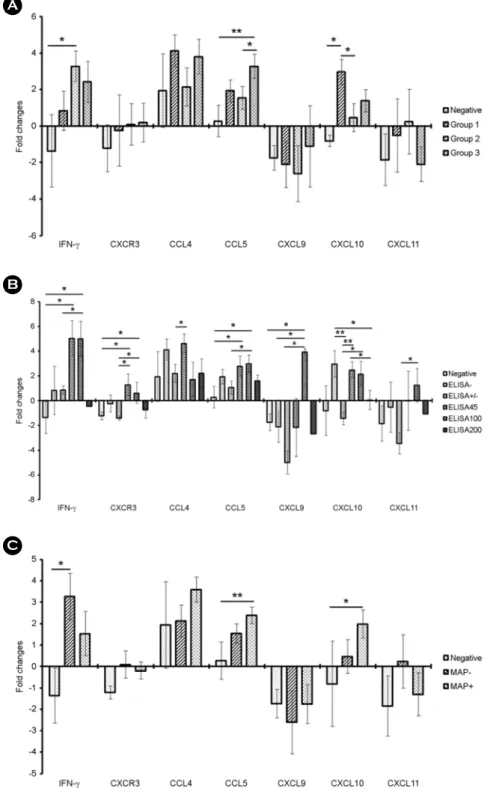
As shown in Fig. 1A, IFN-γ, CCL5, and CXCL10 showed a statistically significant expression in all the groups. In particular, CXCL10 showed a significantly high expression in group 1 compared to the negative group or group 2 (p < 0.05). IFN-γ showed a significantly increased expression in group 2 compared to the negative group (p < 0.05). CCL5 was significantly increased in group 3 compared to the negative group (p < 0.01) or group 2 (p < 0.05). CXCR3, CCL4, CXCL9, and CXCL11 showed no significant differences in expression among the groups. Based on ELISA results, the expression of Th1 type chemokine genes was significantly influenced (Fig. 1B). IFN-γ significantly increased in EL45 and EL100 groups (p < 0.05). Rather, the individuals Table 1. Oligonucleotide primers used for quantitative RT-PCR
Gene Sequence (5'-3')
IFN-γ Forward CAAATTCCGGTGGATGATCTGC
Reverse GGCAGGAGGACCATTACGTT
CXCR3 Forward ACATAGCCAAGTCGGTCACG
Reverse CTCGGAACTTGACACCCACA
CCL4 Forward CCCAGCCAGCTGTGGTATTC
Reverse CTCGGAGCAGCTCAGTTCAA
CCL5 Forward CTTCTGCCTCCCCATATGCC
Reverse ACTTCTTCTCTGGGTTGGCG
CXCL9 Forward CTCTGACTGGAGTTCAAGGAGT
Reverse TCCCTTGGCTGGTGTTGATG
CXCL10 Forward CCACGTGTCGAGATTATTGCC
Reverse TGCCTCTTTCCGTGTTCGAG
CXCL11 Forward AAGCATGAGTGTGAAGGGCA
Reverse AGGGAAACCTTGAACAATTGCAG
Figure 1. Gene expression profiles of Th1-type chemokines in whole blood from MAP-infected individuals after MAP stimulation.
(A) Analysis of gene expression of Th1-type chemokines in whole blood culture after 20 h of MAP-stimulation in each group.
Negative control group (n = 5), ELISA and PCR negative; group 1 (n = 7), ELISA negative and PCR positive; group 2 (n = 10), ELISA positive and PCR negative; and group 3 (n = 8), ELISA and PCR positive. (B) Analysis of gene expression of Th1-type chemokine in whole blood culture after 20 h of MAP-stimulation based on the ELISA results. Negative control group (n = 5), ELISA and PCR negative; EL200 group (n = 4), S/P ratio ≥200; EL100 group (n = 5), S/P ratio <200 but ≥100; EL45 group (n
= 4), S/P ratio <100 but ≥45; ELsup group (suspect/weak positive; n = 5), S/P ratio <45 but ≥21; and an ELneg group (same as group 1; n = 7), S/P ratio <20. (C) Analysis of gene expression of Th-1 type chemokine in whole blood culture after 20 hours of MAP-stimulation based on MAP DNA detection in feces. The expression level was determined by the 2-ΔΔCt method in terms of the β-actin and GAPDH expression levels relative to the control group (*, p < 0.05; **, p < 0.01).
JBV
Bacteriology and Virology VOL 48. NO 4. DECEMBER 2018 in the EL200 group exhibited low IFN-γ values, and the IFN-γ levels did not increase in the individuals with MAP-separated but ELISA negative or weak positive. CCL5 gene was also significantly increased in the EL45 and EL100 groups compared to the negative group or the weak positive group (p < 0.05). The ELISA results for other genes did show a significant gene expression pattern, but the fold-change was less than 2 and had no diagnostic value. Finally, expression patterns of Th1-type chemokine genes were analyzed based on whether MAP was isolated from each individual. As shown in Fig. 1C, the expression of IFN-γ was significantly increased in the ELISA-positive individuals without MAP isolation, and the expression of CCL5 and CXCL10 was significantly increased in MAP-isolated individuals.DISCUSSION
JD manifests several stages of infection, such as silent, subclinical, clinical, and advanced cell infections, depending on the symptom evaluation and quantification of intestinal MAP (11, 12). Additionally, the progression of mycobacterial infection has been thought to be related to Th1 / Th2 migration of the host (13). In the early stage of infection, the cell-mediated CD4+ T cell response (Th1 response) to MAP is displayed (13). Th1 response decreases as the disease progresses, but the antibody response (Th2 response) to MAP increases (13). Thus, since the Th1 / Th2 shift is related to disease progression and bacterial excretion, many studies have determined the MAP infection stage based on detection of MAP and MAP-specific antibody in specimen such as feces, milk, and sera (12, 14, 15).
The development of diagnostic methods that can distinguish between different stages of MAP infection can provide a basis for providing effective treatment and defense strategies for MAP infection, since MAP has the distinct infection stages. For development of novel diagnostic methods of JD, it is important to determine the mRNA expression profiles of various immune markers in individuals with infection at every stage. Here, expression levels of Th-1 type chemokines, CXCR3, CCL4, CCL5, CXCL9, CXCL10, and CXCL11 were evaluated by IGRA using whole blood from MAP-infected cattle. We quantitatively analyzed the mRNA expression profiles of these chemokines in different MAP infection stages and compared them with that of IFN-γ, a biomarker used in IGRA, to confirm the diagnostic value of these chemokines in MAP infection. Among the Th1 type chemokines, IFN-γ, CCL5, and CXCL10 were found to be significantly upregulated compared to IFN-γ in the present study. In addition, IFN-γ levels significantly increased in individuals with MAP-specific antibody, and CCL5 and CXCL10 levels significantly increased in those with MAP DNA. Above all, CXCL10 was specifically expressed in individuals of the subclinical stage, which was MAP DNA-detected with ELISA-negative, and was expected to be detectable at the early state of MAP infection.
In the case of TB, CXCL9, CXCL10, and CXCL11 have been identified as potential alternative or complementary biomarkers to improve the performance of IGRA (8, 16). CXCL9 and CXCL10 levels increased in the plasma of active pulmonary TB and tuberculous pleurisy patients compared to healthy controls, and CXCL11 levels increased in the patients with latent TB (8, 16).
In this study, IFN-γ, CCL5, and CXCL10 were considered valuable biomarkers because of their different MAP-specific gene expression patterns at different stages of MAP infection. In particular, IFN-γ could be a significant discriminatory biomarker in individuals at the MAP-specific antibody formation stage, and CCL5 or CXCL10 could be significant discriminatory biomarkers in individuals in which MAP DNA is detected. Moreover, CCL5 levels were found to be significant in individuals in which both MAP-specific antibodies and MAP DNA were detected. It is expected that CXCL10 could be used to diagnose individuals at the subclinical stage, specifically in MAP-detected individuals, although no antibody was detected.
A number of researchers, including those in our team, have studied the changes in gene expression at different stages of infection and have suggested genes that show significant changes at these stages (12, 14, 15). However, as JD is a chronic infectious disease, the host gene expression pattern may vary depending upon the host condition. Therefore, it is not entirely accurate to consider each individual gene whose expression has been changed as a biomarker. Thus, gene expression changes in immune cells in blood culture after MAP-specific stimulation should be considered useful for diagnosis. As far as we know, this study is the first study to evaluate Th1 chemokines as an alternative biomarker for MAP infections using IGRA. It is expected to be applied to more clinical subjects for future study, and it would be promising to apply multiple mRNA detection methods by
ACKNOWLEDGEMENT
This work was supported by Development Fund Foundation, Gyeongsang National University, 2015, by the National Research Foundation of Korea (NRF) grant funded by the Ministry of Science, ICT & Future Planning (2016R1C1B1014322), and by a grant from the Strategic Initiative for Microbiomes in Agriculture and Food, Ministry of Agriculture, Food and Rural Affairs, (as part of the (multi-ministerial) Genome Technology to Business Translation Program) (918020-4), BK21 PLUS, the Research Institute for Veterinary Sciences, Seoul National University, Republic of Korea.
REFERENCES
1) Motiwala AS, Janagama HK, Paustian ML, Zhu X, Bannantine JP, Kapur V, et al. Comparative transcriptional analysis of human macrophages exposed to animal and human isolates of Mycobacterium avium subspecies paratuberculosis with diverse genotypes. Infect Immun 2006;74:6046-56.
2) Coussens PM. Mycobacterium paratuberculosis and the bovine immune system. Anim Health Res Rev 2001;2:141-61.
3)Sechi LA, Scanu AM, Molicotti P, Cannas S, Mura M, Dettori G, et al. Detection and isolation of Mycobacterium avium subspecies paratuberculosis from intestinal mucosal biopsies of patients with and without Crohn's disease in Sardinia. Am J Gastroenterol 2005;100:1529-36.
4) Shin AR, Kim HJ, Cho SN, Collins MT, Manning EJ, Naser SA, et al. Identification of seroreactive proteins in the culture filtrate antigen of Mycobacterium avium ssp. paratuberculosis human isolates to sera from Crohn's disease patients. FEMS Immunol Med Microbiol 2010;58:128-37.
5) Bögli-Stuber K, Kohler C, Seitert G, Glanemann B, Antognoli MC, Salman MD, et al. Detection of Mycobacterium avium subspecies paratuberculosis in Swiss dairy cattle by real-time PCR and culture: a comparison of the two assays. J Appl Microbiol 2005;99:587-97.
6)Leite FL, Stokes KD, Robbe-Austerman S, Stabel JR. Comparison of fecal DNA extraction kits for the detection of Mycobacterium avium subsp. paratuberculosis by polymerase chain reaction. J Vet Diagn Invest 2013;25:27-34.
7) Lalvani A, Pathan AA, McShane H, Wilkinson RJ, Latif M, Conlon CP, et al. Rapid detection of Mycobacterium tuberculosis infection by enumeration of antigen-specific T cells. Am J Respir Crit Care Med 2001;163:824-8.
8) Kim S, Lee H, Kim H, Kim Y, Cho JE, Jin H, et al. Diagnostic performance of a cytokine and IFN-gamma-induced chemokine mRNA assay after Mycobacterium tuberculosis-specific antigen stimulation in whole blood from infected individuals. J Mol Diagn 2015;17:90-9.
9)Park H-T, Shin M-K, Sung KY, Park H-E, Cho Y-I, Yoo HS. Effective DNA extraction method to improve detection of Mycobacterium avium subsp. paratuberculosis in bovine feces. Korean J Vet Res 2014;54:55-7.
10) Cortes Y, Ojeda M, Araya D, Dueñas F, Fernández MS, Peralta OA. Isolation and multilineage differentiation of bone marrow mesenchymal stem cells from abattoir-derived bovine fetuses. BMC Vet Res 2013;9:133.
JBV
Bacteriology and Virology VOL 48. NO 4. DECEMBER 2018 11) Yakes BJ, Lipert RJ, Bannantine JP, Porter MD. Detection of Mycobacterium avium subsp. paratuberculosis by a sonicateimmunoassay based on surface-enhanced Raman scattering. Clin Vaccine Immunol 2008;15:227-34.
12) Shin MK, Park HT, Shin SW, Jung M, Im YB, Park HE, et al. Whole-blood gene-expression profiles of cows infected with Mycobacterium avium subsp. paratuberculosis reveal changes in immune response and lipid metabolism. J Microbiol Biotechnol 2015;25:255-67.
13) Magombedze G, Eda S, Ganusov VV. Competition for antigen between Th1 and Th2 responses determines the timing of the immune response switch during Mycobaterium avium subspecies paratuberulosis infection in ruminants. PLoS Comput Biol 2014;10:e1003414.
14)Park HE, Shin MK, Park HT, Jung M, Cho YI, Yoo HS. Gene expression profiles of putative biomarker candidates in Mycobacterium avium subsp. paratuberculosis-infected cattle. Pathog Dis 2016;74:ftw022.
15) Park HE, Park HT, Jung YH, Yoo HS. Establishment a real-time reverse transcription PCR based on host biomarkers for the detection of the subclinical cases of Mycobacterium avium subsp. paratuberculosis. PloS One 2017;12:e0178336.
16) Yu Y, Zhang Y, Hu S, Jin D, Chen X, Jin Q, et al. Different patterns of cytokines and chemokines combined with IFN-gamma production reflect Mycobacterium tuberculosis infection and disease. PloS One 2012;7:e44944.