Introduction
Resin-based composites are widely used in restora- tive dentistry with the development of more wear- resistant formulations, better adhesives, and improved light-curing and surface-sealing systems.
Nevertheless, because of fractures or failures in
composite restorations, clinicians must decide whether to replace or simply repair these restora- tions. The complete removal of partially defective composite restorations may be time-consuming and highly risky to remove sound tooth substance, and to injure the pulp tissue. Therefore, instead of its removal, the repair of defected restorations would be
The study of fractural behavior of repaired composite
Sang-Soon Park, Wook Nam, Ah-Hyang Eom, Duck-Su Kim, Gi-Woon Choi, Kyoung-Kyu Choi*
Division of Dentistry, Department of Conservative Dentistry, Graduate of Kyung Hee University, Seoul, Korea
Objectives: This study evaluated microtensile bond strength (μTBS) and short-rod fracture toughness to explain fractural behavior of repaired composite restorations according to different surface treatments.
Materials and Methods: Thirty composite blocks for μTBS test and sixty short-rod specimens for fracture toughness test were fabricated and were allocated to 3 groups according to the combination of surface treatment (none-treated, sand blasting, bur roughening). Each group was repaired immediately and 2 weeks later. Twenty-four hours later from repair, μTBS and fracture toughness test were conducted. Mean values analyzed with two-way ANOVA / Tukey’s B test (α= 0.05) and correlation analysis was done between μTBS and fracture toughness. FE-SEM was employed on fractured surface to examine the crack propagation.
Results: The fresh composite resin showed higher μTBS than the aged composite resin (p < 0.001).
Mechanically treated groups showed higher bond strength than non-mechanically treated groups except none-treated fresh group in μTBS (p < 0.05). The fracture toughness value of mechanically treated surface was higher than that of non-mechanically treated surface (p < 0.05). There was no correlation between fracture toughness and microtensile bond strength values. Specimens having high KIC showed toughening mechanism including crack deviation, microcracks and crack bridging in FE-SEM.
Conclusions: Surface treatment by mechanical interlock is more important for effective composite repair, and the fracture toughness test could be used as an appropriate tool to examine the fractural behavior of the repaired composite with microtensile bond strength. [J Kor Acad Cons Dent 2010;35(6):461-472.]
Key words:Aging time; Composite repair; Fractural behavior; Fracture toughness; Microtensile bond strength; Surface treatment
-Received 26 August 2010; revised 11 October 2010; accepted 13 October 2010- ABSTRACT
Park SS, DDS, DMD, Student; Nam W, DDS, PhD, Student; Eom AH, DDS, DMD, Student; Kim DS, DDS, PhD, Clinical instructor; Choi GW, Professor; Choi KK, DDS, PhD, Professor, Division of Dentistry, Department of Conservative Dentistry, Graduate of Kyung Hee University, Seoul, Korea
*Correspondence to Kyoung-Kyu Choi, DDS, PhD.
Professor, Division of Dentistry, Department of Conservative Dentistry, Graduate of Kyung Hee University, 1 Hoegi-dong, Dongdaemun-gu, Seoul, Korea, 130-702
TEL, +82-2-958-9337; FAX, +82-2-960-5108; E-mail: [email protected]
a favorable treatment option.
The oxygen-inhibition layer is not necessary when bonding additional layers of resin composites, at least for the initial 24 hours in which the remaining free radicals within the polymerized composite enables its chemical coupling to the repaired composite.1,2 However, when free radicals are reduced by aging, polishing or abrasion, surface treatment of composite is required for better repair bond strength between the existing composite and the new composite added.3 Various kinds of surface treatments have been sug- gested including diamond bur roughening and air abrasion. While they promote mechanical interlock, use of intermediate coupling agents improves surface wettability and chemical bonding to the cross-linked polymer matrix and/or filler particles of the old com- posite.4,5Previous studies showed that use of unfilled low-viscosity intermediate resin could improve the bonding irrespective of the surface texture created by different surface treatments.6-8Phosphoric acid seems to have just superficial cleaning effect of the compos- ite surface and have no influence on the repair bond strength.1,5,9,10
Up to date, most studies of composite repair have been accomplished regarding the evaluation of bond strength. Especially, microtensile bond strength (μTBS) has been useful because of its better stress distribution at the true interface, the controlled sub- strate variables, and so on. However, although mea- suring the bond strength is one of the most common tests for assessing fractural behavior of repaired com- posite, it is very sensitive to condition and depends on the size of the cracks occurred during processing, producing and handling.11 Moreover, when substrate fractures occur, results of bond strength test become a measurement of the material’s strength properties rather than actual integrity of the bonded interface.
Therefore, the bond strength test alone is not suffi- cient to decide whether a treatment has enhanced the resistance to fracture or not.12,13 A reliable and valid method for the evaluation of bonded interfaces is needed to help predict, understand and assess clinical bonding failures.
Composite is a brittle material to which linear elas- tic fracture mechanics can be applied to analyze the stress state of the material at fracture.14It would be
more reasonable to evaluate fractural behavior by means of fracture toughness, especially when the failure of restoration is related to crack propagation originating from defects of interfacial surface. The plane-strain fracture toughness, determined by stan- dard tests described by ASTM-E399, is defined as the resistance of a material to rapid crack propaga- tion. It is an intrinsic property and is independent of the size of the initiating crack. In addition, it is con- sidered to be a better measure of fracture resistance than other strength parameters, if a linear-elastic deformation to failure can be assumed.15 Fracture toughness can be characterized by one parameter, KIC
which means critical stress intensity factor when ten- sile load is in-plane shear.16
There are various testing methods for examining the fracture toughness (KIC) of dental composites including single-edge notched beam method, compact tension method, short-rod with chevron notch method, double torsion method and so on. Among those, the short-rod fracture toughness test speci- men, proposed by Barker,17 does not require fatigue pre-cracking. Stable crack growth occurs initially, and assessment of KIC is based on measurement of the load required for crack growth instability to occur.15 Its geometry contains a chevron-shaped bonding area designed to develop a significant stress concentration at the interface. Bond failure occurs along the midplane of the specimen where the bond- ed interface is located. This corresponds to the more usually observed failure mode in clinical practice.
Therefore, the short-rod fracture toughness specimen would provide more clinically relevant information than the tensile or shear bond strength specimen. It is well-suited to the investigation of factors such as surface treatment which affects the adhesion of one material to another.16,18
To date, there have been some data about the frac- ture toughness of dentin-composite adhesive inter- face. However, data about fracture toughness of com- posite-composite repair interface have been rare. The objectives of this study were to determine the corre- lation between μTBS and KICin composite repair and to explain fractural behavior of repaired composite depending on different surface treatments.
Materials and Methods 1. Experimental Materials
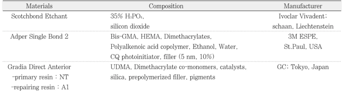
The materials used in this study are listed in Table 1.
2. Microtensile bond strength
1) Specimen preparation
Thirty cylinder-shaped blocks, 8 mm in height and 8 mm in diameter, were fabricated by layering 2- mm-thick increments of Gradia Direct Anterior (shade NT, GC; Tokyo, Japan) using a silicone mold.
Each increment was carefully condensed with a clean plastic filling instrument in order to avoid contami- nation and/or incorporation of voids, then cured for 20s with the tip of the light curing unit (WBL-100 Santafe, S-Denti Co., LTD, Korea) placed in contact with the surface of the mold. The last increment was covered with polyester strip and compressed by slide glass in order to obtain a flat surface of the specimen after light curing. All composite blocks were allocated to 3 groups according to the surface treatment (none- treated, sand blasting and diamond bur roughening) and each group was divided by aging time (fresh and 2 weeks aging). They were stored in artificial saliva at 37℃ before the repair procedure was performed.
2) Surface treatment and repair procedure
Fresh groups were repaired as follows within 30 minutes and aged groups were repaired 2 weeks since blocks had been made.
�Group NF: There was no mechanical surface treatment. 35% Scotchbond Etchant gel (Ivoclar Vivadent; Schaan, Liechtenstein) was applied to the saliva-contaminated composite surface for 30s.
�Group SF: Each composite block was sandblasted for 10s using 25-50 μm Al2O3particles with Basic professional No. 2942 (Renfert GmbH, Hilzingen, Germany) at 5 mm from the surface (pressure of 60 psi) and etched as group 1.
�Group BF: A coarse diamond chamfer bur was used to roughen the surface of each specimen for 10s at high speed with constant water spray.
Then, the surface was etched as group 1.
�Group NA: The same procedure to Group NF was performed on 2 weeks aged composite.
�Group SA: The same procedure to Group SF was performed on 2 weeks aged composite.
�Group BA: The same procedure to Group BF was performed on 2 weeks aged composite.
Each composite block was rinsed for 30s using a stream of oil-free compressed air/water from a syringe tip. An air syringe was then used for 5s to remove excess surface water. Two-step total etch bonding agent, Adper Single bond 2 (SB; 3M ESPE, St. Paul, USA) was applied twice, dried gently with air syringe for 5s to evaporate solvents and light- cured according to the manufacturer’s recommenda- tion. After the respective surface treatment and bonding procedure, each block was inserted in the other mold, 16 mm in height and 8 mm in diameter, leaving 8 mm space to be filled by the repairing com-
Table 1.Materials used in this study
Materials Composition Manufacturer
Scotchbond Etchant 35% H3PO4, Ivoclar Vivadent;
silicon dioxide schaan, Liechtenstein
Adper Single Bond 2 Bis-GMA, HEMA, Dimethacrylates, 3M ESPE,
Polyalkenoic acid copolymer, Ethanol, Water, St.Paul, USA CQ photoinitiator, filler (5 nm, 10%)
Gradia Direct Anterior UDMA, Dimethacrylate co-monomers, catalysts, GC; Tokyo, Japan -primary resin : NT silica, prepolymerized filler, pigments
-repairing resin : A1
Bis-GMA, bisphenylglycidyl dimethacrylate; HEMA, hydroxyethyl methacrylate; UDMA, urethane dimethacrylate.
posite, Gradia Direct Anterior (shade A1, GC;
Tokyo, Japan). For a better assessment of the repair interface, different shade was selected for the repair- ing composite to discern from existing composite. The repairing composite was incrementally inserted and light-cured as previously described (Table 2).
3) Microtensile bond strength test
After a 24-h storage in 37℃ artificial saliva, each composite block was sectioned perpendicular to the bonded repair interface into 1-mm-thick slabs (n = 3 per block) with a low-speed diamond saw (Isomet 1000, Buehler Ltd., Lake Bluff, IL, USA) under con- stant water coolant. Each slab was trimmed along the repair interface to a modified hourglass shaped specimen using a fine diamond bur to concentrate the tensile load on repair interface (1 mm2area of bond- ed surface). Five additional composite resin blocks were fabricated 16 mm in height and 8 mm in diam- eter to test the ultimate tensile strength (UTS).
Specimens were secured at the ends with cyano- acrylate adhesive (Zapit, Dental Ventures of America; Corona, CA, USA) to μTBS testing device.
The test was conducted at a cross-head speed of 1 mm/min until failure. After failure, each repair inter- face area was measured and the bond strength value (MPa) was calculated.
3. Fracture toughness
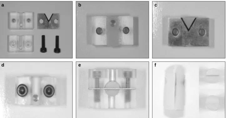
Diagram of short-rod specimen is showed in Figure 1. Ten half specimens, like Figure 2b, were made for each group to conduct short-rod fracture toughness
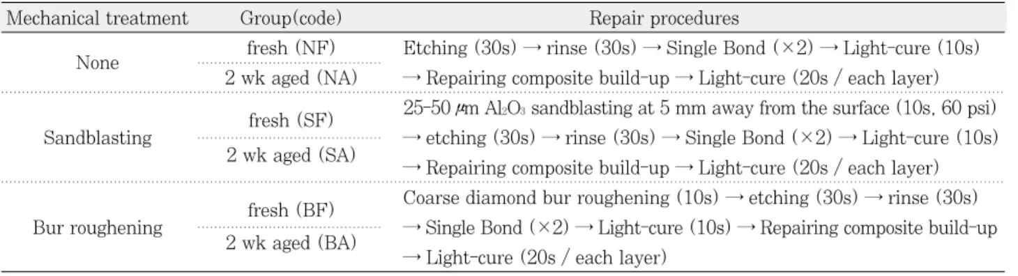
test using acrylic resin mold which is showed in Figure 2a. The surface was covered with slide glass and cured by light. Specimens had been stored in artificial saliva at 37℃ before the repair procedure was performed. Surface treatments and bonding pro- cedures were the same as μTBS test. At each aging time, the half specimen was inserted into acrylic resin mold and covered with 45�beveled spacer (Figure 2c). After the mold was assembled as shown in Figures 2d and 2e, repairing resin was filled into the mold. Light curing was performed at both ends and from upper side for 40s. All specimens were sep- arated from the mold (Figure 2f) and stored in artifi- cial saliva. To compare with bulk fracture toughness of composite resin, additional 10 specimens were fab- ricated in bulk. After 24 hours, specimens were fixed to tensile testing jig by ligature wire. A tensile load was applied to each specimen at an extension rate of 0.5 mm/min until failure. The peak load at the time Table 2.Repair procedures according to mechanical surface treatment
Mechanical treatment Group(code) Repair procedures
None fresh (NF) Etching (30s) → rinse (30s) → Single Bond (×2) → Light-cure (10s) 2 wk aged (NA) → Repairing composite build-up → Light-cure (20s / each layer)
fresh (SF) 25-50 μm Al2O3sandblasting at 5 mm away from the surface (10s, 60 psi) Sandblasting → etching (30s) → rinse (30s) → Single Bond (×2) → Light-cure (10s)
2 wk aged (SA)
→ Repairing composite build-up → Light-cure (20s / each layer) fresh (BF) Coarse diamond bur roughening (10s) → etching (30s) → rinse (30s) Bur roughening → Single Bond (×2) → Light-cure (10s) → Repairing composite build-up
2 wk aged (BA)
→ Light-cure (20s / each layer)
Figure 1.A diagram of short-rod specimen.
P/2
P/2
D
W
of specimen failure was obtained for each specimen and the fracture toughness values (KIC) was calculat- ed with following equation.
4. Statistic analysis
The results of μTBS and interfacial KIC were ana- lyzed by two-way ANOVA / Tukey’s B test using SPSS 16.0 for window, with surface treatment and aging time as main factor (SPSS Inc., Chicago, IL, USA) (p < 0.05). Correlation analysis between μTBS and KICwas conducted.
5. Field emission scanning electron microscope (FE-SEM) examination
The representative specimens in each group were selected. The fractured specimens were mounted in aluminum stubs with carbon tape and sputter-coated with gold using Sputter-coating unit (SC 502 sputter coater, VG MICROTECH, England) and observed with a FE-SEM (Leo SUPRA 55, Carl Zeiss, Germany) at an acceleration voltage of 10 kV.
Results 1. Microtensile bond strength
Means and standard deviations for μTBS of all experimental groups are presented in Table 3. There was a significant difference between fresh resin groups and aged resin groups (p < 0.001). Among the fresh groups, group NF was similar to group SF and BF, while group NA was significantly different from group SA and BA among the aged groups (p < 0.05).
Sand blasting had similar effect to bur roughening regard- less of aging time. The interaction between the factors
“surface treatment”and “aging time”was not significant.
KIC = Pc
Y*m(MPa∙m1/2) D w1/2
Pc: maximum load until specimen is fractured D: diameter w: length
Y*m: minimum stress intensity factor coefficient Y*m= 28.10 + 58.99α0- 122.3α02+ 183.3α03
(when W/D is 2.00)
α0: a0/ W a0: distance to chevron tip
a b c
d e f
Figure 2.Procedure of assembling the mold.
2. Fracture toughness
Means and standard deviations for KICare present- ed in Table 4. The statistical analysis showed that the surface treatment and aging time significantly affected composite to composite fracture toughness (p
< 0.001), with no significant difference between sandblasting groups and diamond bur roughening groups. The interaction between the factors “surface treatment”and “aging time”was statistically signifi- cant (p = 0.001).
There was no correlation between fracture tough- ness and microtensile bond strength values.
3. FE-SEM
In FE-SEM results of none-treated groups (NF, NA) showed low KIC value, adhesive failures were prominent (Figure 3). Sandblasting and diamond bur roughening groups showed mixed failures having broad cohesive failure area (Figures 4 and 5). In these groups, images showed the stress concentrated on the notch tip, transferred along the bevel and ran
into composite matrix through the weakest point existed around the bevel. There were laminations like scale around the notch which mean high resis- tance to fracture (Figure 5b). Figure 6 showed crack deflection and uncracked ligament bridging, which are crack shielding mechanisms. This phenomenon was not observed in specimen having low KICvalue.
Discussion
This study was designed to explain the fractural behavior on repaired composite according to different surface treatments and aging time. To avoid interfer- ence of other factors, bonding and repair materials were standardized. Since the newly added composite does not wet the etched composite surface, mechani- cally treated surfaces in all specimens were coated with a bonding agent. Two-step total etch, SB was selected as a bonding agent to reproduce the common clinical situation, where SB is widely used for conve- nience.
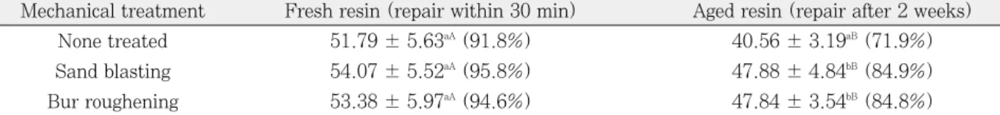
Newly added composite resin was also, Gradia Direct Anterior (GC; Tokyo, Japan), which has Table 3.The mean μTBS and standard deviation for each group (mean ± SD, MPa)
Mechanical treatment Fresh resin (repair within 30 min) Aged resin (repair after 2 weeks)
None treated 51.79 ± 5.63aA(91.8%) 40.56 ± 3.19aB(71.9%)
Sand blasting 54.07 ± 5.52aA(95.8%) 47.88 ± 4.84bB(84.9%)
Bur roughening 53.38 ± 5.97aA(94.6%) 47.84 ± 3.54bB(84.8%)
*Means followed by different letters indicate statistical significant difference (two-way ANOVA/Tukey’s B test, p < 0.05).
Upper case letters compare “aging time”within each “surface treatment”. Lower case letters compare “surface treatment”
within each “aging time”.
*( %): relative percentage of μTBS to ultimate tensile strength.
*Ultimate tensile strength: 56.42 ± 3.71 MPa
Table 4.The mean KICand standard deviation for each (mean ± SD, MPa∙m1/2)
Mechanical treatment Fresh resin (repair within 30 min) Aged resin (repair after 2 wk)
None treated 1.07 ± 0.10aA(72.2%) 0.79 ± 0.15aB(53.7%)
Sand blasting 1.24 ± 0.08bA(84.0%) 1.20 ± 0.10bA(81.4%)
Bur roughening 1.20 ± 0.10bA(81.3%) 1.20 ± 0.10bA(81.2%)
*Means followed by different letters indicate statistical significant difference (two-way ANOVA/Tukey’s B test, p < 0.05).
Upper case letters compare “aging time”within each “surface treatment”. Lower case letters compare “surface treatment”
within each “aging time”.
*( %): relative percentage of FT to bulk fracture toughness.
* Bulk fracture toughness: 1.48 ± 0.03 MPa∙m1/2
Figure 3.FE-SEM observation of none treated groups (×50). Mixed failure was prominent. The area of adhesive failures, dark gray area, was increased after 2weeks aging, (b). (a) Fracture surface of fresh resin. (b) Fracture surface of 2 weeks aged resin.
FE-SEM, Field emission scanning electron microscope.
Figure 4.FE-SEM observation of sandblasting groups (×50). (a) Fracture surface of fresh resin. The area of cohesive failure was prominent (upper: old composite, lower : new composite). (b) Fracture surface of 2 weeks aged resin.
FE-SEM, Field emission scanning electron microscope.
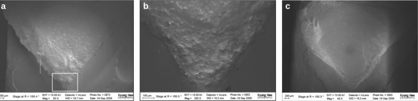
Figure 5.FE-SEM observation of diamond bur roughening groups (a, c: ×50, b: ×250). (a) Fracture surface of fresh resin showed almost cohesive failure. (b) Magnified image at chevron notch (white rectangle) in (a). There were laminations like scale around the notch which mean high resistance to fracture.(c) Fracture surface of 2 weeks aged resin. There was partial cohesive failure.
FE-SEM, Field emission scanning electron microscope.
Figure 6.FE-SEM observation of representative BF specimen. (a) There were crack deflection (white arrow) and microcracks (black arrow). They each are one of crack shielding mechanisms (×20,000). (b) Magnified image of (a). There were another crack shielding mechanisms, uncracked ligament bridgings (white arrow) and crack bridging (black arrow) (×50,000).
FE-SEM, Field emission scanning electron microscope.
a b
a b
a b c
a b
UDMA instead of Bis-GMA. UDMA is an aliphatic molecule which gives the polymer chain great mobili- ty, thereby increasing the degree of conversion than Bis-GMA.19-21The higher degree of conversion is, less- er chemical bonding between methacrylate radicals from old composite and repair composite is. For this reason, UDMA would clearly demonstrate the effect of micromechanical interlock by surface treatment rather than the effect of chemical bonding. That would provide useful information whether the effect of surface treatment surpassed the chemical bonding between resin-based materials or not for repair of old composite restoration.
Old composite can bond with new composite using free radicals in the oxygen inhibition layer or the remaining double bonds within polymerized compos- ite if the oxygen inhibition layer has been removed.
Although it has been shown that different monomers or monomer mixtures are responsible for determining the degree of conversion in dental composite materi- als, all the materials used as the repair substrates have a similar monomer composition.6Degree of con- version of different Bis-GMA based composites ranged between 56% and 68%.22-24 Other study reported that 30% of C=C bonds remained unreact- ed during the copolymerization reaction.25 Aging process diminishes the amount of free radicals and remaining double bonds available and capable of reacting chemically with the new composite.4,26 The likelihood of achieving covalent bonding between resins appears to be negatively correlated to the age of the substrate resin.27
When a composite repair is required, the aging time of the old composite can be various. For example, a patient might return to the dental office with a frac- tured edge of the composite restoration or due to a partially unsatisfactory composite restoration. To simulate this immediate repair, fresh resin groups received rebonding procedure within 30 minutes. It has been assumed that the radical activity of monomer functional groups is the greatest in that time frame. On the other hand, the aged groups sim- ulated the delayed repair condition where composite restoration had been functioning in the oral condition for a long time and there was no oxygen inhibited layer or free radicals available on the surface. There
were few reports regarding to the remaining double bonds following aging time while some reported regarding to the intensity of the light, time of poly- merization, monomer structure and solvent concen- tration.20,28-30 Previous studies on composite repair were conducted at various aging time since the com- posite block had been made, such as immediately,31 24 hours,1,5,32 9 days,3 1 week later32 and 2 weeks later.8Aging condition was set up referring to similar literature storing in 37℃ artificial saliva for 2 weeks.3,8 Further studies are needed to suggest clear standards for the amount of remaining monomer or oxygen inhibition layer by aging time.
In the present study, μTBS of the experimental groups were inferior to UTS, and this finding was in agreement with the previous reports.3,5,9,33 Ferracane and Marker stated that this reduction mainly occurred from softening of the resin matrix, but the cracking within the resin and at the filler/matrix interface might have also contributed to the reduc- tion.33Other authors suggested that the effects of pH changes, salivary enzymes and the wet environment could have resulted in the degradation of composites’
eluting components.3,9
μTBS of fresh groups ranged between 92% and 96%
when compared to UTS, and that of the aged groups ranged between 72% and 85% (Table 3). The aged groups showed significantly lower bond strength than the fresh groups. It seems to be related to the differ- ence of amount of available double bonds between the fresh and the aged composite surfaces as stated above. In addition, the polymerization of composite continued after light cure for 24 hours,34 and the dif- fusion rate of the propagating free radicals under- went a drastic reduction as the polymerization pro- ceeded.29That is, the fresh composites repaired with- in 30 minutes after the light cure have had more free radicals than the aged group have.
The surface treatment of aged resin composite was done to remove the superficial layer altered by the saliva exposing a clean, higher energy composite sur- face and increasing the surface area through creation of surface irregularities. Sandblasting simulated air abrasion in laboratory. Diamond bur roughening is an easy method to roughen the surface in the clinic if there is no microetcher. Different results have been
reported for preparing composite surfaces using air abrasion and bur roughening.1,5,9,10 In the present study, the diamond bur roughening was as effective as sandblasting. μTBS of sandblasting and diamond bur roughening groups were significantly higher than that of untreated groups. Mechanical interlock between the old composite and the newly added com- posite seems to be more effective than chemical bonding. Futhermore, mechanical treatment increas- es surface wettablility.
Fracture toughness values of sandblasting and dia- mond bur roughening groups were also significantly higher than non-mechanically treated groups each aging time. It seems to be related to crack deflection (Figure 6a). Crack deflection is one of the crack shield mechanisms.35Mechanical interlock by surface treatment acts as a filler which provides several toughening mechanisms including crack pinning36and microcrack-induced toughening.37 It can deviate the interfacial plane which occurs crack propagation.
Then, crack tip has more longer path, and linear energy required to fracture is increased.38Futhermore, the energy to make rough fractured surface of com- posite is more than that of smooth fractured surface.
The concept of ‘Craze’also can support the results of SA and BA different from NA group. Craze is the main mechanism of destruction under glass transi- tion temperature. When tensile stress was added to polymer material on a vertical plane of stress direc- tion, microvoids occur. Then, fibrils are orientated in direction of tensile axis and connect both ends of microvoid to make the net structure which controls the enlargement of the microvoids. This net structure which exists at the end of the crack is called craze.38 When there are additional stress concentrators such as defects or voids, the length of orientated fibrils becomes short, and they are easily disconnected at stress and progress to crack. Adhesive agent is weak- er than composite resin and is easy to degrade due to hydrophilic monomers in wet environment. In addi- tion, a defect by technical sensitivity is apt to be in the adhesive layer. Repaired surface without mechanical interlock cannot deviate the crack path and seems to be influenced more by defects in the adhesive layer. As a result, KIC of NA group showed the lowest value.
The interaction between surface treatment and aging time was statistically significant in fracture toughness (p < 0.001), while it was not significant in μTBS. For that reason, the KIC of fresh groups showed somewhat different pattern compared to the μTBS of them. It was diminished approximately 80%
of bulk fracture toughness. It was not restored like bulk fracture toughness after fresh repair and remained relatively unchanged despite aging. These results were similar with previous studies.14,39
It has been proposed that an interfacial fracture mechanics approach which studies the failure of an interface by the initiation and growth of crack using a tool such as the fracture toughness test, would be more appropriate for testing relatively brittle materi- al interface. Nevertheless, this fracture mechanics approach has not received much supports. Soderholm commented that it was so laborious and had time- consuming specimen preparation.39 It means this methodology no longer meets the basic requirements for an easy, fast, and ‘first’product-screening test.
For this reason, there have been many experiments that used bond strength instead. A correlation research between fracture toughness and bond strength, however, is needed to use the bond strength data for a study on fractural behavior.
Unfortunately, there have been only few studies on the correlation between fracture toughness and ten- sile strength of composite-composite repair interface.
In a case of dentin-composite interface, many studies reported there was no significant correlation even though they had a similar pattern.13,15,39 Dhuru and Lloyd have mentioned that the fracture stress of the repair may be a function of both the interfacial strength and defect produced by the operators’repair technique.14 This means composite to composite interface may influence on the fracture toughness test differently from dentin to composite interface due to the matrix discrepancy. Studies on fracture toughness of dentin to composite interface have a limitation to serve as a reference.
In the present study, there was no correlation between fracture toughness and microtensile bond strength values. It seems to be difficult to correlate under the same criteria. First, it is impossible to get same specimen for microtensile bond strength test
and fracture toughness test. Second, the difference between μTBS and KICis thought to be an innate dis- similarity of the test methods. They have different failure mechanisms. The main reason for Fracture mechanics fails more often in the adhesive region, while cohesive failure in the adherend is more com- mon among strength tested specimens.15,39Fracture of tensile bond strength specimens began from some point around the circumference, coincident with the position of stress concentration, and propagated toward the center. On the other hand, the load in short-rod fracture toughness specimen concentrated at the chevron notch. Chevron notch initiates and helps maintain crack propagation along the midplane of the specimen, where the bonded interface is locat- ed. The fracture toughness test could be more appro- priate for testing repaired composite interface.
Specimens of the fracture toughness test showed the fracture mechanism in Fe-SEM observation.
Most specimens of non-mechanically treated groups showed adhesive failure along the interface making smooth surface, while those of mechanically treated groups showed mixed failures in which cohesive fail- ures were dominant making rough surface (Figures 3 and 5). Rough surface has higher KIC than smooth surface. It means more energy is required to fracture the interface as shown in Table 4. The crack propa- gated through composite as well as adhesive layer demonstrating toughening mechanism of the material where the difference of toughness was arisen from (Figure 6). For instance, crack bridging, the most common form of crack-tip shielding, occurred where the dominant crack links with microcracks ahead of the crack tip (Figure 6b). The microcracks provide a mechanism for the formation of uncracked ligament bridges and can act to shield the crack tip toughen- ing the material extrinsically.36,40 These crack shield- ing mechanisms appeared in specimens having high KIC. In other words, fracture toughness test can explain fractural behavior for repaired composite inter- face. It cannot be substituted by bond strength test.
To date, the study on fracture toughness of com- posite to composite repair interface has been scare.
The present study evaluated the effect of surface treatment and aging time on composite repair by a practical test method, fracture toughness. This pro-
vides significant data that can be used for a practical prediction of fractural resistance in the repaired com- posite restorations. However, there is a limitation in that there are many variables yet to be tested including the age at which the repair is made, the interval between fracture and repair, the aging con- dition, and so on. More researches using fracture toughness are necessary for the evaluation of fractur- al behavior of the repaired composite.
Conclusions
Repair of fractured composite can be conducted effectively by means of appropriate surface treat- ment. For the reliable evaluation of fractural behav- ior of the repaired composite, both μTBS test and fracture toughness test were carried out. Within the limitations of the present study, the fresh composite resin showed higher μTBS than the aged composite resin did (p < 0.01). Comparing to μTBS of mechani- cally treated groups, μTBS of non-mechanically treat- ed group was similar in the fresh resin groups, while significantly lower in the aged resin (p < 0.05). The fracture toughness values of mechanically treated groups were higher than that of non-mechanically treated group no matter whether it was sandblasting or bur roughening (p < 0.05). Surface treatment and aging time had a significant influence on the interfa- cial fracture toughness (p < 0.001). There was no correlation between fracture toughness and microten- sile bond strength values. Specimens having high KIC
showed toughening mechanism including crack devi- ation, microcracks and crack bridging.
In conclusion, mechanical surface treatment seems to be an important factor for the effective composite repair and the fracture toughness test could be used as an appropriate tool to examine the fractural behavior of the repaired composite with microtensile bond strength.
References
1. Papacchini F, Dall’Oca S, Chieffi N, Goracci C, Sadek FT, Suh BI, Tay FR, Ferrari M. Composite-to-compos- ite microtensile bond strength in the repair of a micro- filled hybrid resin: effect of surface treatment and oxy- gen inhibition. J Adhes Dent 2007;9(1):25-31.
2. Suh BI. Oxygen-inhibited layer in adhesion dentistry.
J Esthet Restor Dent 2004;16(5):316-323.
3. Rodrigues SA Jr, Ferracane JL, Della Bona A.
Influence of surface treatments on the bond strength of repaired resin composite restorative materials. Dent Mater 2009;23(4):442-451.
4. Fawzy AS, El-Askary FS, Amer MA. Effect of surface treatments on the tensile bond strength of repaired water-aged anterior restorative micro-fine hybrid resin composite. J Dent 2008;36(12):969-976.
5. Cavalcanti AN, De Lima AF, Peris AR, Mitsui FH, Marchi GM. Effect of surface treatments and bonding agents on the bond strength of repaired composites. J Esthet Restor Dent 2007;19(2):90-98(discussion 99).
6. Teixeira EC BS, Thompson JY, Ritter AV, Swift EJ.
Shear bond strength of self-etching bonding systems in combination with various composites used for repairing aged composites. J Adhes Dent 2005;7(2):159-164.
7. Tezvergil A, Lassila LV, Vallittu PK. Composite-com- posite repair bond strength: effect of different adhesion primers. J Dent 2003;31(8):521-525.
8. Brosh T, Pilo R, Bichacho N, Blutstein R. Effect of combinations of surface treatments and bonding agents on the bond strength of repaired composites. J Prosthet Dent 1997;77(2):122-126.
9. Bonstein T, Garlapo D, Donarummo J, Jr., Bush PJ.
Evaluation of varied repair protocols applied to aged composite resin. J Adhes Dent 2005;7(1):41-49.
10. Cesar PF, Meyer Faara PM, Miwa Caldart R, Gastaldoni Jaeger R, da Cunha Ribeiro F.. Tensile bond strength of composite repairs on Artglass using different surface treatments. Am J Dent 2001;14(6):
373-377.
11. Mecholsky JJ Jr. Fracture mechanics principles. Dent Mater 1995;11(2):111-112.
12. Tam LE, Dev S, Pilliar RM. Fracture toughness ov conventional or photopolymerized glass ionomer/dentin interfaces. Oper Dent 1995;20(4):144-150.
13. Ryu GJ, Choi GW, Park SJ, Choi. KK. A Study on Fractural Behavior of Dentin-Resin Interface. J Kor Acad Cons Dent 2007;32(3):208-221.
14. Dhuru VB, Lloyd. CH. The fracture toughness of repaired composite. J Oral Rehabil 1986;13(5):413-421.
15. Tam LE, Pilliar RM. Fracture toughness of dentin/resin-composite adhesive interfaces. J Dent Res 1993;72(5):953-959.
16. Tam LE, Pilliar RM. Effects of dentin surface treat- ments on the fracture toughness and tensile bond strength of a dentin-composite adhesive interface. J Dent Res 1994;73(9):1530-1538.
17. Barker LM. A simplified methods for measuring plane strain fracture toughness. Eng Fract Mech 1977;9:361-369.
18. Tam LE, Pilliar RM. Fracture surface characterization of dentin-bonded interfacial fracture toughness speci- mens. J Dent Res 1994;73(3):607-619.
19. Lee YS, Choi KK, Park SJ. Effect of resin matrix on degree of conversion and fracture toughness of dental composites. J Kor Acad Cons Dent 2002;27(1):77-86.
20. Ruyter IE, Oysaed H. Composites for use in posterior teeth: composition and conversion. J Biomed Mater Res 1987;21(1):11-23.
21. Filho JD, Poskus LT, Guimarães JG, Barcellos AA, Silva EM. Degree of conversion and plasticization of dimethacrylate-based polymeric matrices: influence of light-curing mode. J Oral Sci 2008;50(3):315-321.
22. Sturdevant CM, edited by Roberson TM, Heymann H, Swift EJ. Sturdevant’s art and science of operative
dentistry. 5th ed., 2006. p201-203.
23. Goncalves F, Kawano Y, Pfeifer C, Stansbury JW, Braga. RR. Influence of BisGMA, TEGDMA, and BisEMA contents on viscosity, conversion, and flexural strength of experimental resins and composites. Eur J Oral Sci 2009;117(4):442-446.
24. Eick JD, Gwinnett AJ, Pashley DH, Robinson SJ.
Current concepts on adhesion to dentin. Crit Rev Oral Biol Med 1997;8(3):306-335.
25. Lapc′lk L Jr JJ, Stasko A, Sa′ha P. Electron paramag- netic resonance study of free-radical kinetics in ultra- violet-light cured dimethacrylate copolymers. J Mater Sci Mater Med 1998;9(5):257-262.
26. Miyazaki M, Onose H, Iida N, Kazama H.
Determination of residual double bonds in resin-dentin interface by Raman spectroscopy. Dent Mater 2003;
19(3):245-251.
27. Cavalcanti AN, Lobo MM, Fontes CM, Liporoni P, Mathias P. Microleakage at the Composite-repair Interface: Effect of Different Surface Treatment Methods. Oper Dent 2005;30(1):113-117.
28. Lovell LG, Newman SM, Bowman CN. The effects of light intensity, temperature, and comonomer composi- tion on the polymerization behavior of dimethacrylate dental resins. J Dent Res 1999;78(8):1469-1476.
29. Santerre JP, Shajii L, Leung BW. Relation of dental composite formulations to their degradation and the release of hydrolyzed polymeric-resin-derived products.
Crit Rev Oral Biol Med 2001;12(2):136-151.
30. Ye Q, Spencer P, Wang Y, Misra A. Relationship of solvent to the photopolymerization process, properties, and structure in model dentin adhesives. J Biomed Mater Res A 2007;80(2):342-350.
31. Passos SP, Ozcan M, Vanderlei AD, Leite FP, Kimpara ET, Bottino MA. Bond strength durability of direct and indirect composite systems following surface condition- ing for repair. J Adhes Dent 2007;9(5):443-447.
32. Choi SY, Jeong SW, Hwang YC, Kim SH, Yun C, Oh WM, Hwang. IN. Shear bond strength of repaired com- posite resin restorations. J Kor Acad Cons Dent 2002;
27(6):569-576.
33. Ferracane JL, Marker VA. Solvent degradation and reduced fracture toughness in aged composites. J Dent Res 1992;71(1):13-19.
34. Craig RG, Powers JM. Restorative Dental Materials 11th. Mosby St Louis, 2002.
35. Nalla R K, Kinney J H, Ritchie. RO. Effect of orienta- tion on the in vitro fracture toughness of dentin: the role of toughening mechanisms. Biomaterials 2003;
24(22):3955-3968.
36. Young RJ, Beaumont P. Failure of brittle polymers by slow crack growth. Part 2. Failure processes in a silica particle-filled epoxy resin composite. J Mater Sci 1975;
101:343-1350.
37. Kim KH, Park JH, Imai Y, Kishi. T. Microfracture mechanisms of dental resin composites containing spherically-shaped filler particles. J Dent Res 1994;
73(2):499-504.
38. KIM SC. Polymer Engeneering I. 1994. p287-310.
39. Soderholm KJ. Review of the fracture toughness approach. Dent Mater 2010;26(2):e63-77.
40. Kruzic JJ, Nalla RK, Kinney JH, Ritchie. R. Crack blunting, crack bridging and resistance-curve fracture mechanics in dentin: effect of hydration. Biomaterials 2003;24(28):5209-5221.
국문초록
수리된 복합 레진 수복물의 파괴 거동에 관한 연구
박상순∙남 욱∙엄아향∙김덕수∙최기운∙최경규*
경희대학교 대학원 치의학과 치과보존학교실
연구목적: 본 연구는 미세인장결합강도와 파괴인성을 통해 복합 레진 수복물의 수리 시기와 표면 처리 방법에 따른 파괴 거 동을 알아보고자 시행되었다.
연구 재료 및 방법: Short rod 시편과 composite resin specimen block을 준비하여 표면 처리 방법에 따라 none-treated, sand blasting, bur roughening 군으로 나누고 이를 다시 즉시군과 2주 지연군으로 나누어 수리했다.
결과: 미세인장결합강도와 파괴인성을 측정한 결과, 두 실험 모두에서 즉시군이 지연군보다 높은 값을 보였다. 기계적 표면 처리군이 none-treated군보다 높은 값을 보였고, sand blasting과 bur roughening 사이에 유의한 차이는 없었다. 파괴인 성과 미세인장결합강도는 상관 관계가 없었다. FE-SEM을 보아 수복물의 탈락은 균열 전도와 관계가 있는 것으로 보인다.
결론: 수리된 복합 레진의 파괴 거동 평가에는 파괴인성 실험이 적합하다.
주요단어: 기계적 표면 처리, 미세인장결합강도, 복합 레진 수리, 수리 시기, 파괴 거동, 파괴인성