178
Jaw Thrust Improves the Fiberoptic Laryngeal View during Fiberoptic Nasotracheal Intubation
Teo Jeon Shin, Kwang-Suk Seo, and Hyun Jeong Kim
Department of Dental Anesthesiology and Dental Research Institute, School of Dentistry, Seoul National University Seoul, Republic of Korea
Abstract
하악견인법 적용하 굴곡성 기관지 내시경을 이용한 경비삽관시 내시경하 후두시야의 비교
서울대학교 치과대학 치과마취과학교실 신터전․서광석․김현정
배경: 임상적으로 굴곡성 기관지 내시경을 이용 삽관 시행시 후두경으로 성문부위가 잘 드러나 지 않는 환자의 경우 삽관 시행이 어려운 경우를 경험한다. 하지만 이에 대한 연구는 거의 없는 실정이다. 본 연구에서는 어려운 기도환자에서 굴곡성 기관지경 시행시 후두시야를 확보시 차이가 있는지 확인하고자 하였다.
방법: 전신마취 유도 후 Cormack – Lehane classification을 이용하여 기관 삽관의 어려움을 먼저 평가하였다. 기관지 내시경을 이용하여 내시경하 후두시야의 정도를 평가하였다. 후두경으로 기도 확보가 용이한 그룹(Cormack – Lehane grades 1, 2)과 어려운 그룹(Cormack – Lehane grades 3, 4) 간의 내시경하 후두 시야의 정도가 차이가 나는 지를 확인하였다.
결과: 후두경으로 기도확보가 용이하지 않을 경우에 기관지 내시경으로 후두 시야를 용이하게 (fiberoptic laryngeal view 1, 2) 확보하기가 어려웠다. 반면 하악을 전방으로 견인시 후두시야의 정도 가 통계적으로 유의하게 개선되었다.
결론: 전방하악견인법 (jaw-thrust maneuver)은 기도확보가 어려운 환자에서 기관지 내시경을 이 용한 기관내 삽관 시행시 시야를 개선시켜서 삽관을 용이하게 할 수 있을 것으로 생각된다.
(JKDSA 2010; 10: 178~182)
핵심용어: Endotracheal intubation; Laryngoscopy
Received: December 20, 2010, Revised: December 21, 2010 Accepted: December 24, 2010
Corresponding author: Kwang Suk Seo, Department of Dental Anesthesiology and Dental Research Institute, Seoul National University School of Dentistry, 28 Yeongeon-dong Jongno-gu, Seoul 110-768, Korea Tel: +82-2-2072-0622, Fax: +82-2-766-9427 E-mail: [email protected]
INTRODUCTION
Securing the airway after induction of general ane- sthesia is essential to avoid airway - related serious complication. However, endotracheal intubation is not always possible. In most of cases, poor laryngeal visualization during laryngoscopy makes the intubation difficult. In dental surgery, patients may have potential anatomical abnormalities of the airway. This factor
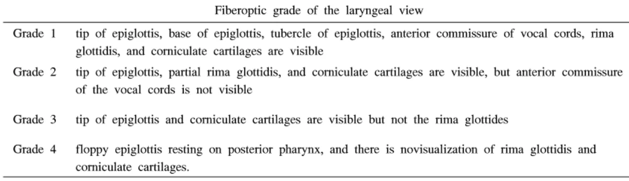
Table 1. The Classification of Fiberoptic Laryngeal View Fiberoptic Grade of the Laryngeal View
Fiberoptic grade of the laryngeal view
Grade 1 tip of epiglottis, base of epiglottis, tubercle of epiglottis, anterior commissure of vocal cords, rima glottidis, and corniculate cartilages are visible
Grade 2 tip of epiglottis, partial rima glottidis, and corniculate cartilages are visible, but anterior commissure of the vocal cords is not visible
Grade 3 tip of epiglottis and corniculate cartilages are visible but not the rima glottides
Grade 4 floppy epiglottis resting on posterior pharynx, and there is novisualization of rima glottidis and corniculate cartilages.
* The classification of fiberoptic laryngeal view is slightly modified based on the grade of the laryngopharyngeal tissue suggested by Cheng KI et al (Cheng et al, 2008)
may contribute to difficulty in laryngeal visualization compared to other types of surgery.
When intubation by direct laryngoscopy fails, the flexible bronchoscope can be effectively used to secure the airway. It seems plausible that the advantage of fiberoptics in difficult airway may arise from the superior quality of laryngeal view. However, we have frequently experienced that laryngeal view is also poor when fiberoptic attempted in patients with difficult laryngoscopy. Interestingly, the poor fiberoptic view is improved by applying jaw thrust maneuver in many cases. These observations may be clinically implicable in that the performance of fiberoptic intubation is usually dependent on the quality of fiberoptic view especially for inexperienced intubator. However, few studies to validate our observations have been per- formed. Therefore, the purpose of study is to investi- gate whether the fiberoptic laryngeal view is related to laryngoscopic laryngeal view and thrusting the jaw is helpful in improving the fiberoptic view in case of nasotracheal intubation.
MATERIAL AND METHODS
After Institutional Review Board was obtained in Seoul National University Dental Hospital, the patients requiring nasotracheal intubation for Oral & Maxillo- facial surgery were recruited. Patients were excluded
from the study if they have recurrent epistaxsis, nasal bleeding, and bleeding tendency.
The same anesthesia protocol was applied to all patients. After monitoring with electrocardiogram, pulse oximetry, capnograph, and noninvasive arterial blood pressure was initiated, 5 mg/kg of thiopental was intravenously injected to the patients. After achieving the loss of consciousness, neuromuscular relaxation was achieved with vecuronium 0.1-0.15 mg/kg IV.
The ventilation of patient’s lung was maintained via a face mask with 5-8 vol % of sevoflurane in 100%
oxygen. After complete muscle relaxation was con- firmed by a nerve stimulator, nasotracheal intubation was attempted through a patent’s nostril.
At first, the same anesthesiologist inserted the layngoscope into the oral cavity and assessed the grade of intubation difficulty using Cormack – Lehane classification (Cormack and Lehane, 1984).
After assessing the grade, the laryngoscope was gently removed and endotracheal tube was inserted through a nostril into the posterior pharynx. The flexible fiberoptic (Olympus LF-2; Olympus Optical Company, Tokyo, Japan) was railroaded onto it. The fiberoptic view was first recorded using videocamera system (OTV-SC, Olympus Optical Company, Tokyo, Japan) as soon as the bronchoscope was placed upper the epiglottis. At the same position, the fiberoptic view was recorded with the jaw thrusted. After obtaining
Table 2. Demographic Data
EI group (n = 25) DI group (n = 25) Age (yr)
Sex (M/F) Height (cm) Weight (kg)
31.6 ± 18.8 11/14 164.8 ± 12.1 57.4 ± 13.3
39.7 ± 15.6 13/12 163.0 ± 11.1 60.6 ± 13.6
* Data are expressed as mean ± SD (standard deviation) for continuous data, ratio for categorical data.
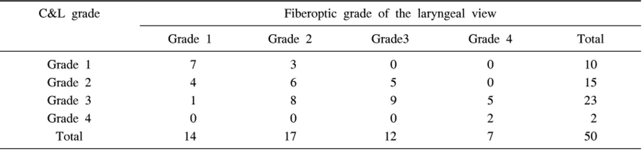
Table 3. Comparison of C&L Grade and Fiberoptic Grade of the Laryngeal View
C&L grade Fiberoptic grade of the laryngeal view
Grade 1 Grade 2 Grade3 Grade 4 Total
Grade 1 Grade 2 Grade 3 Grade 4 Total
7 4 1 0 14
3 6 8 0 17
0 5 9 0 12
0 0 5 2 7
10 15 23 2 50
* C&L grade = Cormack & Lehane grade
Table 4. Comparison of Fiberoptic Grade of the Laryngeal View Before and After Jaw Thrust Maneuver
Fiberoptic grade of the laryngeal view
Jaw thrust (-) Jaw thrust (+)
Easy Airway group (n = 25) 20 (25) 23 (25)
Difficult Airway group (n = 25) 9 (25) 15 (25)*
* The number indicates total number of patients with 1, 2 grade of fiberoptic laryngeal view. The number in parentheses indicates total number of patients in each group. * : P-value < 0.001
the view, the fiberoptic intubation was performed. The anesthesia was maintained with 50% N2O/O2 and sevoflurane. The fiberoptic laryngeal view was ad hoc assessed by the observer blinded to the Cormack – Lehane classification in the same patients. The fiberoptic laryngeal view was graded as follows (Table 1) (Cheng et al, 2008).
Based on pilot study (n = 10), the sample size to detect the difference of the fiberoptic laryngeal view was estimated to be 22 in each group with a power of 0.8 at the level of significance of 0.05. The continuous data was analyzed by t - test. The
categorical data was analyzed by chi – square. A P value less than 0.05 was considered to be statistically significant.
RESULTS
Total 50 patients were included in this study. The patients was classified as either easy airway group (Cormack – Lehane grade 1, 2) or difficult airway group (Cormack – Lehane grade 3, 4), respectively.
No significant differences of sex, age, height and body weight was observed between the groups (Table
2). In easy airway group (n = 25), fiberoptic intu- bation provides 1, 2 grade of the fiberoptic laryngeal view in 20, whereas 9 out of 25 patients in difficulty airway group (n = 25) (Table 3, 4, P < 0.001).
However, the number of patients with 1, 2 grade of the fiberoptic laryngeal view was significantly increased when the jaw thrusted, especially in difficult airway group (Table 4, P < 0.001).
DISCUSSION
Flexible fiberoptic intubation is critical in endo- tracheal intubation with the difficulty laryngoscopy (Benumof, 1991). The literature to review 18,500 fiberoptic intubation cases revealed that the flexible fiberoptic bronchoscope was the most commonly utilized technique in unanticipated difficult intubation (Rose and Cohen, 1994). However, fiberoptic intu- bation has its own failure rates due to inability to visualize the larynx (Ovassapian et al, 1983). Theore- tically, getting the operator’s view into the larynx directly during fiberoptic intubation may be advanta- geous compared with that of a direct laryngoscope in terms of laryngeal visualization. Contrary to our expectation, the laryngeal view was not significantly improved with the use of a fiberoptic bronchoscope via the nostril.
Meanwhile, the most interesting finding in this study is that thrusting the jaw improved the fiberoptic laryngeal view significantly. Consistent with our results, triple airway maneuver improves the fiberoptic laryn- geal view by lifting the epiglottis into anterior direc- tion in patients with limited mouth opening (Cheng et al, 2008). The jaw thrust is known as effective means to open the airway during orotracheal fiberoptic intubation (Durga et al, 2001). It is speculated that upward force of the jaw may drag the epiglottis into anterior direction leading to improvement of fiberoptic laryngeal view. Poor fiberoptic laryngeal view may disturb the performance of nasotracheal fiberoptic intu- bation. Poor exposure of the laryngeal aperture is likely to require anterior displacement of fiberoptic
distal tip so as to advance into the trachea, thus hindering the process of nasotracheal intubation and passage of the tube to the trachea. In this aspect, our results suggest clinical implication when performing nasotracheal intubation especially. Nasotracheal intuba- tion is frequently associated with epistaxis (Tintinalli and Claffey, 1981; Hall and Shutt, 2003). When epistaxis occurs during nasotracheal intubation, the blood regurgitated to oral cavity may obscure fiberop- tic laryngeal view. Performing nasal intubation is very difficult with a fiberoptic brochoscope in the event of unanticipated difficult intubation with concurrent epistaxis. Delayed nasotracheal fiberoptic intubation may also increase the risk of desaturation since the repetitive failure of nasotracheal intubation increase the likelihood of traumatizing the nasal mucosa, and mask ventilation at the intervals of repetitive attempts of fiberoptic intubation could cause the aspiration of blood pooled in the posterior pharynx into the lung.
In this circumstance, jaw-thrust maneuver may be used as an effective technique to secure the airway and improve the laryngeal view, preventing fatal complications from failed fiberoptic intubation.
However, our study has also some limitations. The poor laryngeal view does not necessarily means the difficulty in fiberoptic intubation. The experienced anesthesiologist may easily intubate the patients with poor fiberoptic glottic view. Second, we graded laryn- geal view provided by nasal fiberoptic intubation, not oral fiberoptic intubation. Thus, clinical application of our results in oral fiberoptic intubation is limited.
In conclusion, we shows that fiberoptic intubation do not always permit the excellent laryngeal view in patients with difficult airway although the fiberoptic laryngeal view is improved by thrusting the jaw.
REFERENCES
Benumof J: Management of the difficult adult airway.
Anesthesiology 1991; 75(6): 1087-110.
Cheng KI, Yun MK, Chang MC, Lee KW, Huang SC, Tang CS, et al: Fiberoptic bronchoscopic view change
of laryngopharyngeal tissues by different airway supporting techniques: comparison of patients with and without open mouth limitation. Journal of Clinical Anesthesia 2008; 20(8): 573-9.
Cormack R, Lehane J: Difficult tracheal intubation in obstetrics. Anaesthesia 1984; 39(11): 1105-11.
Durga VK, Millns JP, Smith JE: Manoeuvres used to clear the airway during fibreoptic intubation. Br J Anaesth 2001; 87(2): 207-11.
Hall C, Shutt L: Nasotracheal intubation for head and
neck surgery. Anaesthesia 2003; 58(3): 249-56.
Ovassapian A, Yelich S, Dykes M, Brunner E: Fiberop- tic nasotracheal intubation--incidence and causes of failure. Anesthesia and analgesia 1983; 62(7): 692-5.
Rose D, Cohen M. The airway: problems and predictions in 18,500 patients. Can J Anaesth 1994; 41(5): 372- 83.
Tintinalli J, Claffey J: Complications of nasotracheal intubation. Annals of Emergency Medicine 1981;
10(3): 142-4.