ABSTRACT
Objectives: This study evaluated the amount of remaining root canal filling materials after retreatment procedures performed by undergraduate students using manual, rotary, and reciprocating techniques through micro-computed tomographic analysis. The incidence of instrument fracture and the instrumentation time were also evaluated.
Materials and Methods: Thirty maxillary single rooted teeth were prepared with Reciproc R25 files and filled with gutta-percha and AH Plus sealer by the continuous wave of condensation technique. Then, the specimens were assigned to 3 groups (n = 10), according to the retreatment technique used: manual, rotary, and reciprocating groups, which used K-file, Mtwo retreatment file, and Reciproc file, respectively. Retreatments were performed by undergraduate students.
The sample was scanned after root canal filling and retreatment procedures, and the images of the canals were examined to quantify the amount of remaining filling material. The incidence of instrument fracture and the instrumentation time were recorded.
Results: Remaining filling material was observed in all specimens regardless of the technique used. The mean volume of remaining material was significantly lower in the Reciproc group than in the manual K-file and Mtwo retreatment groups (p < 0.05). The time required to achieve a satisfactory removal of canal filling material and refinement was significantly lower in the Mtwo retreatment and Reciproc groups (p < 0.05) when compared to the manual K-file group. No instrument fracture was observed in any of the groups.
Conclusions: Reciproc was the most effective instrument in the removal of canal fillings after retreatments performed by undergraduate students.
Keywords: Endodontics; Retreatment; X-ray microtomography
INTRODUCTION
Although root canal treatments have a high rate (90%) of success when properly conducted [1,2], failures may occur and are often associated with poorly treated canals [2]. In these situations, nonsurgical root canal retreatment is indicated to improve root canal disinfection
Research Article
Received: Oct 9, 2017 Accepted: Dec 14, 2017
Silva EJ, Belladonna FG, Carapiá MF, Muniz BL, Rocha MS, Moreira EJ
*Correspondence to
Emmanuel João Nogueira Leal Silva, DDS, MSc, PhD
Associate Professor, Department of Endodontics, School of Dentistry, Grande Rio University, Rua Herotides de Oliveira, 61/902 Icaraí - Niterói, RJ 24230-230, Brazil.
E-mail: nogueiraemmanuel@hotmail.com Copyright © 2018. The Korean Academy of Conservative Dentistry
This is an Open Access article distributed under the terms of the Creative Commons Attribution Non-Commercial License (https://
creativecommons.org/licenses/by-nc/4.0/) which permits unrestricted non-commercial use, distribution, and reproduction in any medium, provided the original work is properly cited.
Conflict of Interest
No potential conflict of interest relevant to this article was reported.
Author Contributions
Conceptualization: Silva EJNL, Belladonna FG; Data curation: Silva EJNL, Belladonna FG, Moreira EJL; Formal analysis: Silva EJNL, Belladonna FG; Funding acquisition: Silva EJNL, Moreira EJL; Investigation: Silva EJNL, Belladonna FG, Carapiá MF, Muniz BL, Rocha MS; Methodology: Silva EJNL, Belladonna
Emmanuel João Nogueira Leal Silva ,1* Felipe Gonçalves Belladonna ,2 Marianna Fernandes Carapiá,1 Brenda Leite Muniz,1 Mariana Santoro Rocha,1 Edson Jorge Lima Moreira1
1Department of Endodontics, School of Dentistry, Grande Rio University, Rio de Janeiro, RJ, Brazil
2Department of Endodontics, Fluminense Federal University, Niterói, RJ, Brazil
Micro-computed tomographic evaluation of canal retreatments
performed by undergraduate students
using different techniques
several reports have shown that considerable amounts of root filling materials remain after re-instrumentation [3-5]. This residual material should be removed because it may harbor bacteria, and weaken the quality of seal of the new root canal filling [6,7].
Many techniques have been proposed for removing gutta-percha and sealer during root canal retreatment procedures using various instruments, including hand files, burs, nickel-titanium (NiTi) rotary systems, adjunctive solvents, and/or ultrasonics. Some endodontic systems are specially designed for use in retreatment procedures, such as the Mtwo retreatment system (VDW, Munich, Germany). This system comprises 2 instruments (R15/0.05 and R25/0.05) that have a 2-fluted file with an S-shaped cross-sectional design and a cutting tip in order to facilitate penetration into the obturation mass. Nevertheless, all retreatment systems leave residual debris in the canal walls after endodontic retreatment [2,7-9]. Reciprocating systems, such as Reciproc (VDW) and WaveOne (Dentsply Maillefer, Ballaigues, Switzerland), that were initially developed for root canal preparation can also be used for retreatment purposes. Reciproc instruments have the same cross-sectional design as the Mtwo instruments and are available in 3 different sizes: R25 (25/0.08) for narrow canals, R40 (40/0.06) for medium-volume canals and R50 (50/0.05) for large canals.
There is growing evidence of the safety and benefits of using these instruments during retreatment cases [8-10]. The reciprocating motion relieves stress on the instrument by alternating counterclockwise and clockwise movements, which extends the life span of the NiTi instrument when compared to continuous rotation [11]. Moreover, the idea of a reduced number of NiTi instruments needed to remove root canal obturation materials and to enlarge the canal to a minimum acceptable taper size is indeed appealing due to its safety and technique simplification. From an educational point of view, such procedural simplification and safety can lead to improved outcomes when performed by novice operators [12,13].
The aim of this study was to evaluate the amount of remaining root filling materials after retreatment procedures performed by undergraduate students using manual, rotary, and reciprocating instruments using micro-computed tomographic (micro-CT) analysis. The incidence of instrument fracture and the instrumentation time were also evaluated. The null hypothesis tested was that there would be no differences in the quality of root canal filling material removal performed by undergraduate students using different techniques.
MATERIALS AND METHODS
Teeth selection
This study protocol was approved by the Grande Rio University Ethics Committee (ID:
CAAE 47448315.2.0000.5283). Fifty-three maxillary single rooted teeth presenting one canal and similar root length were selected from a pool of teeth. Buccolingual and mesiodistal radiographs were taken to select teeth with oval canals according to the technique previously described [8]. Canal curvature was determined using Schneider's method [14], and the angle of curvature of each specimen was measured through an image analysis program (AxioVision 4.5, Carl Zeiss Vision, Hallbergmoos, Germany). Access openings were performed using diamond burs and a size 10 K-file (Dentsply Maillefer) was used to initially negotiate the canal. Only straight teeth with root curvature < 10°, approximately 22 ± 1 mm in length, and an initial apical size equivalent to a size 10 K-file were included (n = 30).
FG, Carapiá MF, Muniz BL, Rocha MS; Project administration: Silva EJNL, Belladonna FG;
Resources: Silva EJNL, Moreira EJL; Software:
Silva EJNL, Moreira EJL; Supervision: Silva EJNL; Validation: Silva EJNL; Visualization:
Silva EJNL, Belladonna FG; Writing - original draft: Silva EJNL, Belladonna FG, Moreira EJL; Writing - review & editing: Silva EJNL, Belladonna FG.
ORCID iDs
Emmanuel João Nogueira Leal Silva https://orcid.org/0000-0002-6445-8243 Felipe Gonçalves Belladonna
https://orcid.org/0000-0001-9972-6861
Root canal preparation and filling procedures
Working length (WL) was established introducing a size 10 K-file into the root canal until its tip was visible at the apical foramen under an operating microscope (DFV, Valença, RJ, Brazil) with ×20 magnification, and reducing 1 mm short of this measurement. After WL determination, the apical foramen diameters of all teeth were standardized to a size 15 K-file (Dentsply Maillefer).
The same experienced operator performed all canal shaping and filling procedures. Teeth were instrumented with Reciproc R25 files (VDW) as recommended by the manufacturer. Irrigation was performed using a total volume of 25 mL of 2.5% sodium hypochlorite (NaOCl). Smear layer was removed using 3 mL of 17% ethylenediaminetetraacetic acid (EDTA) for 3 minutes.
Then, canals were irrigated with 2 mL of bi-distilled water, dried with R25 paper points (VDW), and finally filled with R25 gutta-percha cones (VDW) and AH Plus sealer (Dentsply De Trey, Konstanz, Germany). Condensation of the root filling material was achieved by using the continuous wave of condensation technique, and the down pack (EQ-V Endodontic Obturation System, MetaBiomed, Cheongju, Korea) was established at 5 mm from the WL.
Then, the remainders of the canals were filled with the aid of a McSpadden condenser (Dentsply Maillefer). The access cavities were sealed with Cavit G (3M ESPE, Seefeld, Germany). After that, the specimens were radiographed in mesiodistal and buccolingual directions to confirm the quality of the fillings. A lack of voids in all samples was noted in the radiographs; therefore, none of the samples was discarded. The specimens were kept at 100% humidity and 37°C for 4 weeks to allow complete setting of the sealer.
Root canal retreatment
Ten undergraduate dental students of the same semester had 3 fifty-minute lectures presenting and comparing manual, rotary and reciprocating techniques, their main systems and information regarding their properties, and constructional features. After that, demonstrations of endodontic retreatments using the aforementioned techniques were performed. Each participant received a handout defining the sequence of instruments to be used in each technique. Then, each undergraduate student received 3 teeth and retreated them using each of the 3 retreatment techniques: manual, rotary, and reciprocating groups, which used K-file, Mtwo retreatment file, and Reciproc file, respectively. Thus, each retreatment group included ten different specimens.
1. Retreatment with manual K-files (manual K-file group). A size 3 Gates-Glidden drill (Dentsply Maillefer) was used at 5,000 rpm to remove part of the coronal filling material in order to create a reservoir for the solvent. An increment of eucalyptol (0.1 mL) was introduced into the root canal to soften the gutta-percha for 30 seconds before further instrumentation.
Then, all root canals were re-instrumented to the original WL using manual K-files (Dentsply Maillefer) up to size 40 with a push-pull movement alternated with a rotary motion. After that, manual K-files in sizes from 45 to 60 were used in a step-back technique.
2. Retreatment with Mtwo retreatment files (Mtwo retreatment group). A size 3 Gates- Glidden drill and an increment of eucalyptol (0.1 mL) were used at the same manner as described in the manual K-file group. Then, Mtwo retreatment files R15/0.05 and R25/0.05, and Mtwo rotary files in sizes of 30/0.05, 35/0.04, and 40/0.04 (VDW) were used with an electric motor (VDW Silver, VDW). Speed setting and torque for each instrument were used as recommended by the manufacturer. The instruments were applied using in-and-out
movement, associated to a short stroking/brushing motion in a coronal direction to the full original WL. After 3 gentle in-and-out motion strokes, the instruments were removed from the canal, cleaned off by inserting into a clean stand with sponge until the WL was reached.
3. Retreatment with Reciproc files (Reciproc group). A size 3 Gates-Glidden drill and an increment of eucalyptol (0.1 mL) were used at the same manner as described in the manual K-file group. Reciproc files were moved in an apical direction in a reciprocating motion using in-and-out pecking motion of about 3 mm in amplitude with a light apical pressure.
Brushing motion against the lateral canal walls was also used. After 3 pecking motions, the instrument was removed from the canal, and its flutes were cleaned off. An R25 file was used to remove the filling materials until WL was reached. Afterward, the R40 file (VDW) was used with the same protocol.
Each set of instruments was used in one tooth and then discarded. The criteria for the completion of retreatment procedures were smooth canal walls and no evident filling material on the files. The same irrigation protocol was applied in all groups. A 2.5% NaOCl was delivered using a 30-gauge side-vented needle (Max-i-Probe, Dentsply Rinn, Elgin, IL, USA) between each instrument change, and a total of 25 mL of 2.5% NaOCl was delivered per canal. After final instrumentation, all canals were dried with paper points (Dentsply Maillefer) and the access openings were sealed with Cavit G.
Micro-CT scanning procedures and evaluation
The sample was scanned using a micro-CT device (SkyScan 1174v2, Bruker microCT, Kontich, Belgium) after root canal filling and retreatment procedures using the following parameters:
50 kV, 800 mA, isotropic resolution of 21.8 μm, 360° rotation around the vertical axis, rotation step of 0.5°, and frame averaging of 3, using a 1-mm thick aluminum filter. Then, the NRecon v.1.6.9 software (Bruker microCT) was used to reconstruct the acquired images into cross-sectional slices, and the volume of interest was selected to extend from the cemento- enamel junction to the apex of the root, resulting in the acquisition of 700–800 transverse cross sections per tooth. CTAn v.1.16.1 software (Bruker microCT) was used for measuring initial and residual volumes of filling materials (mm3). The percentage of residual volume relative to the initial volume of the filling material was calculated.
Statistical analysis
The preliminary analysis of the raw pooled data revealed a bell-shaped distribution
(D'Agostino and Person omnibus normality test), and statistical analysis was performed using parametric methods. One-way analysis of variance (ANOVA) was followed by post hoc pair-wise comparisons, which were performed using Tukey multiple comparisons. The alpha-type error was set at 0.05, and SPSS 11.0 (SPSS Inc., Chicago, IL, USA) was used as the analytic tool.
RESULTS
Remaining filling materials were observed in all specimens. The mean volume (%) of the remaining filling material is shown in Table 1. All techniques left an average of 3.67%–16.21%
of root filling materials in the root canal after retreatment procedures. The mean volume of remaining material was significantly lower in the Reciproc group when compared to manual K-file and Mtwo retreatment groups (Figure 1, p < 0.05).
The mean operating time is also presented in Table 1. The time required to achieve a satisfactory removal of root canal filling material and refinement was significantly lower in the Mtwo retreatment and Reciproc groups (p < 0.05). No instrument fracture or accident was observed in any of the groups.
Table 1. Remaining filling material (%) and the time required to complete endodontic retreatments (minutes)
Group Remaining filling material (%) Time (min)
Manual K-file 13.86 ± 17.83a 23.33 ± 15.70a
Mtwo retreatment file 16.21 ± 17.08a 10.11 ± 6.15b
Reciproc file 3.62 ± 5.52b 10.11 ± 5.51b
The values are shown as mean ± standard deviation. Different superscript letters demonstrate significant differences in the column (p < 0.05).
Manual
A B C
Reciprocating
Rotary
c
m
a
c
m
a
c
m
a
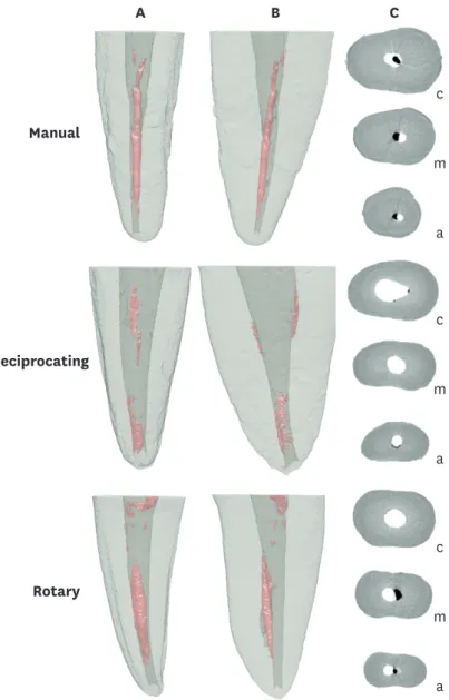
Figure 1. Reconstructed 3-dimensional micro-computed tomographic (micro-CT) images of a given specimen of each group showing the presence of remaining root canal fillings in (A) bucco-lingual and (B) mesio-distal views.
(C) Cross-sections at the (c) cervical, (m) middle, and (a) apical thirds showing the presence of remaining root canal fillings.
DISCUSSION
Although it has not been shown that there is a correlation between the presence of remaining material and retreatment failure [15], removing as much filling material as possible would minimize pulp tissue or any bacteria that may remain hidden in the canal from prior
treatment [16,17]. Therefore, the present study evaluated the amount of remaining root filling materials after retreatment procedures performed by undergraduate students using manual K-files, Mtwo retreatment files, and Reciproc files. Although several techniques have been used to assess the remaining filling material inside the canal walls [3,4,9,10,17], micro-CT imaging was used because this highly accurate, noninvasive, and reproducible technology enables a precise 3-dimensional quantitative evaluation of residual filling material [7,8].
Moreover, this technique not only overcomes the limitations of 2-dimensinal images, but it may also limit potential operator bias in the interpretation of the results [18].
Previous studies have suggested that undergraduate dental students can successfully prepare root canals with rotary instruments, and these instruments are increasingly being integrated into clinical courses [13,19-20]. Up to now, no study evaluated the quality of endodontic retreatment performed with manual, rotary, and reciprocating techniques by undergraduate students. Students enrolled in this study received a standardized introduction to the use of the 3 different instrumentation systems that were used in retreatment cases. Prior to treating the samples in this study, retreatment demonstrations using manual K-files, Mtwo retreatment, and Reciproc instruments were shown to each student.
In the current study, residual filling materials were seen in all specimens, regardless of the retreatment technique used. Similar findings have also been reported in previous studies [3,4,7-10]; however, the Reciproc technique exhibited the lowest percentage of remaining obturation material. Thus, the null hypothesis tested was rejected. Two previous studies presented controversial results regarding filling material removal when comparing Mtwo retreatment and Reciproc systems [9,21]. Zuolo et al. [9] showed that Reciproc files left the lowest mean percentage (4.57%) of remaining filling material, whereas Alves et al. [21]
showed that the percentage of filling material removed with Mtwo instruments (96%) was significantly higher than Reciproc instruments (89%). This discrepancy in the results may be explained by differences in the methodologic design. While maxillary central incisors were analyzed based on photographs taken after splitting the teeth in the former study [9], a micro-CT tool was used to evaluate mesial canals of mandibular molars in the latter one [21]. Mtwo and Reciproc systems were chosen because they are similar in design, therefore allowing a well-controlled comparison between a single-file reciprocating system and a multi-file rotary system. Although these systems are similar in design, it was not possible to standardize the tapers because Reciproc has a variable taper along the shaft. This feature combined with the reciprocating movement can partly explain the improved performance of Reciproc instruments in the removal of root fillings herein. Moreover, contrary to the previous studies where retreatments were always performed by only one operator with experience in all tested techniques, all retreatment procedures were performed by undergraduate students in the present study. None of them had any type of experience using the 3 retreatment techniques. Thus, the present results showed the reduced learning curve of Reciproc instruments due to its simple usability.
Regarding the operating time, both Mtwo retreatment and Reciproc systems required
with others who reported that mechanical instrumentation was significantly more rapid than hand files [22-24]. It can be hypothesized that the active tip and the cutting blades of Mtwo retreatment system and the special design of Reciproc instruments as well as its reciprocating motion positively influenced the time required for the retreatment procedures [24,25].
It is well known that solvents can be used to soften and dissolve gutta-percha in the root canals to facilitate its penetration and removal [4,26]. In the present study, a small (0.1 mL) increment of eucalyptol was used only at the beginning of the retreatment procedures in all groups to soften the coronal filling material, and to improve the penetration of the files.
According to the results of the scans after canal obturation, no statistical difference was observed between the mean percentage volumes of the filling materials in all groups before retreatment procedures. Therefore, it allowed a better comparison of the amount of remaining filling material after the second scans because of standardized samples at the beginning of the retreatment procedures. In this study, maxillary single rooted teeth were used; in fact, in most of the studies evaluating retreatment techniques, straight root canals have been used to simplify the standardization of the specimens [3,4,9,10,17,19]. Some previous retreatment studies have filled teeth with lateral condensation; however, the continuous wave of condensation technique was used in this study because it produces a better root canal filling adaptation to the root canal walls than lateral condensation [27].
Two important methodological aspects should be discussed in the present study. First, since the students enrolled were not experienced, the order of the retreatment (manual, rotary or reciprocating) could affect the results. Moreover, depending on the student, the results could be affected, e.g., one skillful student could show better results. Nevertheless, it is important to emphasize that even an unskilled student would take benefits from a procedural simplification.
CONCLUSIONS
Under the conditions of this study, it can be concluded that Reciproc was the most effective instrument in the removal of canal filling materials during retreatments performed by undergraduate students. Mtwo retreatment and Reciproc techniques required less time to perform the retreatment procedures. No instrument fracture was observed in any of the groups.
REFERENCES
1. Imura N, Pinheiro ET, Gomes BP, Zaia AA, Ferraz CC, Souza-Filho FJ. The outcome of endodontic treatment: a retrospective study of 2000 cases performed by a specialist. J Endod 2007;33:1278-1282.
PUBMED | CROSSREF
2. Siqueira JF Jr, Rôças IN, Ricucci D, Hülsmann M. Causes and management of post-treatment apical periodontitis. Br Dent J 2014;216:305-312.
PUBMED | CROSSREF
3. Taşdemir T, Er K, Yildirim T, Celik D. Efficacy of three rotary NiTi instruments in removing gutta-percha from root canals. Int Endod J 2008;41:191-196.
PUBMED | CROSSREF
4. Gu LS, Ling JQ, Wei X, Huang XY. Efficacy of ProTaper Universal rotary retreatment system for gutta- percha removal from root canals. Int Endod J 2008;41:288-295.
5. Duncan HF, Chong BS. Removal of root filling materials. Endod Topics 2008;19:33-57.
CROSSREF
6. Ricucci D, Siqueira JF Jr, Bate AL, Pitt Ford TR. Histologic investigation of root canal-treated teeth with apical periodontitis: a retrospective study from twenty-four patients. J Endod 2009;35:493-502.
PUBMED | CROSSREF
7. Ma J, Al-Ashaw AJ, Shen Y, Gao Y, Yang Y, Zhang C, Haapasalo M. Efficacy of ProTaper Universal Rotary Retreatment system for gutta-percha removal from oval root canals: a micro-computed tomography study. J Endod 2012;38:1516-1520.
PUBMED | CROSSREF
8. Bernardes RA, Duarte MA, Vivan RR, Alcalde MP, Vasconcelos BC, Bramante CM. Comparison of three retreatment techniques with ultrasonic activation in flattened canals using micro-computed tomography and scanning electron microscopy. Int Endod J 2016;49:890-897.
PUBMED
9. Zuolo AS, Mello JE Jr, Cunha RS, Zuolo ML, Bueno CE. Efficacy of reciprocating and rotary techniques for removing filling material during root canal retreatment. Int Endod J 2013;46:947-953.
PUBMED | CROSSREF
10. Silva EJ, Sá L, Belladonna FG, Neves AA, Accorsi-Mendonça T, Vieira VT, De-Deus G, Moreira EJ.
Reciprocating versus rotary systems for root filling removal: assessment of the apically extruded material.
J Endod 2014;40:2077-2080.
PUBMED | CROSSREF
11. De-Deus G, Moreira EJ, Lopes HP, Elias CN. Extended cyclic fatigue life of F2 ProTaper instruments used in reciprocating movement. Int Endod J 2010;43:1063-1068.
PUBMED | CROSSREF
12. Steffen H, Löw A, Rosin M, Welk A. Comparison of K hand files and ProFiles 0.06/0.04 in simulated curved root canals prepared by students. Quintessence Int 2006;37:811-817.
PUBMED
13. Abu-Tahun I, Al-Rabab'ah MA, Hammad M, Khraisat A. Technical quality of root canal treatment of posterior teeth after rotary or hand preparation by fifth year undergraduate students, The University of Jordan. Aust Endod J 2014;40:123-130.
PUBMED | CROSSREF
14. Schneider SW. A comparison of canal preparations in straight and curved root canals. Oral Surg Oral Med Oral Pathol 1971;32:271-275.
PUBMED | CROSSREF
15. Nair PN. On the causes of persistent apical periodontitis: a review. Int Endod J 2006;39:249-281.
PUBMED | CROSSREF
16. Stabholz A, Friedman S. Endodontic retreatment--case selection and technique. Part 2: treatment planning for retreatment. J Endod 1988;14:607-614.
PUBMED | CROSSREF
17. Saad AY, Al-Hadlaq SM, Al-Katheeri NH. Efficacy of two rotary NiTi instruments in the removal of gutta- percha during root canal retreatment. J Endod 2007;33:38-41.
PUBMED | CROSSREF
18. Solomonov M, Paqué F, Kaya S, Adigüzel O, Kfir A, Yiğit-Özer S. Self-adjusting files in retreatment: a high-resolution micro-computed tomography study. J Endod 2012;38:1283-1287.
PUBMED | CROSSREF
19. Gluskin AH, Brown DC, Buchanan LS. A reconstructed computerized tomographic comparison of Ni-Ti rotary GT files versus traditional instruments in canals shaped by novice operators. Int Endod J 2001;34:476-484.
PUBMED | CROSSREF
20. Abu-Tahun I, Al-Rabab'ah MA, Hammad M, Khraisat A. Technical quality of root canal treatment of posterior teeth after rotary or hand preparation by fifth year undergraduate students, The University of Jordan. Aust Endod J 2014;40:123-130.
PUBMED | CROSSREF
21. Marending M, Biel P, Attin T, Zehnder M. Comparison of two contemporary rotary systems in a pre- clinical student course setting. Int Endod J 2016;49:591-598.
PUBMED | CROSSREF
22. Alves FR, Marceliano-Alves MF, Sousa JC, Silveira SB, Provenzano JC, Siqueira JF Jr. Removal of root canal fillings in curved canals using either reciprocating single- or rotary multi-instrument systems and a supplementary step with the XP-Endo Finisher. J Endod 2016;42:1114-1119.
PUBMED | CROSSREF
23. Sae-Lim V, Rajamanickam I, Lim BK, Lee HL. Effectiveness of ProFile. 04 taper rotary instruments in endodontic retreatment. J Endod 2000;26:100-104.
PUBMED | CROSSREF
24. Betti LV, Bramante CM. Quantec SC rotary instruments versus hand files for gutta-percha removal in root canal retreatment. Int Endod J 2001;34:514-519.
PUBMED | CROSSREF
25. Hülsmann M, Bluhm V. Efficacy, cleaning ability and safety of different rotary NiTi instruments in root canal retreatment. Int Endod J 2004;37:468-476.
PUBMED | CROSSREF
26. Barrieshi-Nusair KM. Gutta-percha retreatment: effectiveness of nickel-titanium rotary instruments versus stainless steel hand files. J Endod 2002;28:454-456.
PUBMED | CROSSREF
27. Wilcox LR. Endodontic retreatment with halothane versus chloroform solvent. J Endod 1995;21:305-307.
PUBMED | CROSSREF
28. Tagger M, Tamse A, Katz A, Korzen BH. Evaluation of the apical seal produced by a hybrid root canal filling method, combining lateral condensation and thermatic compaction. J Endod 1984;10:299-303.
PUBMED | CROSSREF