INTRODUCTION
Malignant peripheral nerve sheath tumors (MPNSTs) are a very rare neoplasm, and they represent only 5-10%
of all the malignant soft tissue sarcomas. These tumors may arise sporadically in adult patients, but these tumors frequently occur in patients suffering with neurofibro- matosis Type I (NF 1). The mean age at the time of diag- nosis of MPNST is in the thirties, but the patients with NF 1 are about 10 yr younger than the patients without NF 1.(1) MPNSTs are also known to have an association with previous irradiation. MPNSTs are known to arise within the field of irradiation, 9 to 36 yr after radiation therapy administered for treating previous malignan- cies.(2) MPNSTs are commonly found in the trunk and the extremities (51% and 45% of the patients, respectively) and in the head and neck in 4% of patients.(3) Most pri-
mary tumors of the breast have an epithelial origin, and a MPNST arising in the breast is an extremely rare find- ing. In our review of the literature on MPNSTs, only 9 cases have been reported from 1983 to the present. We report here on our clinical experience of a patient who presented with a solitary MPNST of the breast, and we treated this tumor with surgical excision and radiation therapy.
CASE REPORT
A 59-yr-old woman presented to our hospital com- plaining of a mass in the right breast. The mass was non-tender, it was firm-to-hard in consistency, approx- imately 3 cm in size and it was located at the 10 o’clock position in the right breast. She had no history of a prior breast mass, trauma, nipple discharge, mastalgia or a family history of breast cancer, and did not present any features of neurofibromatosis. Mammography demon- strated a well defined ovoid mass shadow that was just onto the pectoralis major muscle (Figure 1). Ultrasono- graphy of the breast showed a macrolobulating hypo- Malignant peripheral nerve sheath tumors (MPNSTs) are
malignant variants of peripheral nerve sheath tumors that develop at major or minor peripheral nerve branches or at the sheaths of peripheral nerve fibers. These tumors are derived from Schwann cells or pluripotent cells of a neural crest origin. Malignant tumors of the peripheral nerve sheath are most commonly seen in deeper soft tissues, and usually in the proximity of a nerve trunk. MPNSTs of the breast are very uncommon and they have rarely been reported on. We report here on a case of MPNST of the breast in a 59-year-
old female who presented with a painless breast lump for two months. The excisional biopsy revealed a malignant peripheral nerve sheath tumor based on the microscopic findings and immunohistochemical staining. We performed wide excision of breast tissue around the biopsy site and there- after the patient underwent radiation therapy. The patient remains well without signs of recurrence 1 year following surgery.
Key Words: Breast, Nerve sheath neoplasms
Journal of Breast Cancer
C A S E R E P O R T
Malignant Peripheral Nerve Sheath Tumor of the Breast in a Patient without Neurofibromatosis: A Case Report
Jeong Min Yi, Eun Jeong Moon, Se Jeong Oh, Ahni Lee
1, Young Jin Suh, Jong Min Baek, Seung Hye Choi, Sang Seol Jung
Departments of Surgery and 1Pathology, The Catholic University of Korea, Seoul, Korea
Correspondence: Se Jeong Oh
Department of Surgery, Incheon St. Mary’s Hospital, 665-8 Bupyeong 6-dong, Bupyeong-gu, Incheon 403-720, Korea Tel: 032-510-5798, Fax: 032-510-5816
E-mail: [email protected]
Received: April 10, 2009 Accepted: July 2, 2009
223
J Breast Cancer 2009 September; 12(3): 223-6 DOI: 10.4048/jbc.2009.12.3.223
echoic mass in the upper outer quadrant of the right breast, and its size measured 2.8×1.0 cm (Figure 2). She was admitted for excisional biopsy and she underwent the operation under general endotracheal anesthesia. At the time of surgery, the tumor was found in the deep portion of the right breast, and it was firmly attached to the pec- toralis major muscle. The mass was completely excised.
The excised surgical specimen consisted of a well-circum- scribed, firm, solid mass measuring 2.5×1.2×1.1 cm. Its cut surface was whitish gray, smooth and myxoid and it had prominent fibrous strands. No necrosis, hemor- rhage or cystic degeneration was grossly identified. His- tologically, the neoplasm consisted of spindle cells sur-
rounded by a fibrous stroma (Figure 3). The mitotic count revealed 19 mitosis/10 high-power fields. Immunoche- mical staining was performed on the tissue section with S-100, vimentin, desmin, actin and cytokeratin. The immunohistochemical staining of S-100 protein was diffusely positive in the tumor cells (Figure 4). Smooth muscle actin, CD 34 and CD 68 were non-reactive in the tumor cells. So we could exclude the possibility of other spindle cell tumors including smooth muscle tumor, soli- tary fibrous tumor, vascular tumor and malignant fibrous histiocytoma. On the basis of the cytohistologic appear- ance and the immunohistochemical pattern, this tumor was interpreted to be a spindle cell neoplasm with neural differentiation, and this was suggestive of a MPNST. A wide excision around the previous operation site was per- formed and the patient postoperatively received radiation therapy to the right breast. She had no complication dur- ing the treatment, and she remains well without signs of local recurrences or distant metastases 1 yr following her surgery.
DISCUSSION
Peripheral nerve tumors are infrequently encounter- ed soft tissue lesions that can affect any organ of the body and so they have a myriad of differential diagnoses.
MPNST is classified into the primary malignant subtype of the peripheral nerve.(4) The incidence of MPNSTs is
224 Jeong Min Yi, et al.
Figure 1. The mediolateral oblique view of mammo- graphy demonstrates an ovoid mass shadow just onto the pectoral muscle.
The mass is well defined from the surrounding tis- sue and there is no calci- fication in the mass.
Figure 2. The ultrasonographic image of the right breast showed a well-demarcated mass. The mass is lobulated and ovoid, and it shows a hypoechoic mass density with posterior enhancement.
Figure 3. Microscopic finding of H&E staining. Note densely cel- lular areas alternating with less cellular areas and vague whorled structures (H&E stain, ×40).
0.001% in the general population, but 5% to 42% of the cases have an association with neurofibromatosis type 1 (NF 1). MPNSTs arise in adult patients who range in age from 20 to 50 yr of age. They originate from a major or minor peripheral nerve branch or its sheath. The most common sites of presentation of MPNSTs are the trunk, followed by the extremities and the head and neck.(1,3,5) MPNST of the breast as a primary tumor is very rare and has been only eight such reported cases in the scope of our search,(6-13) and two cases among them were asso- ciated with neurofibromatosis.(7,9)
There are no specific symptoms or signs other than a palpable lump in the breast, and making the correct pre- operative diagnosis of MPNST tumor is difficult. The ini- tial diagnoses by fine needle aspiration cytology in the cases of Catania et al.(6) and Dhingra et al.(9) were a mesenchymal tumor and a spindle cell tumor, respec- tively. The initial diagnosis by excisional biopsy in the case of Malas et al.(7) was fibrous histiocytoma. In the cases of Medina-Franco et al.,(8) the diagnosis of MPNST was made by excision and immunohistochemical staining. The clinical relevance of these cases shows the importance of harvesting enough tissue for histologic analysis and immunohistochemical staining. MPNST have to be dis- tinguished from malignant phyllodes tumor, fibrosarcoma and leiomyosarcoma. We could rule out these other tu- mors by the immunohistochemical staining. The definite treatment for this tumor is only complete surgical resec-
tion and the prognosis of patients with MPNST is signif- icantly determined by whether or not complete resec- tion has been achieved.(1) The principles for the manage- ment of MPNSTs are similar to those for the management of all types of soft tissue sarcomas.(14) The goal of oper- ation is complete removal of the tumor with histologi- cally clear resection margins. Although some authors recommended a mastectomy for the primary therapy of MPNST of the breast,(6,7) the extent of surgery remains uncertain due to its rarity. But axillary dissection is not indicated as the regional lymph nodes are not usually affected.(7) There are no reports in the literature on the role of radiotherapy or chemotherapy for the treatment of MPNST of the breast. In the present case, we performed wide excision and radiotherapy as there was thought to be a risk of recurrence related to the highly mitotic fea- tures of the tumor. The prognosis for patients with MPNST remains poor. The reported 5-yr survival rates were 34- 40% in two studies,(1,3) and the unfavorable features for recurrence are the tumor size, the site of origin, and the margin status.(3) MPNSTs associated with NF 1 are more aggressive and they have a worse prognosis following local recurrence than do the tumors without NF 1.(15)
In conclusion, MPNST of the breast is often unsus- pected and the diagnosis may be missed unless clinicians have awareness of this disease. Clinicians should pay special attention when a patient with the stigmata of neurofibromatosis type 1 (i.e., multiple cutaneous nodules,
Malignant Peripheral Nerve Sheath Tumor in the Breast 225
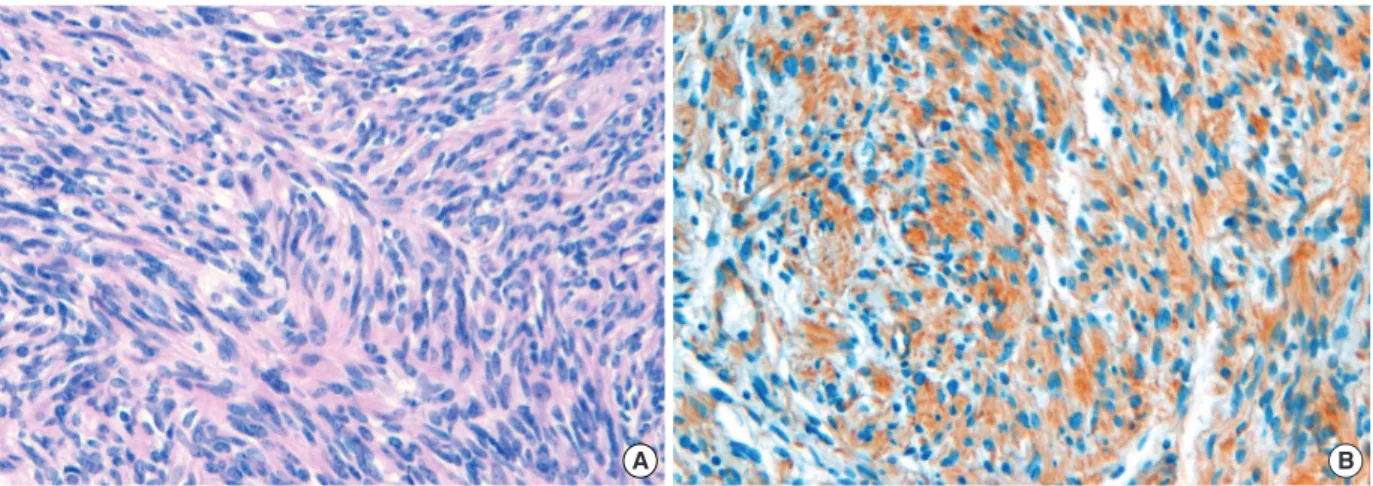
Figure 4. Microscopic finding of H&E and immunohistochemical staining. (A) This area shows more dense fascicles of spindle cells (H&E stain, ×200). (B) S-100 protein shows focal weak staining to diffuse immunoreactive areas. These figures from areas of diffuse immunoreactive for S-100 protein (Immunohistochemical staining, ×200).
A B
cafe@au lait spots, freckling in the axilla or groin) presents with a mass in the breast, clinicians should use special attention for it. Although we performed wide excision and radiotherapy in this case, the optimum treatment is still not clear as the experience with this rare tumor is limited.
REFERENCES
1. Ducatman BS, Scheithauer BW, Piepgras DG, Reiman HM, Ilstrup DM. Malignant peripheral nerve sheath tumours: a clinicopathologic study of 120 cases. Cancer 1986;57:2006-21.
2. Baehring JM, Betensky RA, Batchelor TT. Malignant peripheral nerve sheath tumour: the clinical spectrum and outcome of treatment.
Neurology 2003;61:696-8.
3. Anghileri M, Miceli R, Fiore M, Mariani L, Ferrari A, Mussi C, et al. Malignant peripheral nerve sheath tumors: prognostic factors and survival in a series of patients treated at a single institution. Cancer 2006;107:1065-74.
4. Bhattacharyya AK, Perrin R, Guha A. Peripheral nerve tumors: ma- nagement strategies and molecular insights. J Neurooncol 2004;69:
335-49.
5. Ferner RE, Gutmann DH. International consensus statement on ma- lignant peripheral nerve sheath tumors in neurofibromatosis. Cancer Res 2002;62:1573-7.
6. Catania S, Pacifico E, Zurrida S, Cusumano F. Malignant schwan- noma of the breast. Eur J Surg Oncol 1992;18:80-1.
7. Malas S, Krawitz HE, Sur RK, Uijs RR, Nayler SJ, Levin CV. Von Recklinghausen’s disease associated with a primary malignant sch- wannoma of the breast. J Surg Oncol 1995;59:273-5.
8. Medina-Franco H, Gamboa-Dominguez A, de La Medina AR. Malig- nant peripheral nerve sheath tumor of the breast. Breast J 2003;9:
332.
9. Dhingra KK, Mandal S, Roy S, Khurana N. Malignant peripheral nerve sheath tumor of the breast: case report. World J Surg Oncol 2007;5:142.
10. Hauser H, Beham A, Steindorfer P, Schmidt F, Smola MG. Malignant schwannoma of the breast. Langenbecks Arch Chir 1995;380:350-3.
11. Berrada R, Chahtane A, Lakhdar A, Elhanchi Z, Ferhati D, Baidada, et al. Malignant schwannoma of the breast. A case report. J Gynecol Obstet Biol Reprod (Paris) 1998;27:441-4.
12. Besznya@k I, Dubecz S, Pe@ter I. Malignant schwannoma of the breast.
Orv Hetil 1998;139:137-9.
13. Thanapaisal C, Koonmee S, Siritunyaporn S. Malignant peripheral nerve sheath tumor of breast in patient without Von Recklinghausen’s neurofibromatosis: a case report. J Med Assoc Thai 2006;89:377-9.
14. Angelov L, Davis A, O’Sullivan B, Bell R, Guha A. Neurogenic sar- comas: experience at the University of Toronto. Neurosurgery 1998;
43:56-64.
15. Sordillo PP, Helson L, Hajdu SI, Magill GB, Kosloff C, Golbey RB, et al. Malignant schwannoma: clinical characteristics, survival, and response to therapy. Cancer 1981;47:2503-9.
226 Jeong Min Yi, et al.