70 https://e-jcvi.org
Congenital malformation of the mitral valve is considered to be a rare and complex entity.
1)2)Singular leaflet mitral valve is the least prevalent form and has been mostly observed in infants, associated with symptomatic mitral regurgitation (MR) and tending to be incompatible with life beyond the neonatal period.
1)2)We report herein an unusual cause of unileaflet mitral valve in a 76-year-old man who presented with 5-day history of dyspnea. His cardiac background comprises repair of atrial septal defect (ASD) and percutaneous coronary intervention. He had clinical signs of fluid overload and a pan-systolic murmur in keeping with MR.
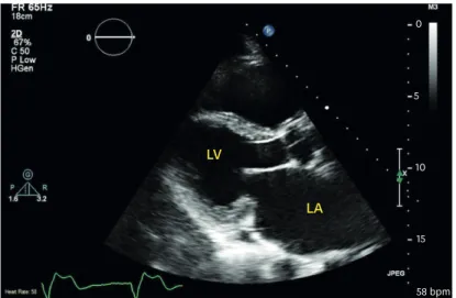
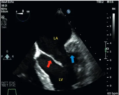
Transthoracic echocardiography revealed moderate to severe MR due to coaptation deficit of unidentified etiology (Figure 1). Two and three-dimensional transesophageal echocardiography demonstrated elongated anterior mitral valve leaflet and hypoplastic posterior leaflet with shortened chordae (Figure 2, 3; Movie 1, Movie 2, Movie 3) causing prominent MR (Figure 4; Movie 4, Movie 5). The left and right ventricles were dilated with systolic impairment with elevated pulmonary systolic pressure.
J Cardiovasc Imaging. 2020 Jan;28(1):70-73 https://doi.org/10.4250/jcvi.2019.0077 pISSN 2586-7210·eISSN 2586-7296
Images in
Cardiovascular Disease
Received: Aug 12, 2019 Accepted: Aug 27, 2019 Address for Correspondence:
Ahmed Salem, MBBS
Department of Cardiology, Milton Keynes University Hospital NHS Foundation Trust, Standing Way Milton Keynes MK6 5LD, UK.
E-mail: [email protected] Attila Kardos, PhD
Department of Cardiology, Milton Keynes University Hospital NHS Foundation Trust, Standing Way Milton Keynes MK6 5LD, UK.
E-mail: [email protected] Copyright © 2020 Korean Society of Echocardiography
This is an Open Access article distributed under the terms of the Creative Commons Attribution Non-Commercial License (https://
creativecommons.org/licenses/by-nc/4.0/) which permits unrestricted non-commercial use, distribution, and reproduction in any medium, provided the original work is properly cited.
ORCID iDs Ahmed Salem
https://orcid.org/0000-0003-2207-170X Georgia Zachou
https://orcid.org/0000-0001-5792-4747 Kanchani Makuloluwa
https://orcid.org/0000-0002-5402-3502 Attila Kardos
https://orcid.org/0000-0002-0231-7605 Conflict of Interest
The authors have no financial conflicts of interest.
Ahmed Salem , MBBS
1Georgia Zachou , MBBS
1Kanchani Makuloluwa , MBBS
1, and Attila Kardos , PhD
1,21Department of Cardiology, Milton Keynes University Hospital, Milton Keynes, UK
2School of Sciences and Medicine, University of Buckingham, Buckingham, UK
Never Too Old for a Congenital Heart Disease: A Case of a Unileaflet Mitral Valve in a 76-year-old Man
LV
LA
0
5
10
15
58 bpm
Figure 1. Transthoracic echocardiogram in para-sternal long axis showing coaptation deficit of the mitral valve.
LA: left atrium, LV: left ventricle.
The patient was stabilised on heart failure therapy and considered for mitral valve replacement.
Unileaflet mitral valves among adults are extremely rare and described mainly in case reports. In our case we propose that the congenital agenesis of posterior mitral leaflet and its functional importance has become apparent as a result of structural changes of the mitral anulus due to left ventricular dilatation secondary to ischemic heart disease.
Although, different forms of ASD could have genetic basis and are associated with anomalous pulmonary venous drainage or mitral valve cleft
3); the link between ASD and unileaflet mitral valve has not been reported thus far.
71 https://e-jcvi.org https://doi.org/10.4250/jcvi.2019.0077
An Unileaflet Mitral Valve in a 76-year-old Man
LA
LV
1 2 3 4 5 6 7 8
9 60 bpm
Figure 2. Transesophageal echocardiography mid-esophageal view, 0°, showing the mitral valve with a rudimental posterior mitral leaflet adherent to the LV posterior wall (blue arrowhead) and elongated anterior mitral leaflet (red arrowhead). LA: left atrium, LV: left ventricle.
AMVL
Figure 3. Three-dimensional short-axis view of the mitral valve showing hypoplastic posterior mitral valve leaflet (red arrowhead) and elongated myxomatous anterior mitral valve leaflet (AMVL).
SUPPLEMENTARY MATERIALS
Movie 1
2D TOE - mid-oesophageal 4 chambers view of the unileaflet mitral valve Click here to view
Movie 2
2D TOE - X-plane mid-oesophageal view of the unileaflet mitral valve Click here to view
Movie 3
3D rendering TOE image of the mitral valve showing the posterior leaflet agenesis Click here to view
Movie 4
2D TOE mid oesophageal view showing the unileaflet mitral leaflet and colour Doppler image of the mitral regurgitation
Click here to view Movie 5
2D TOE - X-plane mid-oesophageal view of the unileaflet mitral valve and mitral regurgitation Click here to view
72 https://e-jcvi.org https://doi.org/10.4250/jcvi.2019.0077
An Unileaflet Mitral Valve in a 76-year-old Man
1 2 3 4 5 6 7 8 60 bpm
MR Alias Vel MR Radius MR Flow rate MR ERO MR Volume
* 37.1 cm/s0.6 cm 83.8 mL/s 0.16 cm2 27 mL
Figure 4. Two-dimensional transesophageal echocardiography mid esophageal view showing showing the unileaflet mitral leaflet and color Doppler image of the mitral regurgitation.
REFERENCES
1. Shah J, Jain T, Shah S, Mawri S. Anathasubramaniam k. Rare case of Unileafflet mitral valve. J Cardiovasc Ultrasound 2016;24:168-9.
PUBMED | CROSSREF
2. Bezgin T, Elveran A, Karagöz A, Çanga Y, Yılmaz F. Mitral valve with a single leaflet. Turk Kardiyol Dern Ars 2014;42:80-2.
PUBMED | CROSSREF
3. Pourafkari L, Baghbani-Oskouei A, Toufan M, Ghaffari S, Nader ND. Hypoplastic posterior mitral valve leaflet: A case report and review of the literature. Echocardiography 2018;35:1052-5.
PUBMED | CROSSREF
73 https://e-jcvi.org https://doi.org/10.4250/jcvi.2019.0077
An Unileaflet Mitral Valve in a 76-year-old Man