J Korean Soc Radiol 2017;76(1):78-82 https://doi.org/10.3348/jksr.2017.76.1.78
INTRODUCTION
Myeloid sarcoma (MS), also known as chloroma, granulocyt- ic sarcoma or extramedullary myeloblastoma, is a collection of myeloid precursor cells that include myeloblasts, promyelocytes, and myelocytes (1, 2). MS usually occurs in the course of acute or chronic myeloid leukemia or leukemic transformation in my- eloproliferative or myelodysplastic disorders. But it has rarely been reported as an isolated manifestation in non-leukemic pa- tients (1). MS has been found in almost every anatomic location, and it more commonly occurs in the skin, bone, soft tissue, lymph nodes and central nervous system (CNS) (1-3). However, in- volvement of the gastrointestinal tract, especially the stomach, is uncommon, and radiologic findings of stomach involvement have been rarely reported in the literature (4). We report the im-
aging features, including those of ultrasonography, CT and posi- tron emission tomography (PET)-CT, in an unusual case of iso- lated MS of the stomach in a 15-year-old girl without evidence of any hematologic disorder.
CASE REPORT
A 15-year-old girl was admitted to our hospital with intermit- tent epigastric pain for 3 months. She had no specific medical or surgical history. Physical examination revealed mild tender- ness in the epigastric and left upper quadrant areas of the abdo- men. There was no hepatosplenomegaly or lymphadenopathy.
Laboratory evaluations showed a white blood cell count of 6300/
mm3, a hemoglobin level of 10.9 g/dL, and a platelet count of 337000/mm3. Serum lactate dehydrogenase level was 498 IU/L,
Imaging Findings of Isolated Myeloid Sarcoma of the Stomach in a Nonleukemic Child: A Case Report and Literature Review
비 백혈병 환아에서 위의 단일 과립세포육종의 영상소견: 증례 보고Yong Kyun Kim, MD
1, Jung Hyun Kim, MD
1, Hee Jo Baek MD
1, Jin Woong Kim, MD
2, Sang Soo Shin, MD
2, Suk Hee Heo, MD
1*
1Department of Radiology, Chonnam National University Hospital, Chonnam National University Medical School, Hwasun, Korea
2Department of Radiology, Chonnam National University Hwasun Hospital, Chonnam National University Medical School, Hwasun, Korea
Myeloid sarcoma is an extramedullary solid neoplasm composed of myeloid precur- sor cells. This tumor usually occurs simultaneously with or following the onset of acute leukemia. Rarely, it can be the first manifestation of acute myeloid leukemia.
The tumor can occur anywhere in the body. However, primary involvement of the stomach without evidence of leukemia is exceedingly rare, and to the best of our knowledge, imaging features of isolated myeloid sarcoma of the stomach have not been reported in children. This case illustrates the imaging appearances of isolated myeloid sarcoma that initially manifested as gastric submucosal wall thickening and discusses the differential diagnosis, in a 15-year-old girl without evidence of hema- tologic malignancy.
Index terms Myeloid Sarcoma Stomach
Multimodal Imaging
Received January 29, 2016 Revised June 11, 2016 Accepted August 1, 2016
*Corresponding author: Suk Hee Heo, MD
Department of Radiology, Chonnam National University Hwasun Hospital, 322 Seoyang-ro, Hwasun-eup, Hwasun-gun 58128, Korea.
Tel. 82-61-379-7112 Fax. 82-61-379-7133 E-mail: [email protected]
This is an Open Access article distributed under the terms of the Creative Commons Attribution Non-Commercial License (http://creativecommons.org/licenses/by-nc/3.0) which permits unrestricted non-commercial use, distri- bution, and reproduction in any medium, provided the original work is properly cited.
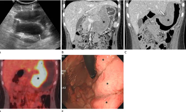
An abdominal ultrasonogram (GE LOGIQ 9; GE Healthcare, Milwaukee, WI, USA) showed marked hypoechoic wall thick- ening of the gastric body (Fig. 1A). Abdominal CT (LightSpeed VCT; GE Healthcare) revealed marked and eccentric wall thickening with homogeneous contrast enhancement in the stomach, particularly the lesser curvature of the body portion (Fig. 1B, C). Mucosa of the thickened gastric wall and perigas- tric fat planes were smooth and preserved. There was no peri- gastric or distant lymphadenopathy. 18F-fluorodeoxyglucose (FDG) PET-CT (GE Discovery 600; GE Healthcare) was per- formed for further staging because the presumptive diagnosis was a lymphoma. On the PET-CT images, increased 18F-FDG was found only in the lesser curvature side of the gastric body with a maximal standardized uptake value (SUV) of 15.2, sug-
normal FDG uptake lesion. Upper GI endoscopy was performed for biopsies, which showed extensive enlarged and hyperemic fold thickening with scattered hemorrhagic spots in the lesser curvature of the gastric body (Fig. 1E). Histopathological find- ings from the endoscopic biopsy specimens were consistent with MS based on the presence of cells of the myeloid lineage with positive immunohistochemical staining for myeloperoxidase and myeloid markers (CD 34, CD 43, CD 117), and negative immu- nohistochemical staining for B-cell (CD 20) and T-cell markers (CD 3 and CD 30) (Fig. 2). Bone marrow examination demon- strated no evidence of marrow involvement with acute myeloid leukemia (AML) blasts.
The patient received cytarabine-based induction and consoli- dation chemotherapy. Unfortunately, 6 months later, the patient
Fig. 1. Multimodal imaging of isolated myeloid sarcoma in a nonleukemic child.
A. Oblique axial gray-scale ultrasonogram shows marked and hypoechoic thickening of the stomach wall (arrows) with loss of stratification.
B, C. Coronal reformatted, contrast-enhanced CT images demonstrate eccentric, marked and homogeneous wall thickening (asterisks) in the lesser curvature of the gastric body. Mucosa of the thickened gastric wall (thin arrows) and perigastric fat planes (thick arrows) are smooth and preserved, which are suggestive of submucosal disease involvement. There is no regional or distant lymphadenopathy.
D. Coronal 18F-FDG PET-CT image shows a diffuse hypermetabolic area (maximal standardized uptake value of 15.2) corresponding to the thick- ened gastric wall (asterisk).
E. Upper GI endoscopy reveals extensive enlarged and hyperemic fold thickening (asterisks) with scattered hemorrhagic spots.
GI = gastrointestinal, 18F-FDG PET-CT = 18F-fluorodeoxyglucose PET-CT
A B
D E
C
noted relapse in the stomach, breast and peritoneum and showed AML M4 type with a blast count of 33% on bone marrow ex- amination. Second-line chemotherapy and radiation therapy were applied quickly, but the patient died due to disease dis- semination and septic shock.
DISCUSSION
MS is a rare, malignant localized solid tumor composed of im- mature granulocytic precursor cells occurring in extramedul- lary sites. In the majority of cases, it is diagnosed at presentation or during the course of AML. The overall prevalence of MS was 2.5–8% in one autopsy series of acute leukemia (1). In a multi- center study of 1832 children with AML, 199 (10.9%) patients had MS and only 13 (0.7%) patients had isolated MS without involvement of the bone marrow (2). Another literature has re- ported that isolated MS without any blood or bone marrow in- volvement at the time of diagnosis is a rare disease with an inci- dence of 2/1000000 in adults (5). MS can occur virtually any- where in the body; the most common sites are skin, orbit, head and neck, bone and CNS (1, 3). MS involving the gastrointestinal tract is relatively rare and it occurs mostly in the small bowel (1, 4). The involvement of the stomach is very rare, and to the best of our knowledge, the imaging features of isolated MS of the stomach have not been reported in children.
There are a few reports on the radiological features of MS in the gastrointestinal tract and the imaging features of MS are variable; lesions can appear as an intramural or exophytic pol- ypoid mass or bowel wall thickening or a combination of these manifestations (4, 6). In our case, the wall of the stomach was markedly thickened and showed homogeneous contrast en- hancement. The gastric wall thickening was eccentric, and it was not circumferential. Overlying mucosa of the thickened gastric wall and perigastric fat planes were smooth on CT and gastric fold was preserved on gastroscopy, suggestive of submucosal disease involvement. There was no regional or distant lymph- adenopathy. In 18F-FDG PET-CT images, the thickened gastric wall showed diffuse hypermetabolism with a maximal SUV of 15.2. These imaging features of the gastric lesion were quite sim- ilar to those of a primary gastric lymphoma and these features cannot be distinguished from those of a primary gastric lympho- ma, although circumferential wall thickening and regional or mesenteric lymphadenopathy are frequently seen in the major- ity of lymphomas (7). Inflammatory myofibroblastic tumor and gastrointestinal stromal tumor (GIST) should also be included in the differential diagnosis because both gastric lesions may show a well-defined exophytic or submucosal mass (8, 9). The gastric inflammatory myofibroblastic tumor shows various ap- pearances, but it usually shows aggressive features such as ul- ceration, adjacent wall infiltration and extraluminal extension, Fig. 2. Histopathologic examination of isolated myeloid sarcoma in a nonleukemic child.
A. Photomicrograph (hematoxylin-eosin stain, × 100 magnification) shows diffuse infiltrates of leukemic blasts and some maturing granulocytes (asterisks) in the submucosal tissue and intact overlying mucosa (M) of the stomach.
B. Microscopic image (hematoxylin-eosin stain, × 400 magnification) demonstrates blastic, hyperchromatic and pleomorphic cells of the granu- locytic series, consistent with granulocytic sarcoma.
A B
common mesenchymal tumor in adolescents and children and it appears as a submucosal mass of varying size with normal overlying mucosa. But, GIST usually shows central necrosis, cavitation, cyst formation, and hemorrhage, when it presents as a large tumor (9).
In an unusual manifestation like our case, in which the tumor precedes the hematologic evidence of leukemia, it may be impos- sible to diagnose MS of the stomach initially and it can lead to di- agnostic challenges. This case illustrates the radiologic findings including those of ultrasonography, CT, and PET-CT of isolated MS that presented as eccentric and smooth wall thickening of the stomach without regional or distant lymphadenopathy.
REFERENCES
1. Meis JM, Butler JJ, Osborne BM, Manning JT. Granulocytic sarcoma in nonleukemic patients. Cancer 1986;58:2697- 2709
2. Dusenbery KE, Howells WB, Arthur DC, Alonzo T, Lee JW, Kobrinsky N, et al. Extramedullary leukemia in children with newly diagnosed acute myeloid leukemia: a report from the Children’s Cancer Group. J Pediatr Hematol On- col 2003;25:760-768
3. Guermazi A, Feger C, Rousselot P, Merad M, Benchaib N, Bourrier P, et al. Granulocytic sarcoma (chloroma): imag-
4. Choi EK, Ha HK, Park SH, Lee SJ, Jung SE, Kim KW, et al.
Granulocytic sarcoma of bowel: CT findings. Radiology 2007;243:752-759
5. Yilmaz AF, Saydam G, Sahin F, Baran Y. Granulocytic sar- coma: a systematic review. Am J Blood Res 2013;3:265- 270
6. Huang XL, Tao J, Li JZ, Chen XL, Chen JN, Shao CK, et al.
Gastric myeloid sarcoma without acute myeloblastic leu- kemia. World J Gastroenterol 2015;21:2242-2248
7. Toma P, Granata C, Rossi A, Garaventa A. Multimodality imaging of Hodgkin disease and non-Hodgkin lymphomas in children. Radiographics 2007;27:1335-1354
8. Patnana M, Sevrukov AB, Elsayes KM, Viswanathan C, Lub- ner M, Menias CO. Inflammatory pseudotumor: the great mimicker. AJR Am J Roentgenol 2012;198:W217-W227 9. Levy AD, Remotti HE, Thompson WM, Sobin LH, Miettinen
M. Gastrointestinal stromal tumors: radiologic features with pathologic correlation. Radiographics 2003;23:283- 304, 456; quiz 532
10. Kim SJ, Kim WS, Cheon JE, Shin SM, Youn BJ, Kim IO, et al.
Inflammatory myofibroblastic tumors of the abdomen as mimickers of malignancy: imaging features in nine chil- dren. AJR Am J Roentgenol 2009;193:1419-1424
비 백혈병 환아에서 위의 단일 과립세포육종의 영상소견: 증례 보고
김용균
1· 김정현
1· 백희조
1· 김진웅
2· 신상수
2· 허숙희
1*
골수성육종은 골수성 전구세포들로 구성된 골수 외 고형암이다. 이는 대부분 급성 백혈병과 병발하거나, 급성 백혈병 발 병 후에 발생하며 급성 골수성 백혈병 발병 전에 선행하여 첫 번째 징후로 나타나는 경우는 매우 드물다. 골수성육종은 우 리 몸 어느 곳에서나 발생할 수 있으며 피부, 뼈, 연부조직과 중추신경계에 주로 발생한다. 위장관계의 침범, 특히 위의 원 발성 침범은 극히 드물고 특히 소아에서 급성 골수성 백혈병에 선행하는 단일 위 골수성육종에 대한 영상소견은 지금까지 잘 알려져 있지 않다. 이 증례는 기저질환으로 혈액종양이 없는 15세 여자 환자에서 위벽의 비대칭적 점막하 비후로 나타 난 단일성 위 골수성육종의 초음파, 전산화단층촬영 및 PET-CT를 포함하는 영상소견과 감별진단에 대해 기술하고자 한다.
1전남대학교 의과대학 전남대학교병원 영상의학과, 2전남대학교 의과대학 화순전남대학교병원 영상의학과