INTRODUCTION
Atopic dermatitis (AD) is a common, chronic and relapsing inflammatory skin disease characterized by itchiness, pruritis, erythema, scaling and papulovesicles.1 AD is the most common skin disease worldwide, affecting up to 30% of children and 3%
of adults.2 A number of familial studies and twin studies have also demonstrated that AD is a highly heritable disease.3
AD belongs to a group of atopic diseases with common char- acteristics including allergen sensitization, epithelial barrier ab- normalities and Type 2 immune responses.4 It is widely assumed that atopy is related to AD in children because immunoglobu- lin E (IgE) plays a pivotal role in its pathogenesis. Hence, mea- surements of the total IgE levels have been used to evaluate al- lergic subjects in clinical practice and to determine the risk fac- tors for severe AD.5
It is now known that AD is a complex disease affected by in- teractions among multiple genetic factors and environmental
components. Several genome-wide association studies (GWASs) on AD in European, Chinese and Japanese populations have identified numerous potential susceptibility loci.6-9 The first meta-analysis of AD in subjects of European descent identified 3 additional risk loci (rs479844 at 11q13.1, rs2164983 at 19p13.2, rs2897442 on KIF3A at 5q31).10 More recently, a multi-ancestry meta-analysis in the largest number of samples studied to date from European, African, Japanese and Latino populations iden- tified 31 susceptibility loci, including 10 new risk loci, for AD.11 Studies of immune-related candidate genes (such as IL23R, IL- Allergy Asthma Immunol Res. 2018 July;10(4):397-405.
https://doi.org/10.4168/aair.2018.10.4.397 pISSN 2092-7355 • eISSN 2092-7363
Association of Genetic Polymorphisms with Atopic Dermatitis, Clinical Severity and Total IgE: A Replication and Extended Study
Jeong-Hyun Kim,
1So-Yeon Lee,
2Mi-Jin Kang,
3Jisun Yoon,
2Sungsu Jung,
2Hyun-Ju Cho,
4Hyo-Bin Kim,
5Soo-Jong Hong
2*
1Department of Medicine, University of Ulsan Collage of Medicine, Seoul, Korea
2 Department of Pediatrics, Childhood Asthma Atopy Center, Environmental Health Center, Asan Medical Center, University of Ulsan Collage of Medicine, Seoul, Korea
3Asan Institute for Life Sciences, University of Ulsan Collage of Medicine, Seoul, Korea
4Department of Pediatrics, Mediplex Sejong Hospital, Incheon, Korea
5Department of Pediatrics, Inje University Sanggye Paik Hospital, Seoul, Korea
This is an Open Access article distributed under the terms of the Creative Commons Attribution Non-Commercial License (http://creativecommons.org/licenses/by-nc/4.0/) which permits unrestricted non-commercial use, distribution, and reproduction in any medium, provided the original work is properly cited.
Purpose: Atopic dermatitis (AD) is a common and chronic inflammatory skin disease affecting up to 20% of children and 3% of adults worldwide.
Although previous reports including genome-wide association study (GWAS) approaches have identified several risk factors that appear to be asso- ciated with AD development, replication studies are lacking. In our current study, we replicated the associations between candidate susceptibility loci and AD. Methods: A total of 885 Korean subjects (425 AD patients and 460 unaffected controls) were genotyped for 17 single nucleotide poly- morphisms (SNPs) from previous GWASs and meta-analyses of AD and from immune-related genes. Results: Several SNPs showed significant as- sociations with AD in the case-control analysis (minimum P = 0.005 at rs17389644), suggesting that these polymorphisms may be related to this disease. In addition, several SNPs showed significant signals (minimum P = 0.004 at rs6473227) in severe AD compared to unaffected controls. In additional linear regression analysis, a few genotypes appeared to have potential effects on the SCORing AD (SCORAD) values (minimum P = 0.003 at rs13361382 on TMEM232) and immunoglobulin E (IgE) levels (minimum P < 0.0001 at rs4713555 near HLA-DRB1 and HLA-DQA1) in AD patients.
Conclusions: Our replication and extended study provide additional supporting information on the genetic associations (especially, variants in TMEM232 and nearby to IL21 and HLA-DRB1/HLA-DQA1) related to AD, its clinical severity and IgE involvement.
Key Words: Atopic dermatitis; single nucleotide polymorphism, severity
Correspondence to: Soo-Jong Hong, MD, PhD, Department of Pediatrics, Childhood Asthma Atopy Center, Environmental Health Center, Asan Medical Center, University of Ulsan Collage of Medicine, 88 Olympic-ro 43-gil, Songpa- gu, Seoul 05505, Korea.
Tel: +82-2-3010-3379; Fax: +82-2-2045-4915; E-mail: [email protected] Received: December 22, 2017; Revised: April 11, 2018;
Accepted: April 13, 2018
•There are no financial or other issues that might lead to conflict of interest.
5RA, and IL2) have also reported the associations between ge- netic variants and immune-mediated diseases including atopic eczema.12-14 For instance, a promoter single nucleotide poly- morphism (SNP; rs2069762) of IL2 was found to be significant- ly associated with allergic disease through the mediation of the type 1 T helper (Th1)/type 2 T helper (Th2) cells balance.14 How- ever, although they are important for substantiating previously identified GWAS and candidate genes, replication studies in different AD cohorts are lacking, and therefore the need for a comprehensive etiology of AD still remains.
The aim of our present study was to investigate whether the genetic variants previously identified by genome-wide and can- didate genes studies would be replicated in our own AD cohort and to perform an extended analysis of the association of these variants with the clinical severity and IgE in AD.
MATERIALS AND METHODS Subjects
AD subjects (n = 425) were recruited solely from our tertiary referral hospital, having been examined at the Childhood Asth- ma Atopy Center of Asan Medical Center in Seoul, Korea and diagnosed according to the criteria of Hanifin and Rajka.15 The severity of AD was assessed in these cases using the SCOring AD (SCORAD) classification as follows: mild < 15, 15 ≤ mod- erate < 40, severe ≥ 40.16 Patients with moderate to severe AD were included in the AD group. A control group (n = 460) was also recruited from 9 primary schools and 16 kindergartens in the Seoul area. Subjects in the control group had no history of AD, food allergy, allergic rhinitis, asthma or any parental AD history, and all gave a negative skin prick test result.
The total serum IgE levels in the peripheral blood were mea- sured in all subjects using a fluorescent enzyme immunoassay (ImmunoCAP system; Phadia AB, Uppsala, Sweden). Subjects were also tested for their sensitivity to the following 16 allergens:
Dermatophagoides pteronyssinus, Dermatophagoides farinae, dog epithelium, cat epithelium, cockroach, grass, mixed tree pollen 1 and 2, Alternaria, Aspergillus, ragweed, mugwort, milk, egg white, peanut and soybean. A positive skin reaction was de- fined as a wheal size ≥ 3 mm after subtraction of a negative control. The Korean version of the International Study of Asth- ma and Allergies in Childhood (ISAAC) questionnaire was used.17 Approval to conduct the study was obtained from the human ethics committees of Asan Medical Center and from the princi- pals of the schools attended by the children (Institutional Re- view Board No. 2015-1031). Written informed consent was ob- tained from the parents of all children who participated.
SNP genotyping
Seventeen SNPs with a previously reported AD association were selected from a panel of immune-related genes, and from the results of previous GWASs and meta-analysis. More detailed
information on each SNP was shown in Supplementary Table 1.
DNA was isolated from the peripheral blood of our 885 study participants (425 AD patients and 460 unaffected controls) us- ing the WizPrepTM gDNA Mini Kit (Wizbiosolutions, Seong- nam, Korea). Genotyping of these blood samples was performed using the high-throughput Fluidigm EP1 system (Fluidigm, South San Francisco, CA, USA) with a Fluidigm SNP TypeTM assay platform. According to the manufacturer’s instructions, a spe- cific target amplification reaction was used to increase the copy number of targeted genomic regions using Qiagen 2X Multiplex PCR Master Mix (Qiagen, Hilden, Germany). Following the am- plification reactions on the dynamic array Integrated Fluidic Circuits (IFCs; Fluidigm), fluorescence intensities were mea- sured with the EP1 reader. Genotypes were determined using the Fluidigm SNP Genotyping Analysis program. Visual inspec- tions were performed for all SNP determinations, and call rate over 95% was applied to further SNP analysis.
Statistical analysis
Logistic and linear regression analyses using Statistical Analy- sis System software (SAS v9.4; SAS Inc., Cary, NC, USA) were performed to determine associations. For the multivariate anal- ysis, age and sex were adjusted as covariates. A P value of < 0.05 was considered statistically significant. In silico analyses includ- ing the Signal Scan program (http://www-bimas.cit.nih.gov/
molbio/signal/) and the SNP functional predictions program (http://rulai.cshl.edu/cgi-bin/tools/ESE3/esefinder.cgi?process
=home) were performed to investigate the potential functions of any SNPs found to be significantly associated with AD. The SNPSpD program (http://gump.qimr.edu.au/general/daleN/
SNPSpD) was used to correct for multiple testing errors.
RESULTS
Characteristics of the study subjects
A cohort comprising 425 AD patients and 460 unaffected con- trols was recruited. The mean age was lower in the AD patients than in the control subjects (6.95 ± 3.58 vs 11.84 ± 2.09, P < 0.01).
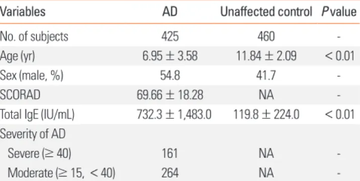
Table 1. Clinical profiles of the study subjects
Variables AD Unaffected control P value
No. of subjects 425 460 -
Age (yr) 6.95 ± 3.58 11.84 ± 2.09 <0.01
Sex (male, %) 54.8 41.7 -
SCORAD 69.66 ± 18.28 NA -
Total IgE (IU/mL) 732.3 ± 1,483.0 119.8 ± 224.0 <0.01 Severity of AD
Severe (≥ 40) 161 NA -
Moderate (≥ 15, < 40) 264 NA -
AD, atopic dermatitis; SCORAD, SCORing AD; IgE, immunoglobulin E; NA, not applicable.
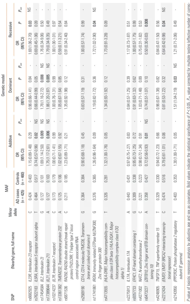
Table 2. Association analysis between genetic polymorphisms and AD SNP(Nearby) gene, full name Minor allele
MAFGenetic model AdditiveDominantRecessive
AD cases (n425)=
Controls (nOR OR OR PPPPPP..corcor.cor (95% CI)(95% CI)(95% CI)460)= NS0.031.69 (1.06-2.70)-0.840.96 (0.66-1.41)0.29-1.15 (0.89-1.48)0.4030.424CIL23R, Interleukin 23 receptorrs6682925 -0.090.69 (0.45-1.06)-0.050.66 (0.44-0.99)0.02NS0.74 (0.57-0.96)0.5170.464TIL5RA, Interleukin 5 receptor subunit alphars7622183 -0.501.76 (0.34-9.06)NS0.0081.78 (1.16-2.72)0.009NS1.69 (1.14-2.50)0.1010.127G(IL2, Interleukin 2)rs17454584 -0.491.79 (0.34-9.35)NS0.0051.86 (1.20-2.87)0.006NS1.75 (1.17-2.61)0.1030.127A(IL21, Interleukin 21)rs17389644 -0.971.00 (1.00-1.01)-0.991.00 (0.76-1.31)0.99-0.1790.173C(IL7R, Interleukin 7 receptor)rs102142371.00 (0.71-1.41) --0.540.59 (0.11-3.21)-0.851.05 (0.67-1.64)1.02 (0.67-1.55)0.920.1060.125Aransmembrane protein 232TMEM232, Trs13361382 rs6871536RAD50, RAD50 double strand break repair protein/TH2LCRR, T helper type 2 locus control region associated RNA
C0.2110.1851.23 (0.89-1.71)0.21-1.35 (0.92-1.98)0.12-0.91 (0.34-2.40)0.84- rs2569190CD14, CD14 molecule/TMCO6, Transmem- brane and coiled-coil domains 6G0.3640.3840.90 (0.68-1.18)0.45-0.83 (0.57-1.19)0.31-1.00 (0.57-1.74)0.99- rs11741861IRGM, Immunity related GTPase M/ZNF300, Zinc finger protein 300G0.3760.3651.26 (0.96-1.64)0.09-1.19 (0.82-1.72)0.36-1.72 (1.02-2.90)0.04NS rs4713555(HLA-DRB1, Major histocompatibility com- plex class II DR beta 1/HLA-DQA1, Major histocompatibility complex class II DQ alpha 1)
T0.3260.2811.32 (1.00-1.76)0.05-1.34 (0.93-1.92)0.12-1.73 (0.91-3.28)0.09- rs2275913IL17A, Interleukin 17AA0.4430.4310.97 (0.75-1.27)0.83-0.84 (0.57-1.25)0.39-1.17 (0.73-1.87)0.51- rs9357733EFHC1, EF-hand domain containing 1G0.3090.3380.95 (0.73-1.25)0.72-0.91 (0.63-1.32)0.62-1.00 (0.57-1.75)0.99- rs4271002NAT2, N-acethyltransferase 2C0.2210.2230.98 (0.72-1.34)0.91-1.03 (0.71-1.48)0.89-0.75 (0.31-1.82)0.53- rs6473227(ZBTB10, Zinc finger and BTB domain con- taining 10)A0.3580.4270.72 (0.56-0.93)0.01NS0.74 (0.51-1.07)0.10-0.50 (0.30-0.83)0.008 rs4246905TNFSF15, TNF superfamily member 15T0.3290.3300.94 (0.72-1.23)0.66-0.96 (0.67-1.38)0.82-0.84 (0.47-1.50)0.56- rs2212434(EMSY, EMSY BRCA2 interacting transcrip- tional repressor)C0.4740.5160.79 (0.61-1.01)0.06-0.81 (0.53-1.22)0.32-0.64 (0.42-0.98)0.04NS rs2143950(PPP2R3C, Protein phosphatase 2 regulatory subunit B’ gamma)T0.4030.3531.30 (1.00-1.71)0.05-1.51 (1.04-2.19)0.03NS1.21 (0.71-2.06)0.49- P value of logistic regression analysis by adjusting age and sex as covariates. Bold values indicate the statistical significance of P<0.05. Pcor. value corrected for multiple testing (effective number of correc- tion=15.98) using the SNPSpD program. AD, atopic dermatitis; SNP, single nucleotide polymorphism; MAF, minor allele frequency; OR, odds ratio; CI, confidence interval; NS, not significant.
The total IgE levels were significantly higher in the AD group (732.3 ± 1,483.0 IU/mL vs 119.8 ± 224.0 IU/mL, P < 0.01). The detailed characteristics of these subjects are summarized in Ta- ble 1.
Association analysis with AD development and severity
To conduct a replication study of previously described sus- ceptible loci in AD, we selected a total of 17 SNPs (10 located at gene regions and 7 at nearby genes) that had been identified by previous GWASs and meta-analyses of AD and also from a pan- el of immune-related genes (Supplementary Table 2). These SNPs were then successfully genotyped in our AD and control populations and were found to be common variants in these subjects with a minor allele frequency (MAF) above 0.05.
A case-control analysis adjusted for age and sex as covariates was performed. The results replicated significant associations between 8 SNPs (rs6682925, rs7622183, rs17454584, rs17389644, rs11741861, rs6473227, rs2212434 and rs2143950) and AD de- velopment (minimum P = 0.005 at rs17389644 under a domi- nant model, Table 2). In addition, most of these significant SNPs found in the case-control analysis and an additional polymor- phism, rs11741861, that is co-located at IRGM and ZNF300 show- ed significant signals in the association analysis with AD severi-
ty (minimum P = 0.004 at rs6473227 under a dominant model, Table 3).
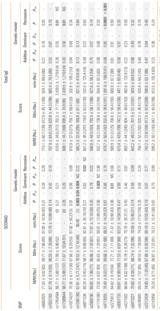
Association analysis with SCORAD and total IgE
Additional linear regression analysis using SCORAD and total IgE, as important indices of AD assessment, was performed. In SCORAD association analysis in AD patients (n = 189), 4 SNPs showed significant signals (rs13361382 on TMEM232, rs6871536 co-located at RAD50 and TH2LCRR, rs11741861 co-located at IRGM and ZNF300, rs2275913 on IL17A; minimum P = 0.003 at rs13361382 under additive model, Table 4) even after correc- tion for multiple testing (Pcor=0.04). Analysis using the total IgE level revealed that rs17454584 near IL2, rs17389644 near IL21 and rs4713555 near HLA-DRB1 and HLA-DQA1 had a potential association in AD patients (minimum P < 0.0001 at rs4713555 under a recessive model, Pcor<0.001, Table 4).
Meta-analysis and in silico analyses of significant SNPs
Based on the SNPs identified from previous studies of AD, a meta-analysis was performed. Among the variants that were replicated with significance in our analysis, 4 SNPs (rs17389644, rs6473227, rs2212434 and rs2143950) appeared to have positive associations with AD (minimum overall P = 8.85 × 10⁻¹⁰ for Table 3. Associations between genetic polymorphisms and AD severity
SNP (Nearby) gene Minor
allele
MAF AD severity
Control (n = 460)
AD cases Control vs
Moderate Control vs
Severe Moderate vs
Severe Moderate
(n = 264) Severe
(n = 161) P Pcor. P Pcor. P Pcor.
rs6682925 IL23R C 0.403 0.407 0.453 0.33 - 0.19 - 0.21 -
rs7622183 IL5RA T 0.517 0.471 0.451 0.05 - 0.15 - 0.66 -
rs17454584 (IL2) G 0.101 0.128 0.124 0.02 NS 0.07 - 0.58 -
rs17389644 (IL21) A 0.103 0.126 0.129 0.02 NS 0.03 NS 0.81 -
rs10214237 (IL7R) C 0.179 0.183 0.156 0.98 - 0.99 - 0.26 -
rs13361382 TMEM232 A 0.106 0.118 0.137 0.97 - 0.48 - 0.23 -
rs6871536 RAD50/TH2LCRR C 0.185 0.198 0.232 0.39 - 0.26 - 0.34 -
rs2569190 CD14/TMCO6 G 0.384 0.374 0.346 0.90 - 0.38 - 0.52 -
rs11741861 IRGM/ZNF300 G 0.365 0.362 0.399 0.60 - 0.03 0.45 -
rs4713555 (HLA-DRB1/HLA-DQA1) T 0.281 0.322 0.331 0.12 - 0.18 - 0.90 -
rs2275913 IL17A A 0.431 0.447 0.436 0.98 - 0.83 - 0.87 -
rs9357733 EFHC1 G 0.338 0.322 0.287 0.50 - 0.52 - 0.32 -
rs4271002 NAT2 C 0.223 0.232 0.203 0.68 - 0.41 - 0.28 -
rs6473227 (ZBTB10) A 0.427 0.367 0.343 0.14 - 0.004 NS 0.58 -
rs4246905 TNFSF15 T 0.330 0.324 0.338 0.60 - 0.77 - 0.54 -
rs2212434 (EMSY) C 0.516 0.500 0.430 0.29 - 0.03 NS 0.06 -
rs2143950 (PPP2R3C) T 0.353 0.405 0.399 0.01 NS 0.47 - 0.86 -
P value of logistic analysis under additive model by adjusting age and sex as covariates. Bold values indicate the statistical significance of P < 0.05. Pcor. value cor- rected for multiple testing (effective number of correction = 15.98) using the SNPSpD program.
AD, atopic dermatitis; SNP, single nucleotide polymorphism; MAF, minor allele frequency; NS, not significant.
Table 4. Associations of genetic polymorphisms with SCORAD and total IgE SNP
SCORADTotal IgE ScoreGenetic model ScoreGenetic model AdditiveDominantRecessiveAdditiveDominantRecessive M/M (No.)M/m (No.)m/m (No.)PPcor.PPcorPPcor.M/M (No.)M/m (No.)m/m (No.)PPcorPPcorPPcor. rs668292571.03±9.02 (62)69.77±7.66 (83)67.00±10.34 (42)0.21-0.34-0.26-715.1±60.7 (139)612.9±45.1 (185)624.7±69.8 (80)0.21-0.16-0.55- rs762218367.78±9.14 (55)68.55±7.39 (86)73.79±10.88 (46)0.14-0.42-0.10-707.9±64.6 (120)620.8±44.3 (196)665.4±70.5 (89)0.52-0.87-0.19- rs1745458468.77±5.88 (137)71.99±9.98 (52) - (0)0.21-0.21---610.6±34.6 (312)702.5±71.7 (96)2,439.5±1,219.8 (4)0.28-0.55-0.01NS rs1738964468.77±5.88 (137)71.67±10.04 (51) - (0)0.26-0.26---608.1±34.7 (308)695.0±70.9 (96)2,439.5±1,219.8 (4)0.30-0.58-0.01NS rs1021423770.36±6.17 (130)67.66±9.29 (53)87.00±50.23 (3)0.91-0.56-0.09-610.9±37.0 (273)759.7±69.4 (120)332.6±105.2 (10)0.94-0.69-0.34- rs1336138267.81±5.71 (141)76.52±12.10 (40) 90.40 (1)0.0030.040.004NS0.22-565.3±32.9 (295)769.8±81.1 (90)821.5±410.8 (4)0.13-0.13-0.64- rs687153669.20±6.45 (115)68.77±8.88 (60)82.81±26.19 (10)0.21-0.62-0.03NS601.2±38.3 (247)718.6±63.0 (130)532.0±125.4 (18)0.39-0.22-0.63- rs256919069.85±7.96 (77)66.86±7.43 (81)77.01±15.10 (26)0.35-0.79-0.02-568.0±44.5 (163)792.4±58.1 (186)427.6±58.2 (54)0.75-0.57-0.14- rs1174186173.50±8.07 (83)65.63±7.29 (81)69.97±13.99 (25)0.06-0.009NS0.96-552.7±43.2 (164)736.8±54.3 (184)654.9±82.5 (63)0.98-0.68-0.53- rs471355570.49±8.03 (77)69.66±7.51 (86)68.51±14.29 (23)0.61-0.66-0.70-679.7±50.4 (182)532.6±39.6 (181)1,081.6±168.9 (41)0.08-0.95-<0.0001<0.001 rs227591364.62±8.79 (54)72.71±7.71 (89)71.47±10.90 (43)0.06-0.01NS0.66-569.9±51.0 (125)700.6±50.4 (193)600.9±67.2 (80)0.81-0.91-0.59- rs935773369.87±6.99 (100)71.53±8.67 (68)63.57±14.58 (19)0.47-0.99-0.11-615.4±43.6 (199)742.2±59.4 (156)447.1±65.9 (46)0.50-0.91-0.20- rs427100268.84±6.62 (108)70.78±8.28 (73)70.70±26.72 (7)0.43-0.40-0.87-683.7±44.1 (240)604.1±48.8 (153)482.8±133.9 (13)0.22-0.23-0.59- rs647322770.60±8.38 (71)68.74±7.02 (96)70.59±15.40 (21)0.74-0.59-0.89-643.2±49.2 (171)701.5±51.3 (187)503.5±69.8 (52)0.98-0.49-0.33- rs424690569.24±7.51 (85)69.53±7.59 (84)74.53±17.57 (18)0.35-0.59-0.24-704.1±52.2 (182)620.3±46.4 (179)420.9±64.2 (43)0.40-0.78-0.18- rs221243474.00±11.03 (45)67.88±6.93 (96)69.18±10.55 (43)0.19-0.07-0.77-742.2±72.8 (104)617.4±42.8 (208)590.8±63.7 (86)0.47-0.64-0.48- rs214395070.24±8.71 (65)68.87±7.22 (91)70.21±12.41 (30)0.87-0.73-0.89-599.2±50.1 (143)647.1±45.9 (199)812.5±102.4 (63)0.22-0.38-0.24- P value of linear regression analysis under additive model by adjusting age and sex as covariates. Pcor. value corrected for multiple testing (effective number of correction=15.98) using the SNPSpD program. M/M, M/m, and m/m indicate the homozygote of the major allele, heterozygote, and homozygote of the minor allele, respectively. Bold values indicate the statistical significance of P<0.05. AD, atopic dermatitis; SCORAD, SCORing AD; IgE, immunoglobulin E; NS, not significant; n, number of subjects.
rs2212434 and rs2143950, Figure). However, among the positive SNPs, only rs6473227 still retained positive significance in the Cochran’s Q test established by the presence of heterogeneity (PCochrane’s Q=0.018).
We then investigated the potential functions of the significant SNPs using in silico analyses. Intriguingly, the minor C allele of rs6682925T>C in the regulatory upstream region was predict- ed to be a putative binding site for the GATA-1 and NF-E tran- scription factors, but not the sequence including the major T allele of this SNP (Supplementary Table 3). In the analysis to predict the functions of these SNPs, rs6682925, rs7622183 and rs6473227 were estimated to be potential exonic splicing en- hancer (ESE) sites that were dependent on the major and mi- nor alleles of each SNP (Supplementary Table 3).
DISCUSSION
AD is a complex disease caused by a combination of multiple genetic and interacting environmental factors. The identifica- tion of the genetic factors that contribute to AD is therefore im- portant developing of new therapeutic and prevention strate- gies for this condition. Although GWASs and meta-analyses have been performed in a number of studies and identified many sus- ceptibility loci for AD,6-11 replication studies for these findings are still lacking and reliable markers for AD thus remain to be iden- tified. In our current replication and additional analysis, several previously described genetic variants again showed significant associations with AD, with the top signals found at rs13361382 in TMEM232, rs17389644 near IL21, and rs4713555 near HLA-
DRB1/HLA-DQA1 for SCORAD, AD development and the total IgE level, respectively. These results suggest that these genetic variants may contribute to a predisposition for AD.
A recent multi-ancestry meta-analysis of the largest number of samples yet analyzed (21,399 AD cases and 95,464 controls) identified 31 susceptibility loci including 10 novel loci related to innate immune signaling and T cell function.11 Variants of im- mune system genes (such as IL23R, IL5RA, and IL2) have also been reported as candidates for immune-mediated disorders.12-14 However, many previous studies have not fully replicated the associations of SNPs with AD described by previous reports, in- cluding variants identified by GWASs or meta-analyses for rea- sons such as small sample sizes, differences in study designs and an inappropriate reliance on standard significance thresh- olds.18,19 Hence, replication studies are needed to provide more accurate estimates of the association of genetic variations with the development of diseases and related phenotypes. In our current study, the rs6473227 and rs2143950 SNPs, which were previously highlighted in a meta-analysis,11 were found to have a significant association with AD and its severity, respectively, suggesting that they could serve as genetic markers for AD.
In our present replication study in an AD and control popula- tion, 8 previously identified SNPs again showed significant as- sociations with AD in our case-control analysis (minimum P = 0.005 at rs17389644 under dominant model, Table 2). However, several known SNPs (rs6682925, rs7622183, rs11741861, rs2212434, and rs2143950) showed only nominal association with AD. On the other hand, despite higher MAFs of a few SNPs (rs17454584, rs17389644, and rs2143950) in both moderate and severe AD Figure. Result of meta-analysis. Plots of the replicated SNPs of (A) rs17389644, (B) rs6473227, (C) rs2212434 and (D) rs2143950 with significance in this study in re- lation to the published studies on AD are calculated using the software package PLINK. The association of each SNP with AD is evaluated through the fixed-effect meta-analysis P value. Additional significance of statistical heterogeneity measured using the χ2-based Cochran’s Q test is considered (PCochrane’s Q<0.05). Bold values indicate the statistical significance of P < 0.05. AD, atopic dermatitis; SNP, single nucleotide polymorphism.
0 1 2 3 Odds ratio
Ellinghaus D et al., 2013 Current study Overall
A rs17389644 (overall P = 1.70 × 10⁻⁹, PCochrane’s Q=0.06)
0.5 0.6 0.7 0.8 0.9 1.0 Odds ratio
Paternoster L et al., 2015 Current study Overall
B rs6473227 (overall P = 1.07 × 10⁻⁹, PCochrane’s Q=0.018)
0 0.2 0.4 0.6 0.8 1.0 Odds ratio
Paternoster L et al., 2015 Current study Overall
C rs2212434 (overall P = 8.85 × 10⁻¹⁰, PCochrane’s Q=0.08)
0 0.5 1.0 1.5 2.0 2.5 Odds ratio
Paternoster L et al., 2015 Current study Overall
D rs2143950 (overall P = 8.85 × 10⁻¹⁰, PCochrane’s Q=0.09)
cases than those of controls, the significance of these SNPs was not increased depending on AD severity. This may be due to the insufficient sample size (in particular, the low number of se- vere AD cases) and/or involvement of other regulators; there- fore, further replication and studies are needed.
The rs17389644 variant near the IL21 locus showing the most significant signal among our AD subjects and notably was pre- viously identified as a novel susceptibility locus for AD using genome-wide immune chip analysis and meta-analysis.20 Sev- eral previous studies have reported that IL21 contributes to the pathogenesis of allergic diseases. The serum IL21 levels were found to increase during the acute exacerbation of asthma and to fall again after treatment.21 In other recent studies, it was found that both IL21 and IL21R expression was higher in acute skin lesions of AD patients,22 and that IL21 levels were are higher in adult AD patients than in unaffected controls.23 Although little is known about the biological mechanism of IL21 on IgE pro- duction in allergic diseases, additional recent studies have sug- gested that IL21 may suppress serum IgE production, with pos- sible involvement of Th2 cells that produce IL4 and/or IL13.24-26 Our current results have indicated that the minor A allele of rs17389644 near IL21 is associated with higher levels of IgE in AD patients (P = 0.01 under recessive model, Table 4), again suggesting that this variant plays a role in altering the serum IgE level in AD with hope of further functional studies elsewhere.
Additional functional studies that investigate the effects of the minor A allele of rs17389644 on IL21 expression or on its trans- acting activity in relation to AD are needed to elucidate this mech- anism.
There has been some conflicting evidence as to whether pre- viously reported susceptibility markers on candidate genes are in fact associated with AD. For instance, some previous studies have reported that SNPs (rs2040704 on RAD50, rs3091307 co-lo- cated within TH2LCRR and near RAD50) at chromosome 5q31.1 are significantly associated with AD.8,27 However, another study reported only a nominal association between this rs3091307 lo- cus and an AD patient group and no association at all with the allergic type of AD.28 In our result, the rs6871536 SNP co-locat- ed at the TH2LCRR and RAD50 was found to have no associa- tion signal, with the exception of a nominal signal only in the association with SCORAD These conflicting results may be due to insufficient sample sizes (in particular, the low number of AD cases with SCORAD) and/or different genetic backgrounds among populations that have been used thus far. Further replication studies of large cohorts comprising different populations are warranted.
SCORAD is an important AD assessment index, and the IgE level is one of the central players in allergic diseases including AD. The serum total IgE level, as a useful endophenotype, is generally increased in patients with AD.29 Different associations of genetic variants with the SCORAD and IgE parameters have been reported in AD cohorts: for instance, no associations have
previously been reported between the FLG R501X mutation and the SCORAD or IgE levels in AD, whereas significant associations have been described between the -1112 C/T SNP of IL13 with both SCORAD and IgE.30 Our current findings also identified significant association signals for several SNPs with SCORAD (at rs13361382 on TMEM232) and IgE (at rs4713555 near HLA- DRB1 and HLA-DQA1). Another recent study has also reported a potential association between genetic variations of TMEM232 and AD in a Chinese population.31 In addition, considering the association between SNPs located near to the HLA-DRB1 locus and total IgE in asthma and the involvement between human leucocyte HLA class II and IgE responses,32,33 rs4713555 may also play a role in regulating the IgE levels in AD. Hence, although further validation is needed, it is possible that SCORAD or IgE- associated SNPs could be predictive markers for AD.
We performed a meta-analysis of 4 SNPs (rs17389644, rs6473227, rs2212434, and rs2143950) that have been identified as risk loci for AD in previous genome-wide studies.11,20 As shown in Figure, rs2212434 near to EMSA and rs2143950 near to PPP2R3C showed the most significant signal. However, when Cochran’s Q test was used to assess heterogeneity of the estimated effect-sizes from the individual studies,34 only rs6473227 near to ZBTB10 still retained positive significance (PCochrane’s Q=0.018). There are few clues in the current literature that might explain the direct rela- tionship between the potential SNP and allergic/immune re- sponses. Therefore, further evaluation will be needed to identi- fy whether these 2 SNPs could indeed be useful markers for AD.
To estimate the potential functions of the SNPs we evaluated in AD, we employed in silico analysis of the variants found to be significantly associated with this disorder. Using the Signal Scan program to identify the putative transcription factor’s binding sites, the CTATCA and CTATC sequences including the ‘C’ al- lele of rs6682925 in the promoter region of IL23R were estimat- ed to be putative binding elements for the GATA-1 and NF-E reg- ulators. However, the major ‘T’ allele of this SNP did not show any results in this regard, suggesting that that the minor ‘C’ al- lele of rs6682925 may affect gene expression (Supplementary Table 3). In our additional search for potential ESE sites for splic- ing machinery using the SNP Function Prediction program, 3 SNPs (rs6682925, rs7622183, and rs6473227) were observed to have different binding scores for splicing factors depending on the major and minor alleles of each (Supplementary Table 3).
In additional search of the expression quantitative trait loci (eQTL) based on the conditional eQTL analysis (https://eqtl.onderzoek.
io/index.php?page = info), rs7622183 was also found to act as cis-acting eQTL, suggesting that this variant might act as a po- tential cis-regulator for the gene. In the case of functional rele- vance for rs2275913, this variant is positioned in the promoter region of IL17A and within a binding motif for the critical regu- lator of nuclear factor-activated T cells (NFAT), leading to a high- er promoter activity and production of IL17A in the minor A al- lele than the major G allele of rs2275913.35,36 In addition, IL17A
rs2275913 has been reported to be associated with the risk of several diseases including inflammatory diseases (for instance, rheumatoid arthritis), allergic asthma and rhinitis.37,38 There- fore, further functional studies are required.
In conclusion, we have replicated the associations between previously identified SNPs and AD in our current study and per- formed extended analyses of these variants to better understand the clinical phenotypes of this condition. Although this study has limitations of small number of samples and lack of func- tional evaluation, our results suggest that several genetic vari- ants may indeed be associated with AD and its clinical pheno- types (SCORAD and total IgE). However, further studies that in- clude functional evaluations of the significantly associated SNPs identified herein will be needed.
ACKNOWLEDGMENTS
This research was supported by Basic Science Research Pro- gram through the National Research Foundation of Korea (NRF) funded by the Ministry of Science, ICT and future Planning (NRF- 2014R1A2A1A10050687).
ORCID
Jeong-Hyun Kim http://orcid.org/0000-0003-4879-0075 So-Yeon Lee https://orcid.org/0000-0002-2499-0702 Mi-Jin Kang https://orcid.org/0000-0001-8510-3268 Jisun Yoon https://orcid.org/0000-0002-4904-9118 Sungsu Jung https://orcid.org/0000-0002-0559-4982 Hyun-Ju Cho https://orcid.org/0000-0003-4282-4000 Hyo-Bin Kim https://orcid.org/0000-0002-1928-722X Soo-Jong Hong https://orcid.org/0000-0003-1409-2113 REFERENCES
1. Leung DY, Bieber T. Atopic dermatitis. Lancet 2003;361:151-60.
2. Weidinger S, Novak N. Atopic dermatitis. Lancet 2016;387:1109-22.
3. van Beijsterveldt CE, Boomsma DI. Genetics of parentally reported asthma, eczema and rhinitis in 5-yr-old twins. Eur Respir J 2007;29:
516-21.
4. Spergel JM. From atopic dermatitis to asthma: the atopic march.
Ann Allergy Asthma Immunol 2010;105:99-106.
5. Stone SP, Muller SA, Gleich GJ. IgE levels in atopic dermatitis. Arch Dermatol 1973;108:806-11.
6. Esparza-Gordillo J, Weidinger S, Fölster-Holst R, Bauerfeind A, Rus- chendorf F, Patone G, et al. A common variant on chromosome 11q13 is associated with atopic dermatitis. Nat Genet 2009;41:596- 601.
7. Hirota T, Takahashi A, Kubo M, Tsunoda T, Tomita K, Sakashita M, et al. Genome-wide association study identifies eight new suscep- tibility loci for atopic dermatitis in the Japanese population. Nat Genet 2012;44:1222-6.
8. Schaarschmidt H, Ellinghaus D, Rodríguez E, Kretschmer A, Bau- recht H, Lipinski S, et al. A genome-wide association study reveals
2 new susceptibility loci for atopic dermatitis. J Allergy Clin Immu- nol 2015;136:802-6.
9. Sun LD, Xiao FL, Li Y, Zhou WM, Tang HY, Tang XF, et al. Genome- wide association study identifies two new susceptibility loci for atop- ic dermatitis in the Chinese Han population. Nat Genet 2011;43:
690-4.
10. Paternoster L, Standl M, Chen CM, Ramasamy A, Bønnelykke K, Duijts L, et al. Meta-analysis of genome-wide association studies identifies three new risk loci for atopic dermatitis. Nat Genet 2011;
44:187-92.
11. Paternoster L, Standl M, Waage J, Baurecht H, Hotze M, Strachan DP, et al. Multi-ancestry genome-wide association study of 21,000 cases and 95,000 controls identifies new risk loci for atopic derma- titis. Nat Genet 2015;47:1449-56.
12. Capon F, Di Meglio P, Szaub J, Prescott NJ, Dunster C, Baumber L, et al. Sequence variants in the genes for the interleukin-23 receptor (IL23R) and its ligand (IL12B) confer protection against psoriasis.
Hum Genet 2007;122:201-6.
13. Cheong HS, Kim LH, Park BL, Choi YH, Park HS, Hong SJ, et al. As- sociation analysis of interleukin 5 receptor alpha subunit (IL5RA) polymorphisms and asthma. J Hum Genet 2005;50:628-34.
14. Christensen U, Haagerup A, Binderup HG, Vestbo J, Kruse TA, Bør- glum AD. Family based association analysis of the IL2 and IL15 genes in allergic disorders. Eur J Hum Genet 2006;14:227-35.
15. Hanifin JM, Rajka G. Diagnostic features of atopic dermatitis. Acta Derm Venereol Suppl (Stockh) 1980;92:44-7.
16. Kunz B, Oranje AP, Labrèze L, Stalder JF, Ring J, Taïeb A. Clinical validation and guidelines for the SCORAD index: consensus report of the European Task Force on Atopic Dermatitis. Dermatology 1997;
195:10-9.
17. Asher MI, Keil U, Anderson HR, Beasley R, Crane J, Martinez F, et al.
International Study of Asthma and Allergies in Childhood (ISAAC):
rationale and methods. Eur Respir J 1995;8:483-91.
18. Bush WS, Moore JH. Chapter 11: Genome-wide association stud- ies. PLOS Comput Biol 2012;8:e1002822.
19. Kraft P, Zeggini E, Ioannidis JP. Replication in genome-wide associ- ation studies. Stat Sci 2009;24:561-73.
20. Ellinghaus D, Baurecht H, Esparza-Gordillo J, Rodríguez E, Mata- novic A, Marenholz I, et al. High-density genotyping study identi- fies four new susceptibility loci for atopic dermatitis. Nat Genet 2013;
45:808-12.
21. Geng S, Gao S, Hu H, Hu Y, Yu S, Zhao S. The differentiation and clinical significance of follicular helper T cells during acute exacer- bation in asthma patients. Xi Bao Yu Fen Zi Mian Yi Xue Za Zhi 2015;
31:1383-6.
22. Jin H, Oyoshi MK, Le Y, Bianchi T, Koduru S, Mathias CB, et al. IL- 21R is essential for epicutaneous sensitization and allergic skin in- flammation in humans and mice. J Clin Invest 2009;119:47-60.
23. Mizutani H, Tamagawa-Mineoka R, Nakamura N, Masuda K, Ka- toh N. Serum IL-21 levels are elevated in atopic dermatitis patients with acute skin lesions. Allergol Int 2017;66:440-4.
24. Lin PY, Jen HY, Chiang BL, Sheu F, Chuang YH. Interleukin-21 sup- presses the differentiation and functions of T helper 2 cells. Immu- nology 2015;144:668-76.
25. Tamagawa-Mineoka R, Kishida T, Mazda O, Katoh N. IL-21 reduc- es immediate hypersensitivity reactions in mouse skin by suppress- ing mast cell activation or IgE production. J Invest Dermatol 2011;
131:1513-20.
26. Wood N, Bourque K, Donaldson DD, Collins M, Vercelli D, Gold-
man SJ, et al. IL-21 effects on human IgE production in response to IL-4 or IL-13. Cell Immunol 2004;231:133-45.
27. Esparza-Gordillo J, Schaarschmidt H, Liang L, Cookson W, Bauer- feind A, Lee-Kirsch MA, et al. A functional IL-6 receptor (IL6R) vari- ant is a risk factor for persistent atopic dermatitis. J Allergy Clin Im- munol 2013;132:371-7.
28. Namkung JH, Lee JE, Kim E, Kim HJ, Seo EY, Jang HY, et al. Associ- ation of polymorphisms in genes encoding IL-4, IL-13 and their re- ceptors with atopic dermatitis in a Korean population. Exp Derma- tol 2011;20:915-9.
29. Furue M. Atopic dermatitis--immunological abnormality and its background. J Dermatol Sci 1994;7:159-68.
30. Trzeciak M, Gleń J, Rębała K, Bandurski T, Sikorska M, Nowicki R.
Coexistence of 2282del4 FLG gene mutation and IL-18 -137G/C gene polymorphism enhances the risk of atopic dermatitis. Postepy Dermatol Alergol 2016;33:57-62.
31. Wu YY, Tang JP, Liu Q, Zheng XD, Fang L, Yin XY, et al. Scanning in- dels in the 5q22.1 region and identification of the TMEM232 sus- ceptibility gene that is associated with atopic dermatitis in the Chi- nese Han population. Gene 2017;617:17-23.
32. Gould HJ, Sutton BJ. IgE in allergy and asthma today. Nat Rev Im-
munol 2008;8:205-17.
33. Moffatt MF, Gut IG, Demenais F, Strachan DP, Bouzigon E, Heath S, et al. A large-scale, consortium-based genomewide association study of asthma. N Engl J Med 2010;363:1211-21.
34. Hoaglin DC. Misunderstandings about Q and ‘Cochran’s Q test’ in meta-analysis. Stat Med 2016;35:485-95.
35. Espinoza JL, Takami A, Nakata K, Onizuka M, Kawase T, Akiyama H, et al. A genetic variant in the IL-17 promoter is functionally associ- ated with acute graft-versus-host disease after unrelated bone mar- row transplantation. PLoS One 2011;6:e26229.
36. Liu XK, Lin X, Gaffen SL. Crucial role for nuclear factor of activated T cells in T cell receptor-mediated regulation of human interleu- kin-17. J Biol Chem 2004;279:52762-71.
37. Eskandari-Nasab E, Moghadampour M, Tahmasebi A. Meta-anal- ysis of risk association between interleukin-17A and F gene poly- morphisms and inflammatory diseases. J Interferon Cytokine Res 2017;37:165-74.
38. Resende EP, Todo-Bom A, Loureiro C, Mota Pinto A, Oliveiros B, Mesquita L, et al. Asthma and rhinitis have different genetic pro- files for IL13, IL17A and GSTP1 polymorphisms. Rev Port Pneumol (2006) 2017;23:10-6.