Factors Associated with the Presence and Severity of Diabetic Ketoacidosis at Diagnosis of Type 1 Diabetes in Korean Children and Adolescents
The aim of this study was to identify the risk factors for presence and severity of diabetic ketoacidosis (DKA) at the onset of type 1 diabetes mellitus (T1DM) in Korean children and adolescents. A retrospective chart review of children and adolescents newly diagnosed with T1DM was conducted in seven secondary and tertiary centers in Korea. Eligible subjects were < 20 years of age and had records on the presence or absence of DKA at the time of T1DM diagnosis. DKA severity was categorized as mild, moderate, or severe. Data were collected on age, height, body weight, pubertal status, family history of diabetes, delayed diagnosis, preceding infections, health insurance status, and parental education level. A total of 361 patients (male 46.3%) with T1DM were included. Overall, 177 (49.0%) patients presented with DKA at T1DM diagnosis. Risk factors predicting DKA at T1DM diagnosis were age ≥ 12 years, lower serum C-peptide levels, presence of a preceding infection, and delayed diagnosis. Low parental education level and preceding infection increased the severity of DKA. These results suggest that alertness of the physician and public awareness of diabetes symptoms are needed to decrease the incidence and severity of DKA at T1DM diagnosis.
Keywords: Type 1 Diabetes Mellitus; Diabetic Ketoacidosis; Risk Factors Hye Jin Lee,1 Hyeoh Won Yu,1
Hae Woon Jung,2 Young Ah Lee,1 Jae Hyun Kim,3 Hye Rim Chung,4 Jaeho Yoo,5 Eunyoung Kim,6 Jeesuk Yu,7 Choong Ho Shin,1 Sei Won Yang,1 and Seong Yong Lee8
1Department of Pediatrics, Seoul National University Children’s Hospital, Seoul National University College of Medicine, Seoul, Korea; 2Department of Pediatrics, Kyung Hee University Medical Center, Kyung Hee University College of Medicine, Seoul, Korea; 3Department of Pediatrics, Inje University Ilsan Paik Hospital, Inje University College of Medicine, Goyang, Korea; 4Department of Pediatrics, Seoul National University Bundang Hospital, Seongnam, Korea; 5Department of Pediatrics, Dong-A University Hospital, Dong-A University College of Medicine, Busan, Korea; 6Department of Pediatrics, Chosun University Hospital, Chosun University College of Medicine, Gwangju, Korea;
7Department of Pediatrics, Dankook University Hospital, Dankook University College of Medicine, Cheonan, Korea; 8Department of Pediatrics, Seoul Metropolitan Government-Seoul National University Boramae Medical Center, Seoul, Korea
Received: 22 July 2016 Accepted: 16 October 2016 Address for Correspondence:
Seong Yong Lee, MD
Department of Pediatrics, Seoul Metropolitan Government- Seoul National University Boramae Medical Center, 20 Boramae-ro 5-gil, Dongjak-gu, Seoul 07061, Korea E-mail: [email protected]
https://doi.org/10.3346/jkms.2017.32.2.303 • J Korean Med Sci 2017; 32: 303-309
INTRODUCTION
The incidence of type 1 diabetes mellitus (T1DM) is increasing worldwide (1,2). In particular, regions with a low incidence have experienced a more rapid increase in newly diagnosed T1DM cases (1). The T1DM population is increasing continuously in Korea, the nation with very low prevalence of T1DM in the world (3-5). Diabetic ketoacidosis (DKA), which is caused by absolute insulin deficiency, is the most serious life-threatening acute complication of T1DM. It is related to 0.15%–0.31% of mortali- ties among patients with T1DM (6). Cerebral edema is respon- sible for 0.46%–4.60% of mortalities and is the leading cause of mortality related to DKA (6). In addition, DKA is related to im-
paired cognitive functions, such as short-term memory and long- term intelligence (7,8). The incidence of DKA at the time of T1DM diagnosis ranges from 15% to 67% (2,9,10) depending on the geographic region. Although the incidence of T1DM has increas- ed worldwide, the incidence of DKA at diagnosis has decreased according to several studies (11,12), or has not changed accord- ing to other studies (2,13).
In previous studies, young age, particularly < 2 years, low ac- cessibility to medical care, and absence of a family history of T1DM were identified as risk factors for DKA at T1DM diagno- sis (14). Particular geographic regions (10) and ethnicities (15) with a low background incidence of T1DM are associated with a higher risk of DKA at T1DM diagnosis. The higher incidence Pediatrics
of DKA associated with a lack of T1DM family history or with a low incidence of T1DM in the population suggests that diabetes awareness reduces the incidence of DKA. No study has report- ed the incidence of or risk factors for DKA in Asian children who have a very low incidence of T1DM.
The objective of this multicenter study was to evaluate the in- cidence of DKA at T1DM diagnosis in Korean children and ad- olescents and to determine the risk factors for developing DKA at T1DM diagnosis. We also evaluated the factors affecting DKA severity. If these risk factors are controllable, it would be possi- ble to reduce the rate and severity of DKA, which is a life-threat- ening complication of T1DM.
MATERIALS AND METHODS Subjects
The subjects were recruited from seven secondary and tertiary centers in Korea and were diagnosed with T1DM from January 2000 to May 2015. The participating centers are located through- out the country (Seoul National University Children’s Hospital and Seoul Metropolitan Government-Seoul National University (SMG-SNU) Boramae Medical Center in Seoul; Dong-A Univer- sity Hospital in Busan; Chosun University Hospital in Gwangju;
Dankook University Hospital in Cheonan; Seoul National Uni- versity Bundang Hospital in Seongnam; and Inje University Il- san Paik Hospital in Goyang).
T1DM was defined as insulin dependency with one of the following conditions: 1) serum C-peptide level < 0.6 ng/mL; 2) 24-hour urine C-peptide level < 30 µg/24 hr; 3) DKA at diagno- sis; or 4) positivity for one or more diabetes-associated autoan- tibodies. DKA was defined as hyperglycemia with venous blood pH < 7.3 or a bicarbonate level < 15 mEq/dL. DKA severity was classified by venous blood pH: severe < 7.1; moderate ≥ 7.1 and
< 7.2; mild ≥ 7.2 and < 7.3.
Eligible subjects were children and adolescents diagnosed with T1DM before 20 years of age who also had medical records regarding the presence or absence of DKA at T1DM diagnosis.
Patients diagnosed before 1 year of age were excluded.
Methods
Medical records were reviewed retrospectively. Data were col- lected on age, height, weight, pubertal status, season (spring, March–May; summer, June–August; autumn, September–No- vember; winter, December–February), family history of diabe- tes, delayed diagnosis, presence of preceding infections, health insurance status, and parents’ education level at diagnosis. The date of diagnosis was defined as the first day of insulin admin- istration. Laboratory results at diagnosis, such as serum glucose, C-peptide, glycated hemoglobin (HbA1c), venous pH and bicar- bonate levels, were also investigated.
Weight loss was estimated as the difference between weight
after recovery from acute diabetes and weight at diagnosis. Body mass index (BMI) was calculated as weight (kg) divided by height squared (m2). The BMI standard deviation score (SDS) was cal- culated for each subject based on 2007 Korean National Growth Charts (16). Delayed diagnosis was defined as a diagnosis of a disease other than diabetes at the first medical consultation. A high parental education level was defined if both parents grad- uated college or university. A low parental education level was defined when at least one parent had an education of high school or less.
All Koreans are covered by one of the following three health- care programs depending on their household income: 1) the national health insurance program; 2) copayment subsidies for the project; and 3) the medical aid program (healthcare for the poor). Thus, health insurance status in Korea is considered an indicator of socioeconomic status.
Statistical analysis
Statistical analyses were performed using SPSS ver. 21.0 for Win- dows software (SPSS Inc., Chicago, IL, USA). Variables were as- sessed for a normal distribution. All continuous variables are described as means ± standard deviation. Student’s t-test and the χ2 test were used to evaluate differences between patients with and without DKA at T1DM diagnosis. Analysis of variance and the χ2 test for trend analysis were used to compare variables between DKA severity groups. Multivariate logistic regression analysis was used to evaluate risk factors for 1) the presence of DKA at T1DM diagnosis and 2) DKA severity at T1DM diagno- sis. A P value < 0.05 was considered statistically significant.
Ethics statement
The present study protocol was reviewed and approved by the Institutional Review Board of Seoul National University Hospi- tal (IRB No. 1507-025-686), Ilsan Paik Hospital (IRB No. 2015- 06-024), Seoul National University Bundang Hospital (IRB No.
B-1607-356-401), Dong-A University Hospital (IRB No. 15-127), Chosun University Hospital (IRB No. 2015-07-016-001), Dankook University Hospital (IRB No. DKUH 2016-02-019), SMG-SNU Boramae Medical Center (IRB No.16-2015-104). Informed con- sent was waived by each IRB.
RESULTS
Subject characteristics
A total of 361 patients with T1DM were included in this study.
Overall, 49.0% (177/361) of the patients with T1DM presented with DKA at T1DM diagnosis. Male patients comprised 46.3%
of the study population, and their mean age at T1DM diagnosis was 8.9 ± 4.0 years. In total, 65.1% of the study population was prepubertal. The mean height SDS was 0.48 ± 1.18, mean weight SDS at diagnosis was −0.46 ± 1.16, mean weight SDS after stabi-
lization was 0.20 ± 0.96, and change in the weight SDS was 0.69
± 0.58. The mean BMI SDS at diagnosis was −0.88 ± 1.44, mean BMI SDS after stabilization was 0.08 ± 1.10, and change in the BMI SDS was 1.00 ± 0.82. Of the total patients, 30% were diag- nosed in spring, 26.6% in summer, 22.2% in autumn, and 21.1%
in winter. Approximately 40% had a family history of diabetes.
Of the total study population, 46.6% were not diagnosed with diabetes mellitus (DM) at their first hospital visit (delayed diag- nosis), and 60.9% had parents with a high education level. Most patients were covered by the national health insurance program, 3.1% were receiving copayment subsidies for the near poor proj- ect, and 2.8% were covered by the medical aid program. The
proportion who were receiving copayment subsidies was high- er among patients with T1DM than among the total population (0.6%), and the proportion receiving medical aid was the same as that in the total population (2.8%) (17).
Comparison between patients with and without DKA The comparison between patients with and without DKA at di- agnosis is shown in Table 1. No differences in sex ratio or mean age at diagnosis were detected between the two groups, but more children were aged < 3 years and ≥ 12 years in the DKA group than in the non-DKA group (14.1% and 29.4% in DKA group vs.
6.5% and 21.2%, in non-DKA group, respectively; P = 0.004).
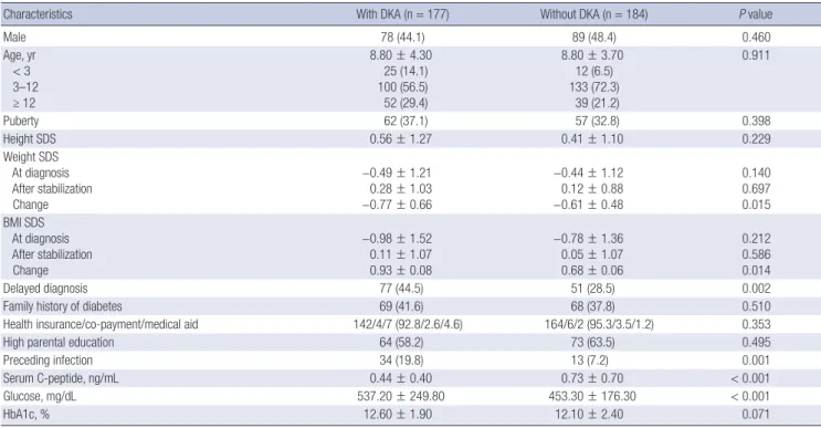
Table 1. Characteristics of patients according to the presence of DKA at diagnosis
Characteristics With DKA (n = 177) Without DKA (n = 184) P value
Male 78 (44.1) 89 (48.4) 0.460
Age, yr < 3 3–12 ≥ 12
8.80 ± 4.30 25 (14.1) 100 (56.5) 52 (29.4)
8.80 ± 3.70 12 (6.5) 133 (72.3) 39 (21.2)
0.911
Puberty 62 (37.1) 57 (32.8) 0.398
Height SDS 0.56 ± 1.27 0.41 ± 1.10 0.229
Weight SDS At diagnosis After stabilization Change
−0.49 ± 1.21 0.28 ± 1.03
−0.77 ± 0.66
−0.44 ± 1.12 0.12 ± 0.88
−0.61 ± 0.48
0.140 0.697 0.015 BMI SDS
At diagnosis After stabilization Change
−0.98 ± 1.52 0.11 ± 1.07 0.93 ± 0.08
−0.78 ± 1.36 0.05 ± 1.07 0.68 ± 0.06
0.212 0.586 0.014
Delayed diagnosis 77 (44.5) 51 (28.5) 0.002
Family history of diabetes 69 (41.6) 68 (37.8) 0.510
Health insurance/co-payment/medical aid 142/4/7 (92.8/2.6/4.6) 164/6/2 (95.3/3.5/1.2) 0.353
High parental education 64 (58.2) 73 (63.5) 0.495
Preceding infection 34 (19.8) 13 (7.2) 0.001
Serum C-peptide, ng/mL 0.44 ± 0.40 0.73 ± 0.70 < 0.001
Glucose, mg/dL 537.20 ± 249.80 453.30 ± 176.30 < 0.001
HbA1c, % 12.60 ± 1.90 12.10 ± 2.40 0.071
Data are expressed as the mean ± standard deviation or number (%).
DKA = diabetic ketoacidosis, SDS = standard deviation score, BMI = body mass index, HbA1c = glycated hemoglobin.
Table 2. Risk factors for DKA at T1DM diagnosis
Variables Univariate analysis Multivariate analysis
OR (95% CI) P value OR (95% CI) P value
Age group, yr < 3 (vs. 3–12) ≥ 12 (vs. 3–12)
2.77 (1.33–5.78) 1.77 (1.09–2.89)
0.007 0.022
1.69 (0.76–3.73) 2.78 (1.57–4.90)
0.197
< 0.001
Puberty (vs. prepuberty) 1.21 (0.78–1.89) 0.398 - -
Preceding infection 3.16 (1.61–6.23) 0.001 3.01 (1.41–6.42) 0.004
Delayed diagnosis 2.01 (1.29–3.13) 0.002 1.75 (1.07–2.84) 0.025
Family history of diabetes 1.17 (0.76–1.80) 0.472 - -
Co-payment/medical care (vs. insurance) 1.59 (0.62–4.06) 0.334 - -
Low parental education 1.25 (0.73–2.14) 0.312 - -
Serum C-peptide 0.26 (0.14–0.46) < 0.001 0.22 (0.11–0.41) < 0.001
Glucose 1.00 (1.00–1.00) < 0.001 - -
HbA1c 1.09 (0.99–1.20) 0.072 - -
DKA = diabetic ketoacidosis, T1DM = type 1 diabetes mellitus, OR = odds ratio, CI = confidence interval, HbA1c = glycated hemoglobin.
Pubertal status, height, and weight at diagnosis did not differ between the two groups. Patients with DKA at diagnosis lost more weight than did patients without DKA. Delayed diagnosis (44.5% vs. 28.5%) and preceding infections (19.8% vs. 7.2%) were more prevalent in the DKA than non-DKA group at diagnosis.
However, a family history of diabetes, health insurance status, and parental education level did not differ between the two groups.
The serum C-peptide level was lower and initial serum glucose level higher in the DKA than the non-DKA group. No difference was found in HbA1c levels at diagnosis.
Risk factors for DKA at T1DM diagnosis
Univariate analysis revealed that age < 3 years or ≥ 12 years in- creased the risks for DKA (odds ratio [OR], 2.77; 95% confidence interval [CI], 1.33–5.78; P = 0.007 and OR, 1.77; 95% CI, 1.09–
2.89; P = 0.022, respectively), compared with the 3–12-year age group (Table 2). A preceding infection (OR, 3.16; 95% CI, 1.61–
6.23; P = 0.001) and a delayed diagnosis (OR, 2.01; 95% CI, 1.29–
3.13; P = 0.002) also increased the risk for DKA. Serum C-pep- tide levels were lower in patients with DKA than in those with- out DKA (OR, 0.26; 95% CI, 0.14–0.46; P < 0.001). Multivariate analysis revealed that age ≥ 12 years (OR, 2.78; 95% CI, 1.57–
4.90; P < 0.001), a preceding infection (OR, 3.01; 95% CI, 1.41–
6.42; P = 0.004), delayed diagnosis (OR, 1.75; 95% CI, 1.07–2.84;
P = 0.025), and a low serum C-peptide level (OR, 0.22; 95% CI, 0.11–0.41; P < 0.001) increased the risk for DKA at T1DM diag- nosis (Table 2).
Comparisons between groups according to DKA severity We analyzed 163 of 177 patients with DKA at diagnosis, exclud- ing 14 patients whose DKA severity could not be determined due to incomplete records. A lower socioeconomic status as- sumed by insurance status, lower parental education level, pre- ceding infection, and lower serum C-peptide level were related to severe DKA (Table 3). The proportion of patients with health insurance (vs. copayment subsidies and the medical aid pro- gram) was 98.4% in the mild DKA group, 93.0% in the moderate DKA group, and 84.8% in the severe DKA group (P for trend = 0.037). Approximately 68% of parents had a high education lev- els in the mild DKA group, 59.4% in the moderate DKA group, and 37.9% in the severe DKA group (P for trend = 0.014). As the DKA severity increased, the rate of preceding infections incre- ased (P for trend = 0.010), and the mean serum C-peptide level decreased (P for trend = 0.024). No differences in mean age, sex, puberty, delayed diagnosis, or family history of DM were observ- ed among the mild, moderate, and severe DKA groups.
Risk factors for severe DKA at T1DM diagnosis
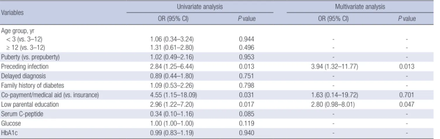
Univariate analysis revealed that preceding infections (OR, 2.84;
95% CI, 1.25–6.44; P = 0.013), low parental education level (OR, 2.96; 95% CI, 1.22–7.20; P = 0.017), and copayment subsidies or medical aid (OR, 4.55; 95% CI, 1.15–18.09; P = 0.031) increased the risk for severe DKA at T1DM diagnosis (Table 4). Multivari- ate logistic regression analysis of the factors determined to be statistically significant in the univariate analysis showed that a Table 3. Characteristics of the patients with DKA according to severity of DKA
Variables Mild (n = 67, 41.1) Moderate (n = 52, 31.9) Severe (n = 44, 27.0) P value
Male 27 (40.3) 28 (53.8) 18 (40.9) 0.281
Age, yr < 3 3–12 ≥ 12
8.81 ± 4.24 8 (11.9) 40 (59.7) 19 (28.4)
8.72 ± 4.37 6 (11.5) 31 (59.6) 15 (28.8)
9.24 ± 4.17 5 (11.4) 24 (54.5) 15 (34.1)
0.827
Height SDS 0.54 ± 1.06 0.59 ± 1.52 0.62 ± 1.36 0.948
Weight SDS At diagnosis After stabilization Change
−0.46 ± 1.26 0.29 ± 1.01 0.74 ± 0.46
−0.52 ± 1.25 0.38 ± 1.18 0.90 ± 0.81
−0.57 ± 1.16 0.17 ± 0.84 0.71 ± 0.75
0.914 0.632 0.358 BMI SDS
At diagnosis After stabilization Change
−0.94 ± 1.51
−1.05 ± 1.68
−1.11 ± 1.44
0.19 ± 0.99 1.13 ± 0.17 1.04 ± 0.18
1.10 ± 0.72 1.27 ± 1.10 1.01 ± 1.05
0.293 0.852 0.448
Puberty 25 (38.5) 19 (37.3) 15 (38.5) 0.989
Delayed diagnosis 31 (47.0) 24 (47.1) 19 (44.2) 0.951
Family history of diabetes 28 (43.1) 19 (39.6) 18 (43.9) 0.903
Co-payment/medical aid (vs. insurance) 1 (1.6) 3 (7.0) 5 (15.2) 0.037
High parental education 28 (68.3) 19 (59.4) 11 (37.9) 0.014
Preceding infection 8 (12.1) 9 (17.6) 14 (32.6) 0.010
Serum C-peptide, ng/mL 0.54 ± 0.53 0.38 ± 0.26 0.35 ± 0.23 0.024
Glucose, mg/dL 464.80 ± 186.30 583.60 ± 338.40 591.10 ± 200.30 0.010
HbA1c, % 12.70 ± 1.70 12.50 ± 2.00 12.60 ± 1.90 0.923
Data are expressed as the mean ± standard deviation or number (%).
DKA = diabetic ketoacidosis, SDS = standard deviation score, BMI = body mass index, HbA1c = glycated hemoglobin.
preceding infection and low parental education level increased the risk for severe DKA (OR, 3.94; 95% CI, 1.32–11.77; P = 0.013 and OR, 2.80; 95% CI, 0.98–8.01; P = 0.047, respectively).
DISCUSSION
In the present study, 49% of patients newly diagnosed with T1DM had DKA. Age ≥ 12, lower serum C-peptide levels, presence of a preceding infection, and delayed diagnosis significantly increas- ed the risk for DKA at T1DM diagnosis. Low parental education level and preceding infection increased the risk of severe DKA.
This incidence of DKA at T1DM diagnosis in this study is rel- atively high compared with that in Western countries where there is a higher prevalence of T1DM (11,14,18,19). This might be explained by ascertainment bias, as this study was conduct- ed at secondary and tertiary centers, which tend to admit pa- tients with more severe symptoms or delayed visits to the hos- pital after the initial presentation. As most patients with T1DM are referred to secondary and tertiary centers, the incidence of DKA in this study may not be exaggerated and may indicate the overall incidence of DKA at diagnosis in Korea.
Previous studies have reported that younger age at T1DM di- agnosis is a risk factor for DKA (13,14). In this study, children aged < 3 years or ≥ 12 years had a higher risk for DKA at T1DM diagnosis. However, after adjusting for serum C-peptide levels, age < 3 years was not a significant risk factor for DKA, suggest- ing that the higher risk of DKA at a younger age may be due to more decreased β-cell function. In fact, aggressive diabetes and delayed detection of diabetes symptoms are more frequent in young children. Participating in a prospective cohort that in- formed the parents about diabetes symptoms decreased the risk of DKA at T1DM diagnosis in young children (19). On the other hand, children and adolescents ≥ 12 years continued to have a significantly higher risk of DKA at T1DM diagnosis after adjusting for C-peptide levels, puberty, and delayed diagnosis.
Children and adolescents at this age have likely escaped paren-
tal control; thus, detection or reporting of symptoms may be delayed. A study in New Zealand reported an increased risk for DKA at age around 11 years (13). Because earlier detection of symptoms in young children is possible through involvement in toileting and provision of water by parents, the authors sug- gested that greater self-awareness and earlier reporting of symp- toms in children older than 11 years are needed to prevent DKA.
C-peptide is a surrogate marker of endogenous insulin secre- tion. A low C-peptide level implies decreased β-cell function.
Thus, patients with DKA due to a significant β-cell failure are likely to have lower C-peptide and higher glucose levels. As ex- pected, our study showed that C-peptide levels of DKA group were lower than those of non-DKA group.
Infection is related to T1DM and an increased risk for DKA.
Infection can accelerate destruction of β-cells through an auto- immune process (20). Therefore, destruction of β-cells may in- crease the risk for DKA and its severity. Infection also results in inflammation, release of pro-inflammatory cytokines, and a counter regulatory response related to insulin resistance and metabolic decompensation (21). On the other hand, symptoms of infection may mask the symptoms of T1DM, delay diagnosis, and increase the risk for DKA.
A delayed diagnosis in this study referred to a missed T1DM diagnosis at the first medical counseling visit for symptoms such as polyuria, polydipsia, and weight loss. As mentioned earlier, some patients were misdiagnosed with gastroenteritis or a re- spiratory infection and diagnosed with T1DM later, after DKA occurred. After adjusting for preceding infections, a delayed di- agnosis still increased the risk for DKA. Thus, initial suspicion and monitoring blood sugar levels could prevent a delayed di- agnosis and reduce development of DKA.
Public education about diabetes symptoms alone can reduce the frequency of DKA through preventing a delayed diagnosis.
An Italian study reported that a decrease in the frequency of DKA at T1DM diagnosis from 78% to 12.5% 8 years after the ed- ucation program for the public and primary physicians (22).
Table 4. Risk factors for severe DKA (vs. mild to moderate DKA)
Variables Univariate analysis Multivariate analysis
OR (95% CI) P value OR (95% CI) P value
Age group, yr < 3 (vs. 3–12)
≥ 12 (vs. 3–12) 1.06 (0.34–3.24)
1.31 (0.61–2.80) 0.944
0.496 -
- -
-
Puberty (vs. prepuberty) 1.02 (0.49–2.16) 0.953 - -
Preceding infection 2.84 (1.25–6.44) 0.013 3.94 (1.32–11.77) 0.013
Delayed diagnosis 0.89 (0.44–1.80) 0.751 - -
Family history of diabetes 1.09 (0.53–2.26) 0.798 - -
Co-payment/medical aid (vs. insurance) 4.55 (1.15–18.09) 0.031 1.63 (0.14–19.72) 0.701
Low parental education 2.96 (1.22–7.20) 0.017 2.80 (0.98–8.01) 0.047
Serum C-peptide 0.34 (0.10–1.16) 0.085 - -
Glucose 1.00 (1.00–1.00) 0.119 - -
HbA1c 0.99 (0.83–1.19) 0.940 - -
DKA = diabetic ketoacidosis, OR = odds ratio, CI = confidence interval, HbA1c = glycated hemoglobin.
Other studies from Sweden (23) and Finland (11) also reported that education reduces the frequency of DKA at T1DM diagnosis.
We found that DKA severity was influenced by socioeco- nomic factors as well as the presence of preceding infection. An education level higher than college in both parents reduced the risk for severe DKA in this study. Furthermore, if the education level of one or both parents was high school or lower, the risk for severe DKA at T1DM diagnosis was 2.8-fold higher compared with parents with more education. This result is consistent with previous studies. A lower education level in the mother was re- lated to a higher risk of DKA at onset in Lithuania (24). A lower parental education level was related to a higher prevalence of DKA according to the US Search for Diabetes in Youth study (18).
Those receiving copayment subsidies and medical aid with low income probably have less time and capacity to take care of their children and may not detect diabetic symptoms early. How- ever, the relationship between DKA severity and insurance sta- tus was not significant after adjusting for other factors, which may be due to the small sample size of the copayment subsidies and medical aid groups.
This study had some limitations, including the retrospective medical record review. Some records contained incomplete data regarding parental education level and previous visits to other hospitals before the T1DM diagnosis. In addition, data from one secondary and six tertiary centers, where more criti- cal patients tend to visit, may have caused selection bias. Fur- ther prospective studies inclusive of more complete data are needed. Despite these limitations, this study is the first nation- wide multicenter study to evaluate the risk factors for DKA, in- cluding socioeconomic factors, at T1DM diagnosis, performed in East Asia, where the incidence of T1DM is very low. In the fu- ture, more studies on T1DM and DKA incidence time trends will be helpful to understand the risk factors for DKA.
In conclusion, the incidence of DKA at T1DM diagnosis in Korean children and adolescents in this study was 49%. The risk factors for DKA at T1DM diagnosis were age ≥ 12 years, low C- peptide levels, delayed diagnosis, and a preceding infection. A low parental education level and preceding infection were more likely to cause severe DKA. Thus, public education and the phy- sician’s alertness are important to reduce the incidence and se- verity of DKA, a life-threatening complication due to T1DM.
DISCLOSURE
The authors have no potential conflicts of interest to disclose.
AUTHOR CONTRIBUTION
Conceptualization: Lee HJ, Jung HW, Lee YA, Shin CH, Lee SY.
Data curation: Yu HW, Kim JH, Chung HR, Yoo J, Kim E, Yu J, Lee SY. Investigation: Lee HJ, Jung HW, Lee YA, Kim JH, Chung
HR, Yoo J, Kim E, Yu J, Shin CH, Yang SW, Lee SY. Writing - orig- inal draft: Lee HJ. Writing - review & editing:Kim JH, Lee YA, Shin CH, Yang SW, Lee SY.
ORCID
Hye Jin Lee http://orcid.org/0000-0002-7925-5238 Hyeoh Won Yu http://orcid.org/0000-0002-7224-400X Hae Woon Jung http://orcid.org/0000-0003-0494-4626 Young Ah Lee http://orcid.org/0000-0001-9179-1247 Jae Hyun Kim http://orcid.org/0000-0002-0203-7443 Hye Rim Chung http://orcid.org/0000-0001-8550-4969 Jaeho Yoo http://orcid.org/0000-0003-3065-944X Eunyoung Kim http://orcid.org/0000-0002-3073-5222 Jeesuk Yu http://orcid.org/0000-0003-0020-2000 Choong Ho Shin http://orcid.org/0000-0002-9813-1134 Sei Won Yang http://orcid.org/0000-0002-5304-5750 Seong Yong Lee http://orcid.org/0000-0002-7077-869X REFERENCES
1. DIAMOND Project Group. Incidence and trends of childhood type 1 dia- betes worldwide 1990-1999. Diabet Med 2006; 23: 857-66.
2. Wojcik M, Sudacka M, Wasyl B, Ciechanowska M, Nazim J, Stelmach M, Starzyk JB. Incidence of type 1 diabetes mellitus during 26 years of obser- vation and prevalence of diabetic ketoacidosis in the later years. Eur J Pe- diatr 2015; 174: 1319-24.
3. Ko KW, Yang SW, Cho NH. The incidence of IDDM in Seoul from 1985 to 1988. Diabetes Care 1994; 17: 1473-5.
4. Shin CH. Epidemiologic characteristics of type 1 diabetes in children aged 14 years or under in Korea, 1985-2000. Korean J Pediatr 2008; 51: 569-75.
5. Kim JH, Lee CG, Lee YA, Yang SW, Shin CH. Increasing incidence of type 1 diabetes among Korean children and adolescents: analysis of data from a nationwide registry in Korea. Pediatr Diabetes 2016; 17: 519-24.
6. Edge JA, Ford-Adams ME, Dunger DB. Causes of death in children with insulin dependent diabetes 1990-96. Arch Dis Child 1999; 81: 318-23.
7. Cameron FJ, Scratch SE, Nadebaum C, Northam EA, Koves I, Jennings J, Finney K, Neil JJ, Wellard RM, Mackay M, et al. Neurological consequenc- es of diabetic ketoacidosis at initial presentation of type 1 diabetes in a prospective cohort study of children. Diabetes Care 2014; 37: 1554-62.
8. Ghetti S, Lee JK, Sims CE, Demaster DM, Glaser NS. Diabetic ketoacido- sis and memory dysfunction in children with type 1 diabetes. J Pediatr 2010; 156: 109-14.
9. Dunger DB, Sperling MA, Acerini CL, Bohn DJ, Daneman D, Danne TP, Glaser NS, Hanas R, Hintz RL, Levitsky LL, et al. ESPE/LWPES consensus statement on diabetic ketoacidosis in children and adolescents. Arch Dis Child 2004; 89: 188-94.
10. Lévy-Marchal C, Patterson CC, Green A; EURODIAB ACE Study Group.
Europe and Diabetes. Geographical variation of presentation at diagno- sis of type I diabetes in children: the EURODIAB study. European and Dibetes. Diabetologia 2001; 44 Suppl 3: B75-80.
11. Hekkala A, Knip M, Veijola R. Ketoacidosis at diagnosis of type 1 diabetes in children in northern Finland: temporal changes over 20 years. Diabe-
tes Care 2007; 30: 861-6.
12. de Vries L, Oren L, Lebenthal Y, Shalitin S, Lazar L, Phillip M. Decrease in frequency of ketoacidosis at diabetes onset over the past two decades - perspectives of a paediatric tertiary care centre. Diabet Med 2012; 29: e170-5.
13. Jefferies C, Cutfield SW, Derraik JG, Bhagvandas J, Albert BB, Hofman PL, Gunn AJ, Cutfield WS. 15-year incidence of diabetic ketoacidosis at onset of type 1 diabetes in children from a regional setting (Auckland, New Zea- land). Sci Rep 2015; 5: 10358.
14. Usher-Smith JA, Thompson MJ, Sharp SJ, Walter FM. Factors associated with the presence of diabetic ketoacidosis at diagnosis of diabetes in chil- dren and young adults: a systematic review. BMJ 2011; 343: d4092.
15. Alvi NS, Davies P, Kirk JM, Shaw NJ. Diabetic ketoacidosis in Asian chil- dren. Arch Dis Child 2001; 85: 60-1.
16. Moon JS, Lee SY, Nam CM, Choi JM, Choe BK, Seo JW, Oh K, Jang MJ, Hwang SS, Yoo MH, et al. 2007 Korean national growth charts: review of develop- mental process and an outlook. Korean J Pediatr 2008; 51: 1-25.
17. National Health Insurance Service (KR). Major health insurance statistics:
2015 [Internet]. Available at http://www.nhis.or.kr [accessed on 27 June 2016].
18. Rewers A, Klingensmith G, Davis C, Petitti DB, Pihoker C, Rodriguez B, Schwartz ID, Imperatore G, Williams D, Dolan LM, et al. Presence of dia- betic ketoacidosis at diagnosis of diabetes mellitus in youth: the search
for diabetes in youth study. Pediatrics 2008; 121: e1258-66.
19. Elding Larsson H, Vehik K, Bell R, Dabelea D, Dolan L, Pihoker C, Knip M, Veijola R, Lindblad B, Samuelsson U, et al. Reduced prevalence of diabetic ketoacidosis at diagnosis of type 1 diabetes in young children participat- ing in longitudinal follow-up. Diabetes Care 2011; 34: 2347-52.
20. Yang Z, Zhou F, Dorman J, Wang H, Zu X, Mazumdar S, LaPorte RE. Asso- ciation between infectious diseases and type 1 diabetes: a case-crossover study. Pediatr Diabetes 2006; 7: 146-52.
21. Rayfield EJ, Ault MJ, Keusch GT, Brothers MJ, Nechemias C, Smith H. In- fection and diabetes: the case for glucose control. Am J Med 1982; 72: 439- 50.
22. Vanelli M, Chiari G, Ghizzoni L, Costi G, Giacalone T, Chiarelli F. Effecti- veness of a prevention program for diabetic ketoacidosis in children. An 8-year study in schools and private practices. Diabetes Care 1999; 22: 7-9.
23. Samuelsson U, Stenhammar L. Clinical characteristics at onset of type 1 diabetes in children diagnosed between 1977 and 2001 in the south-east region of Sweden. Diabetes Res Clin Pract 2005; 68: 49-55.
24. Sadauskaite-Kuehne V, Samuelsson U, Jasinskiene E, Padaiga Z, Urbonaite B, Edenvall H, Ludvigsson J; DEBS Study Group. Severity at onset of child- hood type 1 diabetes in countries with high and low incidence of the con- dition. Diabetes Res Clin Pract 2002; 55: 247-54.