www.jpis.org
pISSN 2093-2278 eISSN 2093-2286 Copyright © 2013 Korean Academy of PeriodontologyThis is an Open Access article distributed under the terms of the Creative Commons Attribution Non-Commercial License (http://creativecommons.org/licenses/by-nc/3.0/).
Efficacy of nonsurgical periodontal therapy on glycaemic control in type II diabetic patients: a randomized controlled clinical trial
Ravishankar Lingesha Telgi, Vaibhav Tandon*, Pradeep Shankar Tangade, Amit Tirth, Sumit Kumar, Vipul Yadav Department of Public Health Dentistry, Kothiwal Dental College & Research Centre, Moradabad, India
Purpose: Diabetes and periodontal disease are two common diseases with high prevalence rates. Recent evidence has shown a bidirectional relationship between diabetes and periodontitis. The aim of this study was to investigate the effects of nonsur- gical periodontal therapy on glycemic control in type 2 diabetes mellitus patients.
Methods: Sixty subjects aged 35–45 years with blood sugar controlled by oral hypoglycaemic agents were randomly divided equally among 3 groups: group A (scaling, mouthwash, and brushing), group B (mouthwash and brushing), and group C (brush- ing only). Glycated haemoglobin (HbA1c), fasting blood sugar (FBS), probing pocket depth (PPD), gingival index (GI), plaque index (PI), and the relevant drug history were recorded at baseline and after 3 months of intervention. Comparison of the mean difference among the variables was performed by parametric and nonparametric tests, which were further evaluated using multiple regression analysis.
Results: The mean differences between the PPD, FBS, HbA1c, GI, and PI in groups A and B were found to be statistically signif- icant (P<0.001). Multiple regression analysis in group A showed that out of all the independent variables, GI and frequency of drug administration independently (b=0.3761 and b=0.598) showed a significantly greater impact on HbA1c (R2=0.832, P<0.05).
Conclusions: Nonsurgical periodontal therapy can effectively decrease HbA1c levels in type 2 diabetes mellitus patients on medication.
Keywords: Glycosylated hemoglobin A, Periodontal debridement, Type II diabetes mellitus.
INTRODUCTION
Diabetes mellitus (DM) is a chronic, noncommunicable disease and also one of the major global public health issues.
It is defined as a clinical syndrome characterized by hyper- glycemia; due to a defect in insulin secretion by pancreatic β cells, a decrease in insulin sensitivity, or a combination of both. The most common form of DM is type 2 diabetes mel- litus (DM2), which accounts for 85% of all diabetes patients [1]. DM currently is the twelfth leading cause of death in the world. The prevalence of DM has risen dramatically in recent
years [2]. Asia, in particular, has the highest prevalence of dia- betes in the world. Countries exhibiting a high rate in growth of the diabetic population include India and China, among many other developing countries [3]. DM2, the most common type of diabetes, is characterized by hyperglycemia, hyperlip- idemia, and associated complications. Clinical and epidemi- ological evidence demonstrates that individuals with diabe- tes tend to have a higher prevalence of and more severe peri- odontitis than nondiabetics [4]. Periodontal disease is the most prevalent oral complication in patients with DM2. It is characterized by gingival inflammation, periodontal pocket
Received: Mar. 20, 2013; Accepted: Aug. 13, 2013
*Correspondence: Vaibhav Tandon
Department of Public Health Dentistry, Kothiwal Dental College & Research Centre, Kanth Road, Moradabad-244001, Uttar Pradesh, India E-mail: [email protected], Tel: +91-9319082837, Fax: +91-5912452996
bone resorption, ultimately resulting in tooth loss.
Furthermore, patients with poor control of diabetes experi- ence more periodontitis than well-controlled diabetics [4,5].
Direct and indirect evidence supports the concept that peri- odontal infection adversely affects glycemic control in peo- ple with diabetes. Southerland et al. [6] proposed a common pathogenesis involving an increased inflammatory response for periodontitis and diabetes. Patients with periodontitis have increased serum levels of inflammatory cytokines, while diabetic patients have hyper-inflammatory immune cells that can aggravate production of inflammatory cytokines [7]. This exacerbation can increase insulin resistance, that is, a physio- logical condition in which cells fail to respond to the normal actions of the hormone insulin and make it more difficult for patients to control their diabetes [7,8].
In a review article by Mealey [8], he cited a number of es- tablished mechanisms by which diabetes can influence the periodontium, which include the following: alteration in the host immune-inflammatory response, altered wound heal- ing, accumulation of advanced glycation end products, and elevated proinflammatory cytokines. When oral hygiene is compromised, oral bacteria may form a plaque biofilm that is resistant to chemicals and immune cells [6,7]. Without me- chanical debridement, the plaque biofilm matures and causes gingivitis in a few days. The infection then leads to formation of pockets between the teeth and gums, signalling a break- down of the periodontal apparatus and bone. The observa- tion that periodontal therapy appears to reduce periodontal infection and inflammation suggests that periodontal thera- py may facilitate metabolic control of diabetes, improving insulin sensitivity by reducing peripheral inflammatory cyto- kine levels [7]. Therefore, we put forward the hypothesis that if periodontitis is causally related to worsening of parameters of diabetic patients, then periodontal treatment should im- prove glycemic control.
Therefore, the present study was performed to investigate the effects of nonsurgical periodontal therapy on glycemic control in DM2 patients.
MATERIALS AND METHODS
Based on a pilot study, a sample size of 15 patients in each group was estimated considering (α=0.05 [95% confidence interval] and β=0.2 [80% power]). The mean difference be- tween the glycated hemoglobin (HbA1c) levels of the untreat- ed group and treated group was observed to be 1.08±0.93. On the basis of this difference, to be clinically significant, the necessary sample size was estimated using sample size and power calculations developed by William D. Dupont and li-
mercial-NoDerivs 3.0 United States License. for a 5% type I error and 20% type II error, and it was found to be 15 subjects.
Due to the longitudinal nature of the study, anticipating the attrition of some participants, a sample size of 20 patients in each group was recruited. Patients who visited the Diabetic Centre in Moradabad (India) for their routine check-up and investigation were screened for DM2 and periodontitis. The inclusion criteria were as follows: patients with DM2, blood sugar controlled only with oral hypoglycemic agents, mild to moderate periodontitis (pocket depth of 4–5 mm), presence of a minimum of 28 teeth, and no systemic antibiotic admin- istration and no periodontal treatment in last six months.
The exclusion criteria were the following: patients with sys- temic diseases other than DM2, tobacco and alcohol users, and patients suffering from oral disease who needed emer- gency treatment.
Before starting the study, the examiner was calibrated so as to achieve a minimum kappa value of 0.80. In order to en- sure intraexaminer consistency, a randomly selected group of 6 patients was examined and re-examined for the plaque index (PI) score, gingival index (GI) score, and probing pocket depth (PPD) in mm. The scores were assessed for reliability by applying the kappa statistic. The kappa value for intraex- aminer reliability ranged from 0.89 to 0.92.
A total of 60 patients aged 35–45 years who fulfilled the eli- gibility criteria and signed the informed consent were re- cruited to the study. The study protocol was reviewed and approved by the Institutional Ethics Review Committee, Mo- radabad. In this study, the examiner and participants were both blinded.
All of the 60 patients were randomly divided into 3 equal groups:
· Group A (n=20): patients, who had undergone scaling, were advised to regularly use 0.12% mouthwash (once daily) and brush (twice daily)
· Group B (n=20): patients who were advised to regularly use 0.12% chlorhexidine mouthwash (once daily) and brush (twice daily)
· Group C (control) (n=20): patients who were advised to brush (twice daily)
The participants were reinstructed on the use of mouth- wash and brushing and regular performance was reinforced every month through telephone calls to ensure compliance.
The oral examination was conducted in the dental clinic wing of the Diabetic Centre. The examination included assessing periodontal pocket depth in mm, gingival status, plaque ac- cumulation, and glycemic status (HbA1c in % and fasting blood sugar in mg/dL) of each individual patient at baseline and after 3 months of the intervention. Periodontal pocket
probe at six points for each tooth and the arithmetic mean value of all the teeth was considered.
The gingival status was assessed by the GI of Loe and Sil- ness [9], and dental plaque by the PI of Silness and Loe [10].
The participants were interviewed for demographic data, and the duration and frequency of drug usage, which was later cross-verified with their hospital records.
Statistical analysis was carried out using SPSS ver. 16.0 (SPSS, Inc., Chicago, IL, USA). Levene’s test for homogeneity of variance (P<0.05) was performed, as we assumed equity of variance was more important than an assumption of normal- ity. A comparison of the mean differences of the PPD, HbA1c, and fasting blood sugar level was analyzed by a paired t-test and between the groups by one-way analysis of variance (ANOVA), while the mean differences in the GI and PI scores were analyzed by the Wilcoxon signed rank test and between the groups by Kruskal-Wallis ANOVA. Statistical significance was considered to be when P≤0.05 (95% confidence interval).
The associations between glycemic control and potential predicting factors were evaluated with multiple linear regres- sion analysis using HbA1c as a dependent variable. Variables that were found to have a greater impact were included in stepwise regression analysis.
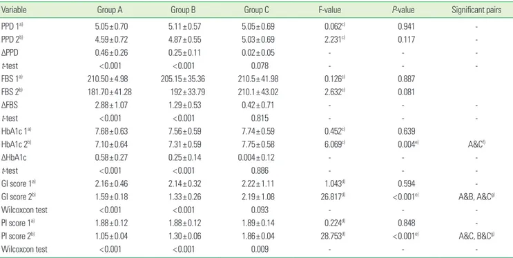
All of the 60 participants completed the study. It was found that the mean changes in the PPD, fasting blood sugar, and HbA1c after intervention in groups A and B were statistically significant (P<0.001), while in group C, they were statistically non significant (P =0.078). On comparison between the groups, the mean PPD and fasting blood sugar level did not show any statistically significant differences. It was found that the HbA1c level (P=0.004), GI score (P=0.001), and PI score (P=0.001) showed a statistically significant difference after 3 months of the treatment regime. A greater reduction in the mean±standard deviation HbA1c level could be seen in group A (0.58±0.27), followed by group B (0.25±0.14) and group C (0.004±0.12). A statistically significant difference was seen in the comparison between the groups, with the significant pair being groups A and C. A marked statistically significant difference in the GI and PI scores was seen (P<0.001) after 3 months of intervention. A statistically signif- icant difference was seen on comparing among the groups, with the significant pairs being groups A and B, and groups A and C for the GI scores; and groups A and C, and groups B and C for the PI scores (Table 1).
Table 2 depicts the association of all of the independent variables with HbA1c in group A through a simple linear re-
Table 1. Comparison of mean changes of PPD, GI, PI, HbA1c, frequency of drug and fasting blood sugar level among all the three groups.
Variable Group A Group B Group C F-value P-value Significant pairs
PPD 1a) 5.05±0.70 5.11±0.57 5.05±0.69 0.062c) 0.941 -
PPD 2b) 4.59±0.72 4.87±0.55 5.03±0.69 2.231c) 0.117 -
ΔPPD 0.46±0.26 0.25±0.11 0.02±0.05 - - -
t-test <0.001 <0.001 0.078 - - -
FBS 1a) 210.50±4.98 205.15±35.36 210.5±41.98 0.126c) 0.887
FBS 2b) 181.70±41.28 192±33.79 210.1±43.02 2.632c) 0.081
ΔFBS 2.88±1.07 1.29±0.53 0.42±0.71 - - -
t-test <0.001 <0.001 0.815 - - -
HbA1c 1a) 7.68±0.63 7.56±0.59 7.74±0.59 0.452c) 0.639
HbA1c 2b) 7.10±0.64 7.31±0.59 7.75±0.58 6.069c) 0.004e) A&Cf)
ΔHbA1c 0.58±0.27 0.25±0.14 0.004±0.12 - - -
t-test <0.001 <0.001 0.886 - - -
GI score 1a) 2.16±0.46 2.14±0.32 2.22±1.11 1.043d) 0.594 -
GI score 2b) 1.59±0.18 1.33±0.26 2.19±1.08 26.817d) <0.001e) A&B, A&Cg)
Wilcoxcon test <0.001 <0.001 0.093 - - -
PI score 1a) 1.88±0.12 1.88±0.12 1.89±0.14 0.224d) 0.848 -
PI score 2b) 1.05±0.04 1.30±0.06 1.86±0.04 28.753d) <0.001e) A&C, B&Cg)
Wilcoxcon test <0.001 <0.001 0.009 - - -
Values are presented as mean±standard deviation.
PPD: probing pocket depth, GI: gingival index, PI: plaque index; HbA1c: glycated haemoglobin, FBS: fasting blood sugar, Group A: scaling, Group B: mouthwash, Group C: control.
a)1, at baseline. b)2, after 3-month intervention. c)Analysis of variance. d)Kruskall Wallis test. e)Significance. f)Bonferroni test. g)Mann-Whitney test.
gression model. The simple linear regression model showed that out of all of the independent variables, the GI score and frequency of drug use independently (b=0.911 and b=0.493) showed a strong relationship with the HbA1c. On stepwise regression analysis, both the GI score and frequency of drug usage showed statistical significance (R2=0.832) at P<0.05.
The B value reflected that with an increase in 1.23 units in the GI score, there would be an increase in 1 unit of the HbA1c level (Table 3).
DISCUSSION
The influence of diabetes on periodontal health has been discussed widely in the dental literature [11]. Periodontal dis- ease has been reported to be the sixth complication of diabe- tes [12].
There is substantial evidence to support considering diabe- tes as a risk factor for poor periodontal health; there is also evidence that periodontal infection adversely affects glyce- mic control in diabetes. This study assessed the effect of nonsurgical periodontal therapy on glycemic control in DM2 patients on oral medication by comparing treatment groups (A, B) with a control group (C). The control group was choos- en so as to overcome the Hawthorne effect [13]. Although the subjects in group C were given no treatment except brushing and oral hygiene instructions, after the completion of study, these subjects were given full non-surgical periodontal treat- ment and supportive treatment if needed. The periodontal parameters measured were PI, GI, and PPD, and were corre- lated with fasting blood sugar and HbA1c. The HbA1c was taken because HbA1c is a reflection of the mean blood glu- cose concentration over the preceeding 1–3 months, while fasting blood sugar reflects differences over a short period of time, which is clinically less relevant.
The healing results of nonsurgical periodontal therapy were assessed after 3 months. Opinions differ in the litera-
ture concerning the appropriate time for assessing the heal- ing response to nonsurgical therapy. Studies done by various authors have shown that in periodontal pockets of 4–7 mm depth, most changes occur in the first 3–5 months, while in deep pockets up to 12 mm, a gradual improvement takes place over a period of 12 months [14]. In our study, the response to nonsurgical periodontal therapy was evaluated after 3 months, as the majority of the examined sites had a PPD up to 3–6 mm only.
The outcome of this study corroborates prior evidence sup- porting an interaction between periodontal status and dia- betic metabolic control [15], and supports the hypothesis that successful periodontal treatment can improve glucose me- tabolism. These findings document a direct interrelationship between periodontal conditions and metabolic parameters in DM2 patients, extending earlier studies reporting a similar relationship that did not consider DM2 specifically [16].
Periodontal disease may affect insulin signalling through proinflammatory mediators. The highly vascularised inflamed periodontium can be a source of inflammatory mediators, such as tumor necrosis factor (TNF), which can affect glucose and fat metabolism [17]. The proinflammatory cytokine TNF impairs insulin signalling by increasing the adipose secre- tion of free fatty acids. There is a consensus that this process weakens glycemic control in diabetic patients by raising in- sulin resistance. Accordingly, periodontal therapy might im- prove glycemic control by decreasing proinflammatory me- diators.
Indirect and direct evidence supports the concept that peri- odontal infection adversely affects glycemic control in peo- ple with diabetes [18]. Indirect evidence supporting the bio- logical plausibility of this link is derived from studies of the relationship between insulin resistance and the response to inflammation. Insulin resistance has been observed in active inflammatory connective tissue diseases [19], other clinical diseases [20], and acute infection [21].
The inflamed periodontium is highly vascular, and the ul- cerated pocket epithelium may serve as a portal to the sys- temic circulation for bacterial products and locally produced inflammatory mediators. Hence, chronic periodontitis, a between independent variables and HbA1c in group A subjects.
Independent variable b Standard error t P-value
PPD 0.097 0.212 0.456 0.656
GI 0.911 0.646 1.410 0.182
PI 0.229 0.414 0.553 0.589
FBS 0.231 0.003 0.421 0.680
Frequency 0.493 0.34 1.448 0.171
Duration 0.044 0.064 0.68 0.509
P<0.05, significant.
HbA1c: glycated haemoglobin, PPD: probing pocket depth, GI: gingival index, PI:
plaque index; FBS: fasting blood sugar, Group A: scaling, Group B: mouthwash, Group C: control.
of the two most independent variables with glycated haemoglobin.
Stepwise regression
Standardized
coefficients Unstandardized
coefficients t P-value
b Standard error b
Frequency 0.754 0.191 0.598 3.955 0.001
Gingival index 1.237 0.498 0.376 2.486 0.024
R2=0.832. P<0.05, significant.
source for sustained entry of bacterially derived lipopolysac- charides and host-produced inflammatory mediators into the systemic circulation [22]. Among the inflammatory me- diators produced in response to the bacterial challenge in chronic periodontitis are interleukin (IL)-1ß, IL-6, and TNF-α.
These mediators have been shown to influence glucose me- tabolism [23], while TNF-α has also been reported to cause insulin resistance [24]. IL-1ß and IL-6 have been reported to antagonize insulin action [25].
The results of this study showed that, following profession- al scaling, there was marked improvement in the HbA1c level by 0.58%, which was similar to the result of a meta-analysis of 0.40% [11]. Moreover, our findings are consistent with pri- or positive responses to nonsurgical periodontal therapy in persons with DM2 reported by Westfelt et al. [26]. On the oth- er hand, studies involving systemic antibiotics accompany- ing mechanical therapy reported an improvement in both periodontal status and glycemic control [27], whereas studies including periodontal treatment alone reported improve- ment in periodontal status only [4]. In this study, group A, fol- lowing periodontal therapy (i.e., scaling), a marked significant reduction in HbA1c was observed along with decreased plaque scores, decreased GI scores, and lower PPD values. The results showed that the frequency of drug use and gingival inflam- mation had a greater impact on HbA1c than the other inde- pendent variables, which was in agreement with the study done by Kiran et al. [28]. The reduced plaque levels, in turn, would reduce the gram-negative bacteria in plaque. This de- creased plaque level would decrease gingival inflammation, which had an effect on reducing insulin resistance. In group B, with the use of 0.12% chlorhexidine, there was a signifi- cant reduction in HbA1c, but it was comparatively less than that of group A. This might be because using mouthwash does not itself completely eliminate the etiological factors like plaque responsible for inducing gingivitis.
It was found that providing professional scaling to the dia- betic patient, as an adjunct to oral hypoglycemic drugs, pro- vided an additive effect in reducing HbA1c levels and thus contributing to the good general health of the patient. Thus, dental professionals, along with physicians, can lend a help- ful hand in improving the glycemic level by controlling the inflammatory cytokines in DM2 patients.
The limitations of this study included the following: The sample size was not large enough to analyze patients with moderate and severe periodontitis separately, the plasma marker of systemic inflammation was not considered and correlated, the long-term effect of periodontal therapy for at least 6 months should have been considered, and the dosage of the drug was also not recorded.
therapy can effectively decrease fasting blood sugar and HbA1c levels in diabetic patients on medication. Preventive peri- odontal regimens for diabetic patients should be sufficiently intense and sustained so as to eliminate periodontal inflam- mation and should be closely coordinated with the patient’s overall clinical diabetic management.
CONFLICT OF INTEREST
No potential conflict of interest relevant to this article was reported.
REFERENCES
1. Mealy B. Diabetes Mellitus. In: Greenberg MS, Glick M, editors. Burket’s oral medicine diagnosis and treatment.
10th ed. New York: B.C. Decker; 2003. p.563-77.
2. Taylor GW. The effects of periodontal treatment on diabe- tes. J Am Dent Assoc 2003;134 Spec No:41S-48S.
3. Chinese Diabetes Society. China guideline for type 2 dia- betes. Chin J Diabetes Mellitus 2010;2(Suppl 2):6-56.
4. Seppala B, Ainamo J. A site-by-site follow-up study on the effect of controlled versus poorly controlled insulin-de- pendent diabetes mellitus. J Clin Periodontol 1994;21:161-5.
5. Tervonen T, Knuuttila M. Relation of diabetes control to periodontal pocketing and alveolar bone level. Oral Surg Oral Med Oral Pathol 1986;61:346-9.
6. Southerland JH, Taylor GW, Offenbacher S. Diabetes and periodontal infection: making the connection. Clin Dia- betes 2005;23:171-8.
7. Dag A, Firat ET, Arikan S, Kadiroglu AK, Kaplan A. The ef- fect of periodontal therapy on serum TNF-alpha and HbA1c levels in type 2 diabetic patients. Aust Dent J 2009;
54:17-22.
8. Mealey BL. Periodontal disease and diabetes: a two-way street. J Am Dent Assoc 2006;137 Suppl:26S-31S.
9. Loe H, Silness J. Periodontal disease in pregnancy: I. prev- alence and severity. Acta Odontol Scand 1963;21:533-51.
10. Silness J, Loe H. Periodontal disease in pregnancy: II. cor- relation between oral hygiene and periodontal condtion.
Acta Odontol Scand 1964;22:121-35.
11. Cohen DW, Friedman LA, Shapiro J, Kyle GC, Franklin S.
Diabetes mellitus and periodontal disease: two-year lon- gitudinal observations. I. J Periodontol 1970;41:709-12.
12. Lowe GD. The relationship between infection, inflamma- tion, and cardiovascular disease: an overview. Ann Peri- odontol 2001;6:1-8.
13. Moeintaghavi A, Arab HR, Bozorgnia Y, Kianoush K, Aliza- deh M. Non-surgical periodontal therapy affects meta-
cal trial. Aust Dent J 2012;57:31-7.
14. Lowenguth RA, Greenstein G. Clinical and microbiologi- cal response to nonsurgical mechanical periodontal ther- apy. Periodontol 2000 1995;9:14-22.
15. da Cruz GA, de Toledo S, Sallum EA, Sallum AW, Ambro- sano GM, de Cassia Orlandi Sardi J, et al. Clinical and lab- oratory evaluations of non-surgical periodontal treatment in subjects with diabetes mellitus. J Periodontol 2008;79:
1150-7.
16. Tervonen T, Karjalainen K, Knuuttila M, Huumonen S. Al- veolar bone loss in type 1 diabetic subjects. J Clin Peri- odontol 2000;27:567-71.
17. Losche W, Karapetow F, Pohl A, Pohl C, Kocher T. Plasma lipid and blood glucose levels in patients with destructive periodontal disease. J Clin Periodontol 2000;27:537-41.
18. Teeuw WJ, Gerdes VE, Loos BG. Effect of periodontal treatment on glycemic control of diabetic patients: a sys- tematic review and meta-analysis. Diabetes Care 2010;33:
421-7.
19. Svenson KL, Lundqvist G, Wide L, Hallgren R. Impaired glucose handling in active rheumatoid arthritis: relation- ship to the secretion of insulin and counter-regulatory hormones. Metabolism 1987;36:940-3.
20. Beck-Nielsen H. Clinical Disorders of Insulin Resistance.
In: Alberti KG, Viberti G. International textbook of diabe-
21. Sammalkorpi K. Glucose intolerance in acute infections. J Intern Med 1989;225:15-9.
22. Grossi SG, Genco RJ. Periodontal disease and diabetes mellitus: a two-way relationship. Ann Periodontol 1998;3:
51-61.
23. Iwamoto Y, Nishimura F, Nakagawa M, Sugimoto H, Shikata K, Makino H, et al. The effect of antimicrobial periodontal treatment on circulating tumor necrosis fac- tor-alpha and glycated hemoglobin level in patients with type 2 diabetes. J Periodontol 2001;72:774-8.
24. Feingold KR, Grunfeld C. Role of cytokines in inducing hyperlipidemia. Diabetes 1992;41 Suppl 2:97-101.
25. Ling PR, Istfan NW, Colon E, Bistrian BR. Differential ef- fects of interleukin-1 receptor antagonist in cytokine- and endotoxin-treated rats. Am J Physiol 1995;268(2 Pt 1):E255-61.
26. Westfelt E, Rylander H, Blohme G, Jonasson P, Lindhe J.
The effect of periodontal therapy in diabetics: results after 5 years. J Clin Periodontol 1996;23:92-100.
27. Alpagot T, Wolff LF, Smith QT, Tran SD. Risk indicators for periodontal disease in a racially diverse urban population.
J Clin Periodontol 1996;23:982-8.
28. Kiran M, Arpak N, Unsal E, Erdogan MF. The effect of im- proved periodontal health on metabolic control in type 2 diabetes mellitus. J Clin Periodontol 2005;32:266-72.