Korean J Gastroenterol Vol. 60 No. 6, 391-393 http://dx.doi.org/10.4166/kjg.2012.60.6.391 pISSN 1598-9992 eISSN 2233-6869
IMAGE OF THE MONTH
Korean J Gastroenterol, Vol. 60 No. 6, December 2012 www.kjg.or.kr
소아에서 이소성 췌장을 동반한 위 중복낭종
이상엽, 고재성
서울대학교 의과대학 소아과학교실
Gastric Duplication Cyst with Ectopic Pancreas in a Child
Sang Youp Lee and Jae Sung Ko
Department of Pediatrics, Seoul National University College of Medicine, Seoul, Korea
CC This is an open access article distributed under the terms of the Creative Commons Attribution Non-Commercial License (http://creativecommons.org/licenses/
by-nc/3.0) which permits unrestricted non-commercial use, distribution, and reproduction in any medium, provided the original work is properly cited.
교신저자: 고재성, 110-744, 서울시 종로구 대학로 101, 서울대학교어린이병원 소아청소년과
Correspondence to: Jae Sung Ko, Department of Pediatrics, Seoul National University Children's Hospital, 101 Daehak-ro, Jongno-gu, Seoul 110-744, Korea. Tel: +82- 2-272-2197, Fax: +82-2-743-3455, E-mail: [email protected]
Financial support: None. Conflict of interest: None.
Fig. 1. Endoscopic findings. The duodenal bulb was narrowed and an ulcer with blurred margin (black arrowheads) was shown.
Fig. 2. CT findings. The stomach was distended and a cystic mass with thick wall (black arrowheads) was attached to the gastric antrum.
증례: 4세 여아가 3개월 동안 지속된 복부팽만감과 복통으 로 인근 병원을 방문하였다. 말초혈액검사에서 혈색소 8.1 g/dL, 대변검사에서 혈색소가 양성이었다. 복부 전산화단층 촬영에서 십이지장염과 십이지장 팽대부 궤양, 십이지장 천공 이 의심되어 지역 대학병원에 의뢰되었다. 상부위장관내시경 에서 십이지장 게실이 발견되어 lansoprazole을 투약하였으
나 복통과 설사가 지속되어 본원으로 전원되었다. 과거력에서 특이소견 없었고 가족력에서 어머니가 십이지장궤양을 앓고 있었다. 신체검진에서 혈압 113/62 mmHg, 심박수 117회/분, 호흡수 24회/분, 체온 36.2oC였고, 전반적으로 창백하고 복부 는 부드러웠고 종괴는 만져지지 않았다. 말초혈액검사에서 혈 색소 10.9 g/dL, 백혈구 9,170/mm3, 혈소판 369,000/mm3였
392
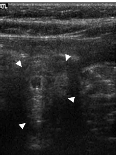
이상엽, 고재성. 소아에서 이소성 췌장을 동반한 위 중복낭종The Korean Journal of Gastroenterology Fig. 3. Abdominal ultrasonography findings. A 2.3 cm sized
protruding mass containing fluid and mucosa (white arrowheads) was noted at anteroinferior portion of the gastric antrum.
Fig. 4. Pathologic findings (H&E). (A) A gastric duplication cyst (×100). The wall had mucus producing glands internally (right lower part), and smooth muscle externally (left upper part). (B) An ectopic pancreas composed of the pancreatic ducts and acini was seen on the one side of the duplication cyst (×200).
다. 일반화학검사에서 철포화도(iron saturation rate)는 6.0%
로 감소되어 있었다. 상부위장관내시경에서 십이지장궤양과 함께 십이지장 팽대부가 좁아져 있었다(Fig. 1). 타 병원에서 시행한 복부 전산화단층촬영을 판독한 결과 위장이 팽만되어 있었고 십이지장 팽대부 벽면이 국소적으로 비대되어 좁아져 있으며 쓸개의 뒤쪽에 관형의 낭성 병변(tubular cystic le- sion)이 관찰되었다(Fig. 2). 복부 초음파검사에서 위의 전면 하부에 2.3 cm 크기의 내부에 액체가 차있는 종괴가 관찰되 었다(Fig. 3). 종괴를 제거하기 위해서 날문부절제술(antrec- tomy)을 시행하였다. 병리검사 결과에서 종괴의 크기는
4.8×4.0×2.2 cm였으며 회백색으로서 점막하층에서 장막하 층까지 위치해 있었다. 내부에 일부 낭성변화가 나타나며 낭 내부에 점액성 물질이 들어 있었다. 종괴는 위에 부착되어 있 었고 낭벽의 내벽이 점액 분비샘들로 내접해 있고 바깥으로는 평활근층이 싸고 있는 중복낭종의 소견을 보였다. 종괴의 점 막하층에서 장막하층에 걸쳐서 췌관(pancreatic duct)과 선 포(acinus)가 갖추어져 있는 이소성 췌장이 관찰되었다(Fig.
4). 수술 후 복통은 재발하지 않았고, 2개월 후 시행한 일반혈 액검사에서 혈색소는 11.7 g/dL으로 상승하였다.
진단: 이소성 췌장을 동반한 위 중복낭종
위 중복낭종(gastric duplication cyst)은 위장관 중복낭종 의 일종인 선천성 기형이다.1 중복낭종은 회장 말단에서 발견 되는 경우가 가장 많고 위에서 발견되는 경우는 드물다. 평활 근과 소화기 상피세포로 싸여 있으며 낭종의 벽면이 위벽과 부착되어 있다.2 위 중복낭종의 내부는 점막 상피세포에서 분 비하는 투명한 점액상 액체로 차 있으며, 액체의 분비량에 따 라 낭종의 크기가 다양하게 나타난다. 여성에서 나타나는 경 우가 남성에 비해 더 많고3 주로 생후 1년 이내에 발견된다.4 증상은 복통, 복부 종괴, 장의 폐색, 위장관 출혈로 나타날 수 있다.5,6 복부 전산화단층촬영에서 위 중복낭종은 조영증강이 되지 않는 낭성 종양으로 나타난다. 초음파에서는 안쪽의 고 에코성 층과 바깥의 저에코성 층으로 이루어진 이중 벽 징후 (double wall sign)가 특징적이다. 이소성 췌장(ectopic pan- creas)은 췌장 조직이 췌장 본체와는 해부학적으로나 혈역학 적으로 연속성이 없이 비정상적인 위치에 있는 것을 일컫는 다. 위장관계의 어느 부위에서나 발생할 수 있으나 위에서 가 장 많이 발생하며 소화관 밖에서 나타나는 경우도 있다.
Lee SY and Ko JS. Gastric Duplication Cyst with Ectopic Pancreas in a Child
393
Vol. 60 No. 6, December 2012
30-50세에 증상이 나타나는 경우가 많으며 남성에서 더 많이 발견된다. 소아에서 이소성 췌장으로 인해 증상이 나타나는 경우는 드물다.7 증상으로는 심와부 동통, 소화불량, 토혈이 나타날 수 있다.8,9 종괴효과에 의해 유문부 폐색, 총담관 폐 색, 장중첩증 등이 나타나는 경우도 보고되어 있다.8-10 전형적 인 이소성 췌장은 점막하 종양의 형태로 제형 함요를 나타낸 다.
위 중복낭종과 이소성 췌장은 그 양상에 따라 보존적인 치 료와 수술적인 치료가 가능하다. 위 중복낭종의 경우 증상이 없는 교통성 중복낭종은 수술을 하지 않을 수 있으나 비교통 성 중복낭종이나 증상이 있는 교통성 중복낭종은 수술을 통해 제거하는 것이 원칙이다.11 이소성 췌장의 경우 증상이 없으면 경과 관찰하지만 증상이 있으면 수술한다.12 이번 증례는 위 중복낭종이 커지면서 십이지장의 부분 폐색과 십이지장 궤양 이 발생하여 4세에 증상이 나타난 것으로 보인다. 소아의 십 이지장 궤양은 H. pylori 감염, 약물, 스트레스 등에 의해 발생 한다.13 중복낭종에 이소성 췌장이 동반된 경우는 드물고, 이 소성 췌장은 병리검사를 통해서 확인되기 때문에 수술 전에는 이소성 췌장의 존재를 모르는 경우가 많다.14,15
이번 증례는 이소성 췌장을 동반한 위 중복낭종이 십이지 장 궤양을 유발한 경우로 절제수술 이후 병리검사를 통해서 확진할 수 있었다.
REFERENCES
1. Taft DA, Hairston JT. Duplication of the alimentary tract. Am Surg 1976;42:455-462.
2. Rowling JT. Some observations on gastric cysts. Br J Surg 1959;46:441-445.
3. Kremer RM, Lepoff RB, Izant RJ Jr. Duplication of the stomach.
J Pediatr Surg 1970;5:360-364.
4. Pruksapong C, Donovan RJ, Pinit A, Heldrich FJ. Gastric duplication. J Pediatr Surg 1979;14:83-85.
5. Kim DH, Park W, Kim JY, et al. A case of gastric duplication cyst.
Korean J Gastrointest Endosc 2006;33:42-45.
6. Soper RT, Selke AC. Duplication cyst of the duodenum: case re- port and discussion. Surgery 1970;68:562-566.
7. Christodoulidis G, Zacharoulis D, Barbanis S, Katsogridakis E, Hatzitheofilou K. Heterotopic pancreas in the stomach: a case report and literature review. World J Gastroenterol 2007;13:
6098-6100.
8. Jung SH, Im EH, Kim YM, et al. A case of gastric ectopic pancreas complicated by chronic pancreatitis. Korean J Gastrointest Endosc 2006;32:409-413.
9. Park BK, Park SJ, Paik YH, et al. Endoscopic findings of ectopic pancreas in the stomach. Korean J Gastrointest Endosc 1999;
19:739-746.
10. Feldman M, Weinberg T. Aberrant pancreas: a cause of duode- nal symptom. J Am Med Assoc 1952;148:893-898.
11. Kuraoka K, Nakayama H, Kagawa T, Ichikawa T, Yasui W. Adeno- carcinoma arising from a gastric duplication cyst with invasion to the stomach: a case report with literature review. J Clin Pathol 2004;57:428-431.
12. McGarity WC, Perry CW 3rd. Complications of gastric heterotopic pancreas: two case reports. Am Surg 1971;37:77-79.
13. Lee NM, Yun SW, Chae SA, Yoo BH, Cha SJ, Kwak BK. Perforated duodenal ulcer presenting with massive hematochezia in a 30-month-old child. World J Gastroenterol 2009;15:4853- 4855.
14. Camoglio FS, Forestieri C, Zanatta C, et al. Complete pancreatic ectopia in a gastric duplication cyst: a case report and review of the literature. Eur J Pediatr Surg 2004;14:60-62.
15. Blais C, Massé S. Preoperative ultrasound diagnosis of a gastric duplication cyst with ectopic pancreas in a child. J Pediatr Surg 1995;30:1384-1386.