서론
치주질환에 이환된 치열에서 치아의 예후를 판단하고 합리 적인 치료계획을 수립하기 위해서는 개개 치아의 예후에 영향 을 미치는 요소들을 정확하게 평가해야 한다. 부착수준은 치 주질환의 연구에서 가장 흔히 사용되는 변수이고 또한 치주질
환에 의한 치아 상실과도 밀접한 관계를 가지고 있다1). 부착수준을 평가하는 표준적인 방법으로 방사선 사진 촬 영과 치주탐침이 사용되어 왔다. 그러나 부착수준 평가 시 치주탐침을 사용하여 부착수준을 측정하는 방법의 한계는 잘 알려져 있다. Magnusson과 Listgarten2)은 탐침을 이용 하여 측정한 부착수준은 치은지수에 따라서 접합상피의 근 단측 말단부가 과대평가 되거나 과소평가 될 수 있다고 하 였다. Hujoel 등3)은 탐침을 이용한 일차원적인 측정값으로 이차원적인 임상정보를 평가하는 과정에서 오류가 발생할 수 있다는 것을 제시하였다. 탐침을 이용한 수직적 부착상 실 측정은 부착상실부위의 폭경에 관한 정보를 제공하지 않
중증 치주염에 의해 발거된 치아의 수평부착상실에 대한 연구
김진숙, 김성조, 최점일, 이주연*
부산대학교 치의학전문대학원 치주과학교실
Horizontal attachment loss in extracted teeth due to severe periodontitis Jin-Suk Kim, Seong-Jo Kim, Jeom-Il Choi, Ju-Youn Lee*
Department of Periodontology, School of Dentistry, Pusan National University
ABSTRACT
Purpose: The attachment level is strongly associated with tooth loss and provides useful information on patterns of destruction of the periodontium. The presence of horizontal attachment loss would not be detected in clinical measurement. Therefore, the purpose of the present study was to estimate the patterns of periodontal destruction based on the attachemnt area and horizontal attachment loss in extracted teeth due to severe periodontitis.
Materials and Methods: 307 teeth satisfied the criteria for assessment. An indirect method, based on digital images obtained from a digital camera and an image analysis program, was used to calculate the area of root surface and attachment loss and the extent of horizontal attachment loss. The data were analysed using SPSS.
Results: No statistically significant differences among root surfaces were observed in anterior teeth on the loss of attachment area. However, in posterior teeth statistically significant differences in palatal surfaces of maxillary and mandibular premolar and molar surfaces compared with buccal surfaces were observed. Horizontal attachment loss was observed in 21.5% of the teeth examined. Frequency of horizontal attachment loss was highest in the maxillary first premolar (34.8%), followed by the maxillary second premolar (27.3%) and maxillary canine (25%). The mean length of horizontal attachment loss was 1.5mm.
Conclusion: More meticulous examination will be needed of the palatal surfaces of maxillary and mandibular premolar and molar teeth. The percentage of teeth with horizontal attachment loss greater than 2.1 mm was 5.2%. Considering the length of curette blades, about 5.2% of teeth were not properly debrided. Therefore, Additional supportive therapy such as local drug delivery has to be considered in treatment of the first maxillary, second premolar and canine due to the high prevalence of horizontal attachment loss. (J Korean Acad Periodontol 2008;38:15-22)
KEY WORDS: horizontal attachment loss; severe periodontitis; attachment level; extracted teeth.
Correspondence: Dr. Ju-Youn Lee
Department of Periodontology, School of Dentistry, Pusan National University, 1-10 Ami-dong, Seo-gu, Pusan, 602-739, Korea e-mail: [email protected], Tel: 82-51-240-7946, Fax: 82-51-243-7606
*본 연구는 2006년도 부산대학병원 임상연구비 지원으로 이루어졌음.
접수일: 2008년 1월 16일; 채택일: 2008년 2월 1일
고, 다근치와 같이 해부학적으로 복잡한 치아에서는 계측이 부정확하며, 치근의 만곡과 같은 해부학적인 형태가 반영되 지 않기 때문에 치주부착을 과소평가할 수 있다고 하였다.
또한 몇몇 연구에서는 치근이 치근단측으로 좁아지는 형태 로 인해 유사한 양의 수직부착상실이 있을 때 치근의 치관 측 1/3보다 치근측 1/3에서의 상실된 부착면적이 적다고 하 였다4,5). 더불어 Klock 등6)은 일차원적인 측정 방법은 골내 낭을 간과할 수 있지만, 부착면적의 측정은 그렇지 않다는 것을 제시하였다.
이러한 일차원적인 부착수준 계측의 문제점을 개선하기 위한 여러 시도가 있어왔다. Brown7)과 Jepsen8)은 latex solution 을 이용해서 치근면적을 계산하였고 Verdonschot 등9)은 치근 의 면적을 측정하기 위하여 digital image processing tech- nique을 사용하였다. Waerhaug10)은 염색된 발거치가 부착수 준 측정에 매우 적당하다고 하였다. Saglie 등11)과 Powell과 Garnick12)은 발거치를 이용한 계측값이 조직절편을 이용하여 얻어진 계측값과 거의 동일하며, 탐침이 결합조직 부착을 관통 할 가능성이 없기 때문에 임상적인 측정값보다 더 정확하다고 하였다.
이전의 부착수준에 대한 많은 연구들은 대부분 수직적 요소 를 평가하고 있다13-17). 하지만 질환의 진행과정 중 상당비율의 수평부착상실이 Yamamoto 등18)에 의해 보고되었다. 수평부착 상실은 진료실에서 측정하는 것이 현실적으로 불가능하고 설사 존재하더라도 형태학적인 장애요인은 기구를 이용한 치주처치 를 힘들게 한다. 존재하는 수평부착상실 부위는 적절한 처치가 이루어지지 못한 경우 지속적인 질환진행의 잠재적 요소가 될 수 있다. 이에 본 연구는 중증의 치주질환에 이환되어 발거된 치아의 수직적 및 수평적 부착상실의 양상을 조사하여 차후 치 주 질환 진행의 잠재적 위험요소가 될 수 있는 부위의 예측과 적절한 처치를 위한 근거를 제시할 목적으로 시행하였다.
재료 및 방법
2006년과 2007년 사이에 부산대학교 병원 치주과에서 ʻ가망없음ʼ으로 진단받고 발거된 영구치가 수집되었다. 백악 법랑경계를 확인할 수 없는 치아는 연구에서 제외하였다.
즉, 백악법랑경계를 넘어서 치근면으로 연장된 우식증이나 수복물, 파절을 가진 치아는 연구 대상에 포함하지 않았다.
또한 발치과정에서 치근면이 손상된 치아와 교정적, 근관치
료적 그리고 보철적 문제와 관련되어 발치한 치아도 연구에 서 제외하였다. 이러한 기준을 만족시키는 총 307개의 치아 가 연구에 이용되었다.
발거된 치아는 Waerhaug19)이 기술한 방법으로 염색되었으 며 염색시료로서 erythrosine 용액을 사용하였다. 치아를 5분 동안 erythrosine 용액(trace®, Lorvic Co., St. Louis, MO, USA)에 담가 치주부착부위를 염색하였다. 그 후 흐르는 물에 15분정도 세척하고, 공기 중에서 건조하였다. 백악법랑경계를 노출시키고 측정을 용이하게 하기 위하여, 초음파 스케일러를 이용하여 치태와 치석을 제거하였다. 그 후 디지털 카메라(EOS 350D, Canon Co., Japan)로 각 치아의 협면, 설면/구개면, 근심면, 원심면을 촬영하였다. 치아의 장축이 지표면과 평행하 도록 위치시켰으며, 촬영되는 치근면과 카메라의 렌즈가 수직이 되도록 하였다. 치아의 디지털 영상을 컴퓨터로 이동하였다. 컴 퓨터 화면상에서 디지털 영상은 확대되었고 영상 분석 소프트 웨어 프로그램(AxioVision, Carl Zeiss Co., UK)을 이용하여 치근 면적, 부착상실 면적 및 수평부착상실을 측정하였다.
1. 치근 면적과 부착상실 면적 측정
치아별 실제 치근면적, 부착상실면적, 수평부착상실을 측정 하기 위하여 치아와 동일한 조건하에서 촬영된 자를 영상 분석 소프트웨어 프로그램 상에서 calibration을 시행하여 디지털 영 상에서의 면적과 길이를 실제 면적과 길이로 변환할 수 있도록 하였다. 각각의 치근면에 대하여 치근면적 및 부착상실면적이 측정되었다. 각 치아의 치근면적은 4면에서 측정된 치근면의 합으로 하였다. 치근면적에 대한 부착상실면적의 비율로서 부착 상실률을 계산하였다.
부착상실률 = 부착상실면적/치근면적 × 100
2. 수평부착상실 측정
수평부착상실이 있는 치아에서는 치아장축에 평행한 선분과 치관측 최대수평풍융점이 만나는 접점과 치아장축에 평행한 선 분과 최대수평함몰점이 만나는 접점간의 수평 거리를 측정 하였다(Fig.1). 치아당 2개 이상의 치면에서 수평부착상실이 관찰되는 경우 그 평균값을 치아의 수평부착상실량으로 하 였다. 치아유형에 따른 수평부착상실빈도, 치면에 따른 수 평부착상실빈도 및 수평부착상실량이 측정되었다.
Figure 1. Method for measuring horizontal attachment loss.
Horizontal attachment loss was assessed by measuring the dis- tance(a) between the tangent of the most convex point at the co- ronal side and the tangent of the most concave point of the stained periodontal membrane.
3. 통계 과정
각 측정항목 당 독립적인 반복 측정 후, 두 측정값의 평균치 를 측정값으로 결정하였다. 통계처리는 SPSS(version 12k for windows, SPSS Inc., Chicago, USA)를 이용하여 시행하였 다. 각 치근면(협면, 설면, 근심면, 원심면)에 대한 평균과 표준 편차가 계산되었다. 치근면간 부착상실률의 차이를 통계적으로 규명하기 위해서 일원배치 분산분석(ANOVA one way)을 이용 하였다. 유의수준은 95% 신뢰구간을 설정하여 유의성을 검정하 였다. 또한 수평부착상실의 빈도가 치아 별, 치면 별로 계산되 었고 수평부착상실량의 평균이 계산되었다.
결과
치아 유형별로 조사된 치아 수 및 측정된 평균 치근면적이 Table 1에 제시되었다. 본 연구에서 발거된 치아의 평균 부착 상실률은 총 72.2±14.7%였다. 각 치아유형과 치근면에 따른 평균부착상실률이 Table 2에 나타나있다. 상악의 경우 상악 제 2소구치를 제외한 소구치와 대구치의 구개면 부착상실률이 협 면에 비해 크게 나타났다. 하악의 경우도 소구치와 대구치의 설 면 부착상실률이 협면에 비해 크게 나타났다.
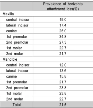
각 치아별 수평부착상실빈도가 Table 3에 제시되었다. 수평 부착상실은 조사된 치아의 21.5%에서 관찰되었다. 수평부착 상실의 빈도는 상악 제1소구치(34.8%)에서 가장 높았으며, 그 다음으로 상악 제2소구치(27.3%), 상악 견치(25%) 순이 었다. 수평부착상실량의 분포가 Fig. 2에 제시되었다. 수평 부착상실의 평균은 1.5 mm였으며 2.1 mm 이상의 수평부착 상실빈도는 5.2%였다. 각 치면별 수평부착상실의 빈도와 양 이 Table 4와 Table 5에 나타나 있다. 수평부착상실은 협설 면에서보다 근원심면에서 높은 빈도로 나타났으며, 근원심 의 수평부착상실량은 협면에서보다 크게 나타났다.
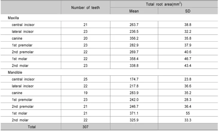
Table 1. Number of teeth and total root area by tooth type
Number of teeth Total root area(mm2)
Mean SD
Maxilla
central incisor 21 263.7 38.8
lateral incisor 23 236.5 32.2
canine 20 356.2 35.8
1st premolar 23 282.9 37.9
2nd premolar 22 269.7 40.6
1st molar 22 358.4 46.7
2nd molar 23 338.8 43.4
Mandible
central incisor 25 174.7 23.8
lateral incisor 22 217.8 36.6
canine 19 283.9 35.2
1st premolar 23 242.0 28.3
2nd premolar 21 246.7 36.4
1st molar 21 371.1 55
2nd molar 22 325.9 33.3
Total 307
SD: standard deviation
Table 3. Prevalence of horizontal attachment loss by tooth type
Prevalence of horizonta attachment loss(%) Maxilla
central incisor 19.0
lateral incisor 17.4
canine 25.0
1st premolar 34.8
2nd premolar 27.3
1st molar 22.7
2nd molar 21.7
Mandible
central incisor 12.0
lateral incisor 13.6
canine 15.8
1st premolar 21.7
2nd premolar 23.8
1st molar 23.8
2nd molar 22.7
Total 21.5
Figure 2. Distribution of horizontal attachment loss. Percentage of horizontal attachment loss of ≥0.1 mm was 21.5% in all ex- tracted teeth examined. Frequency of horizontal attachment loss of ≥2.1 mm was 5.2%.
0.0 1.0 2.0 3.0 4.0 5.0 6.0 7.0
0.1-0.5 0.6-1.0 1.1-1.5 1.6-2.0 2.1-2.5 2.6-3.0 3.1-3.5 3.6-4.0 4.1-4.5 4.6-5.0 Horiz ontal attachment loss(mm)
%
Table 2. Mean rate of attachment area loss by tooth type and surface
Rate of attachment area loss (%)✝
Buccal Lingual(Palatal) Mesial Distal Total
Maxilla
central incisor 60.8(19.3) 71.7(14.9) 61.0(18.6) 67.7(17.2) 65.3(17.9)
lateral incisor 60.5(16.9) 66.0(19.5) 66.1(15.7) 61.8(16.8) 63.6(16.6)
canine 70.6(15.4) 74.3(14.3) 72.6(13.3) 71.4(17.4) 72.2(15.0)
1st premolar 65.6(17.5) 79.2(15.2) 69.7(17.3) 73.7(13.7) 72.1(16.5)
*
2nd premolar 66.0(16.2) 76.8(15.5) 70.0(16.1) 72.5(17.1) 71.3(16.4)
1st molar 69.7(20.1) 84.6(15.4) 75.8(16.2) 70.0(13.8) 75.4(17.5)
* *
2nd molar 70.2(10.8) 82.4(11.8) 77.6(10.7) 73.0(13.3) 75.8(12.4)
*
Mandible
central incisor 73.8(11.4) 79.2(9.3) 74.8(10.9) 76.1(10.0) 76.0(10.5)
lateral incisor 60.5(17.0) 66.0(17.1) 66.2(15.7) 61.8(16.8) 63.6(16.6)
canine 66.9(17.8) 70.6(15.8) 71.4(14.0) 66.7(16.9) 68.9(16.0)
1st premolar 66.3(12.7) 77.9(13.6) 73.1(12.7) 73.8(13.5) 72.8(13.6)
*
2nd premolar 67.7(9.6) 79.1(11.1) 75.8(11.1) 72.7(12.7) 73.8(11.7)
*
1st molar 71.4(12.2) 82.5(8.6) 73.7(13.5) 71.7(13.0) 74.9(12.6)
* *
2nd molar 76.1(9.5) 85.2(10.7) 74.5(10.8) 75.7(10.6) 77.9(11.1)
*
* *
Total 67.9(14.9) 77.4(14.0) 72.0(14.1) 71.4(14.4) 72.2(14.7)
ANOVA one way, *: p<0.05, Scheffe’s method, ✝: means (SD)
고찰
본 연구는 발거치 상에 잔존하는 치주인대로부터 치주낭 기 저부의 2차원적 형태를 평가하고 각 치아유형별 부착상실률과 수평부착상실의 양상을 분석하였다. 치주낭 기저부의 형태에 관 한 정확한 지식은 치주탐침과 스케일러와 같은 기구의 정확한 적용에 사용될 수 있다. 부착면적 및 수평부착상실을 측정하기 위하여 디지털 카메라와 영상 분석 소프트웨어를 이용하였는데, 다른 방법과 비교하여 비교적 간단하게 2차원적인 부착상실 형 태를 측정할 수 있었다.
본 연구에서 측정한 치아유형별 평균 치근면적이 Table 1 에 제시되었다. 이 값은 22개의 연구로부터 얻어진 4,730개 의 치근면적 값을 메타분석한 Hujoel20)의 결과와 비교하였을 때 전치부에서는 다소 컸으며, 구치부에서는 다소 작았다.
전치부에서는 곡면의 치근이 4면에서 촬영되어 치근면적이 다소 중첩되어 계산되었기 때문에 면적이 크게 측정되었고, 반면 구치부에서는 디지털 영상이 치근면의 만곡 등과 같은 3차원적인 형태를 반영하지 못했기 때문에 작게 측정된 것으 로 생각된다. 또한 치근의 외면적만을 측정하고 치근분지부 및 치근내면은 측정되지 않았는데 이것이 다근치에서 면적이
작게 측정된 이유라고 생각된다. 본 연구에서 계측한 발거시 의 부착 상실률은 평균 72.2±14.7%이었다. 이러한 측정값 은 발거 시 부착상실을 50~70%라고 보고한 Splieth 등21)의 연구와 대다수의 치아가 50% 이하의 부착상실에서 발거된 Klock와 Haugejorden22)의 연구보다 더 컸다. 이것은 치주 적인 문제로 발거된 치아만을 대상으로 하였고, 환자와 술자 가 치아를 가능한 한 유지하려는 경향 때문인 것이라고 생각 된다. 치아유형과 치근면에 따른 평균부착상실률이 Table 2 에 나타나 있다. 전치부에서는 치근면간 부착상실률의 차이 는 통계적으로 나타나지 않았다. 구치부의 경우에는 상, 하 악 모두 소구치와 대구치의 설면이 협면과 비교하여 통계적 으로 유의하게 큰 부착상실률을 나타냈다. 이러한 결과는 10,212개의 발거한 치아를 대상으로 치아와 치면에 따른 부 착상실을 연구한 Yamamoto 등18)의 연구 결과와 일치하였 다. 그러나 Okamoto 등15)의 연구와는 다른 경향을 보였는 데, 본 연구에서는 설면(구개면)보다는 인접면에서 더 큰 상 실률을 나타냈다. 그 이유로 Okamoto 등15)의 연구는 평균 부착 수준이 5mm 이하인 치아를 대상으로 하여 측정한 결 과이고 본 연구는 중증 치주염으로 발거된 치아를 대상으로 했기 때문에 평균 부착 수준이 더 컸다는 것을 들 수 있다.
Table 4. Prevalence of horizontal attachment loss by tooth surface
N Prevalence (%)
Buccal 307 3.3
Lingual(Palatal) 307 3.9
Mesial 307 6.8
Distal 307 7.5
Total 1228 5.9
N: number of tooth surface
Table 5. Horizontal attachment loss by tooth surface
Horizonta attachment loss (mm) Mean ± SD
Buccal 0.6 ± 0.4 *,✝
Lingual(Palatal) 0.9 ± 0.7 Mesial 1.9 ± 1.3 *
Distal 1.8 ± 1.4 ✝ Total 1.5 ± 1.3 ANOVA one way, Scheffe's method
* : significantly different (p<0.05) between buccal and mesial
✝ : significantly different (p<0.05) between buccal and distal
이것은 Halazonetis 등23)의 연구에 의해서 설명될 수 있는데, 이 연구에서 평균 부착 수준이 6 mm 이상인 경우에는 치근 면과 치아의 종류에 관계없이 부착상실이 나타났지만, 6 mm 이하인 경우에는 인접면에서 더 큰 부착상실을 나타냈다.
수평부착상실은 조사된 치아의 21.5%에서 관찰되었다. 치 아별로는 상악 제1소구치, 상악 제2소구치, 상악 견치 순으 로 수평부착상실이 높은 빈도로 나타났다. 이러한 결과는 Yamamoto 등18)의 결과와 유사하다. 상악 소구치의 높은 수 평부착상실의 빈도는 해부학적 형태와 관련된 것으로 생각된 다. 인접면측의 치근면 구와 함몰은 상악 소구치와 하악 전 치에서 빈번히 나타나고 그러한 함몰부는 하악에서보다 상악 에서 더 넓고, 조기에 파괴적 질환진행에 노출되는 경향이 있다24). Leknes와 Selvig25)의 연구에서 이 치아들의 치근면 구와 함몰면이 있는 면의 부착상실은 없는 면보다 통계적으 로 유의하게 크며 특히 치근단측으로 갈수록 약해지는 얕은 U자형의 구를 가지는 하악 전치에서보다 치근단 끝까지 연 장되는 V자형의 구를 가지는 상악 소구치에서 그러하다고 하 였다. 치면별 수평부착상실의 빈도 및 양이 Table 4와 Table 5에 나타나있다. 근심, 원심측의 수평부착상실빈도가 협, 설 측의 수평부착상실 빈도보다 더 높았고 근원심의 수평부착상 실량은 협면에서보다 큰 것으로 나타나 여기서도 인접면 구 와 함몰이 수평부착상실에 기여할 것이라는 것을 확인할 수 있었다. 2.1 mm 이상의 수평부착상실의 빈도는 5.2%였다.
큐렛 날이 수평적으로 2 mm 이하로만 접근이 가능하다는 것 을 생각했을 때, 이러한 치아는 수평부착상실이 큐렛 날의 길이보다 커서 세심한 치근 소파술을 시행할지라도 적절히 소파되지 않을 것으로 생각된다. 수평부착상실은 진료실에서 측정하는 것이 현실적으로 불가능하다. 또한 수평부착상실이 큐렛 날의 길이보다 더 깊다면 적절히 소파하기 힘들고 그리 하여 치주질환은 건강한 상태로 회복되지 않고 질환 진행의 잠재적인 위험을 가지고 있게 된다. 그러므로 상악 제1소구 치, 상악 제2소구치, 상악 견치와 같이 수평부착상실의 빈도 가 높게 나타나는 치아에서는 기계적 처치에 부가적으로 국 소적 항생제 사용과 같은 적극적인 보조적 치주치료가 고려 되어야 할 것으로 생각된다.
참고문헌
1. Martha E, Nunn ME. Understanding the etiology of perio- dontitis: an overview of periodontal risk factors. Perio-
dontol 2000 2003;32:11-23.
2. Magnusson I, Listgarten MA. Histological evaluation of probing depth following periodontal treatment. J Clin Periodontol 1980;7:26-31.
3. Hujoel P, Bollen AM, DeRouen TA. Quantification of pe- riodontal attachment at single-rooted teeth. J Clin Perio- dontol 1989;16:224-228.
4. Jeffcoat MK, Williams RC. Relationship between linear and area measurements of radiographic bone levels utilizing simple computerized techniques. J Periodontal Res 1984;19:
191-198.
5. Hujoel P, Bollen AM, DeRouen TA. Quantification of pe- riodontal attachment at multi-rooted teeth. J Clin perio- dontol 1992;19:193-196.
6. Klock KS, Gjerdet NR, Haugejorden O. Periodontal attach- ment loss assessed by linear and area measurements in vitro. J Clin Periodontol 1993;20:443-447.
7. Brown R. A method of measurement of root area. J Can Dent Assoc 1950;16:130-132.
8. Jepsen A. Root surface measurement and a method for x-ray determination of root surface area. Acta Odontol Scand 1963;21:35-46.
9. Verdonschot EH, Sanders AJ, Plasschaert AJ. A com- puter-aided image analysis system for area measurement of tooth root surfaces. J Periodontol 1990;61:275-280.
10. Waerhaug J. A method for evaluation of periodontal prob- lem on extracted teeth. J Clin Periodontol 1975;2:160-168.
11. Saglie R, Johansen JR, Fløtra L. The zone of completely and partially destructed periodontal fibers in pathological pockets. J Clin Periodontol 1975;2:198-202.
12. Powell B, Garnick JJ. The use of extracted teeth to eval- uate clinical measurements of periodontal disease. J Perio- dontol 1978;49:621-624.
13. Anerud KE, Robertson PB, Loe H et al. Periodontal dis- ease in three young adult populations. J Periodontal Res 1983;18:655-668.
14. Baelum V, Fejerskov O, Manji F. Periodontal disease in adult kenyan. J Clin Periodontol 1988;15:445-452.
15. Okamato H, Yoneyama T, Lindhe J, Haffajee A, Socransky S. Methods of evaluating periodontal disease data in epi- demiological research. J Clin Periodontol 1988;15:430-439.
16. Papapanou PN, Wennstrom JL, Grondahl K. Periodontal status in relation to age and tooth type. A cross-sectional radiographic study. J Clin Periodontol 1988;15:469-478.
17. Yoneyama T, Okamoto H, Lindhe J, Sokransky SS, Haffajee AD. Probing depth, attachment loss and gingival recession. Finding from a clinical examination in Ushiku, Japan. J Clin Periodontol 1988;15:581-591.
18. Yamamoto T, Ishikawa A, Watanabe Y et al. Prevalence of horizontal attachment loss in extracted teeth. J Clin Periodontol 2004;31:791-795.
19. Waerhaug J. Healing of the dento-epithelial junction fol- lowing subgingival plaque control Ⅱ: as observed on ex- tracted teeth. J Periodontol 1978;49:119-134.
20. Hujoel PP. A meta-analysis of normal ranges for root sur- face areas of the permanent dentition. J Clin Periodontol 1994;21:225-229.
21. Splieth C, Giesenberg J, Fanghanel J. Periodontal attach-
ment level of extractions presumably performed for perio- dontal reasons. J Clin Periodontol 2002;29:514-518.
22. Klock KS, Haugejorden O. In vitro determination of the forceps level for extraction of teeth for periodontal reasons.
J Clin Periodontol 1993;20:155-160.
23. Halazonetis TD, Haffajee AD, Socransky SS. Relationship of clinical parameters to attachment loss in subsets of sub- jects with destructive peridontal diseases. J Clin Periodontol 1989;16:563-568.
24. Fox SC, Bosworth BL. A morphological survey of prox- imal root concavities: A consideration in periodontal therapy. J Am Dent Assoc 1987; 114:811-814.
25. Leknes KN, Selvig KA. Root groove: a risk factor in pe- riodontal attachment loss. J Periodontol 1994;65:859-863.