설편평상피암에 있어서의 고밀도 SNP Genotyping 어레이를 이용한 전게놈북제수와 헤테로접합성 소실의 분석
黑岩 司·山本信治·恩田健志·別所央城·藥師寺 孝·片倉 朗·野伸夫·柴原孝彦 동경치과대학 구강외과학강좌
Abstract(J Korean Assoc Oral Maxillofac Surg 2011;37:550-5)
Analysis of copy number abnormality (CNA) and loss of heterozygosity (LOH) in the whole genome using single nucleotide polymorphism (SNP)
genotyping arrays in tongue squamous cell carcinoma
Kuroiwa Tsukasa, Yamamoto Nobuharu, Onda Takeshi, Bessyo Hiroki, Yakushiji Takashi, Katakura Akira, Takano Nobuo, Shibahara Takahiko
Department of Oral and Maxillofacial Surgery, Tokyo Dental College
Chromosomal loss of heterozygosity (LOH) is a common mechanism for the inactivation of tumor suppressor genes in human epithelial cancers.
LOH patterns can be generated through allelotyping using polymorphic microsatellite markers; however, owing to the limited number of available microsatellite markers and the requirement for large amounts of DNA, only a modest number of microsatellite markers can be screened. Hybridization to single nucleotide polymorphism (SNP) arrays using Affymetarix GeneChip Mapping 10 K 2.0 Array is an efficient method to detect genome-wide cancer LOH. We determined the presence of LOH in oral SCCs using these arrays. DNA was extracted from tissue samples obtained from 10 patients with tongue SCCs who presented at the Hospital of Tokyo Dental College. We examined the presence of LOH in 3 of the 10 patients using these arrays.
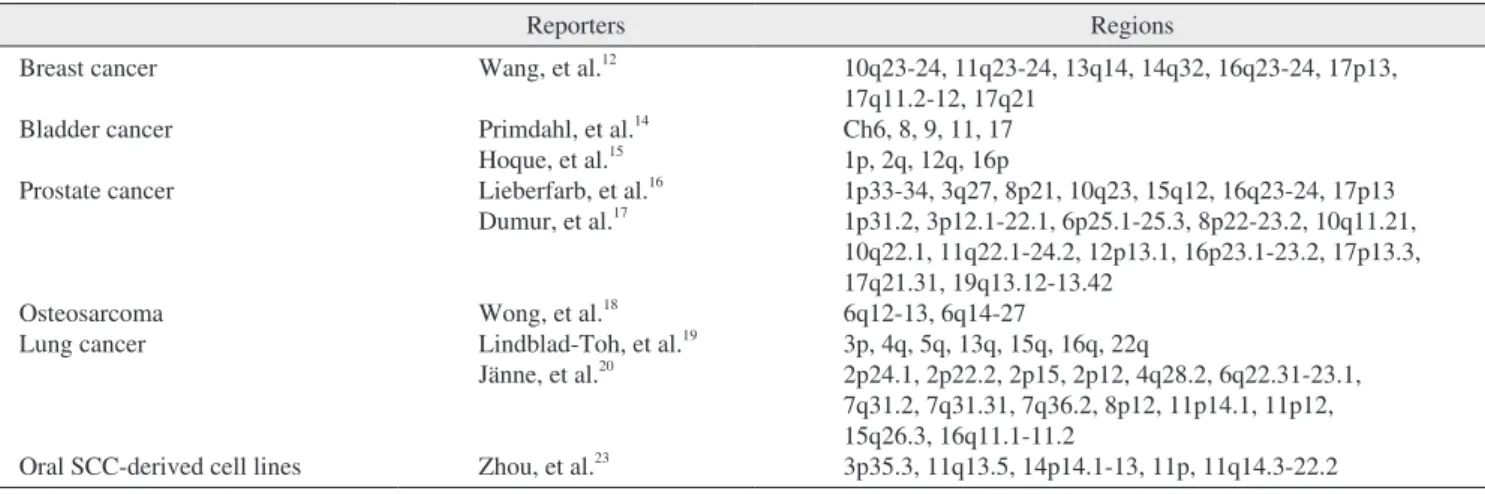
At the locus that had LOH, we examined the presence of LOH using microsatellite markers. LOH analysis using Affymetarix GeneChip Mapping 10K Array showed LOH in all patients at the 1q31.1. The LOH regions were detected and demarcated by the copy number 1 with the series of three SNP probes. LOH analysis of 1q31.1 using microsatellite markers (D1S1189, D1S2151, D1S2595) showed LOH in all 10 patients (100). Our data may suggest that a putative tumor suppressor gene is located at the 1q31.1 region. Inactivation of such a gene may play a role in tongue tumorigenesis.
Key words: Tongue squamous cell carcinoma, Loss of heterozygosity, Copy number abnormality, Single nucleotide polymorphism, tumor suppressor gene
역에 대한
comparative genomic hybridization (CGH)
에한 정되었기때문에작은결실이나증폭의검출은어려운실정이었다
. LOH
해석또한비교적소수의마이크로새틀라이트마커의제노타이핑에한정되어있었다
.
최근대규모 단일염기다형
(single nucleotide poly mor-
phism, SNP)
타이핑용으로개발된고밀도올리고뉴클레오티드
(GeneChip Mapping 10 K Array, Affymetrix)
가개발되 어암세포게놈에발생하는다양한변화를총체적으로해 석가능한어레이로서주목받고있다1.
우리는지금까지구 강편평표피암의발생과정에대해제2
번2-4, 3
번5,6, 21
번7,8염 색체상의결실상황을LOH
법이용을통해대립유전자불 균형의해석을실행해왔다.
그결과40
증례의구강편평표 피암환자로부터2 q
에서2
개소, 3 p
에서3
개소, 21 q
에서4
개소에공통결실영역의동정을 명확히함으로써 이러한 염색체좌위에는이종양의발생에관여하는중요한암억I. 제 언
발암의탐색은게놈복제수의이상
(copy number abnor- mality, CNA)
이나헤테로접합성소실(loss of heterozy gosity,
LOH)
등세포자체가억제불능상태로무질서하게증식하는원인인유전자변화의검출로부터시작된다
.
대부분의 암의경우,
이러한변화는암화를 촉진하는암유전자의활 성화혹은암억제유전자의비활성화에 의해서발생한다.
이러한유전자의복제수측정법은지금까지비교적큰영柴 原 孝 彦
동경치과대학 구강외과학강좌 Shibahara Takahiko
Department of Oral and Maxillo-Facial Surgery, Tokyo Dental Colledge 5-2- 14-605 Inagekaigan, Mihama-ku, Chiba 261-0005, Japan
TEL: +81-43-247-1902 FAX: +81-43-247-1902 E-mail: [email protected]
*본논문은일본구강외과학회지 2008년 54권 5호에게재되었으며한일논문교류목적으로이차출판하기로상호간허용되었음.
proteinase K (100 μg/mL)
를30 μL
더해교반·
분해하여50
oC
의온욕조에서 하룻밤소화하였다.
페놀·
클로로포름추출 법에의해추출·
정제한후에탄올침전법으로세정·
농축하 였다.
이에대응하는정상
DNA
는말초정맥혈로부터채취한 후, Dr. GenTLE (TakaraBIO Inc., Shiga, Japan)
를이용하여DNA
를 추출하였다.
추출된DNA
의 농도를분광광도계(spectorophotometer)
로측정하여각DNA
샘플을50 ng/μL
의농도로조정하여-80
oC
에서동결보존하였다.
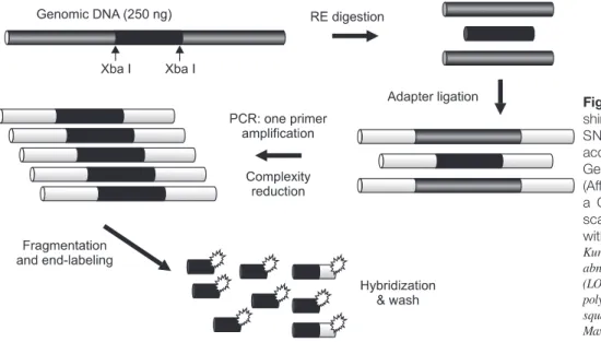
1. GeneChip Mapping 10 K Array를 이용한 DNA Mapping array해석
설편평상피암
10
증례 중3
증례(
증례1, 2, 3)
에 대해GeneChip Mapping 10 K Array
를 이용한DNA Mapping array
해석을실시하였다. SNP Genotyping
어레이를이용한 게놈복제수의원리와개략을Fig. 1
에제시한다.
Affymetrix Single Primer Assay
프로토콜에따라전게 놈DNA (250 ng)
를제한효소Xba I (New England Biolabs) 10 unit
로37
oC, 2
시간소화하였다.
그다음70
oC
로20
분간 가열처리한 후,
소화산물을standard ligation buffer (New England Biolabs)
에서아답터(0.25 μM)
를DNAligase (New England Biolabs)
에의한라이게이션반응을실행하였다.
반응후의 샘플에대해70
oC, 20
분 간의조건으로 열실활 처리한후,
라이게이션반응산물을주형으로한PCR
반응 으로타겟DNA
의증폭을실행하였다.
반응액은0.75 µM
의 프라이머, 2.5 mM MgCl
2·250 μM dNTP·10 unit AmpliTag Gold (Applied Biosystems)
으로조성하였다. PCR
의조건 은 최초95
oC, 3
분간의열변성을실시한후, 95
oC 30
초간, 59
oC 30
초간, 72
oC 30
초간을35
사이클반복한후,
마지막 에72
oC, 7
분간의조건으로신장 반응을실행하였다. PCR
증폭반응액을2%TBE
아가로스겔상에서120 V, 1
시간 의 조건으로전기영동하였다.
그리고샘플을0.24 unit
의Fragmentation Reagent (Affymetrix)
으로37
oC, 30
분간소화 제유전자가존재하고있음을시사하였다.
GeneChip Mapping 10K Array
를이용한DNA mapping array
해석은유방암9-13,
방광암14,15,
전립선암16,17,
골육종18,
폐암10,19,20등에그유용성이 보고되었지만구강암을 대상
으로한연구는거의이뤄지지않았다
.
이런연유로이연구 는GeneChip Mapping 10K Array
를이용하여구강암에서 빈도가높은설편평상피암을대상으로전게놈상의CNA
와LOH
에대하여검토하였다.
II. 대상증례 및 연구방법
2004
년부터2006
년까지2
년간동경치과대학치바병원구 강외과의검진을통해설편평상피암으로진단받은28
명중,
시료채취가가능한10
명의환자를대상으로하였다.(Table
1)
또한이번연구는본대학의윤리위원회의승인을받은후
,
모든증례에환자본인 및환자가족을대상으로동의 를받아실행하였다.
검체는수술시(
모든증례에서화학요 법·
방사선요법등의수술전치료는실시하지않았다)
또는 생검시에채취한원발종양조직10
검체와대응하는정상DNA
로동일환자의말초정맥혈(
약10 mL)
을이용하였다.
절제조직을2
개로나누어,
한쪽은종양조직으로부터정상조직을신중히절제한후즉시동결하여
DNA
추출까지-80
oC
에보존하였다.
다른한편은10%
포르말린으로고정 하여병리조직진단을실시하였다.
병리조직학적진단은 국제종양분류에준하여동경치과대학병리학교실이 실시 하였다21.
임상병리학적stage
평가는UICC TNM Stage
평 가시스템을기준으로하였다22.
DNA
의추출법병리조직학검사를통해모든환자는설편평상피암인것으로확인되어
DNA
추출을위한종양검체는
80%
이상 종양으로구성된 것을 확인하였다.
채취된종양조직
,
혈액은즉시 액체질소에보존하여신선동 결표본으로가공·
조정하였다.
우선조직을액체질소하에 서분쇄한다음,
게놈DNA
를TNES buffer (10 mM Tris-HCl (pH 8.0)·150 mM NaCl·10 mM EDTA·0.1% SDS) 1,000 μL
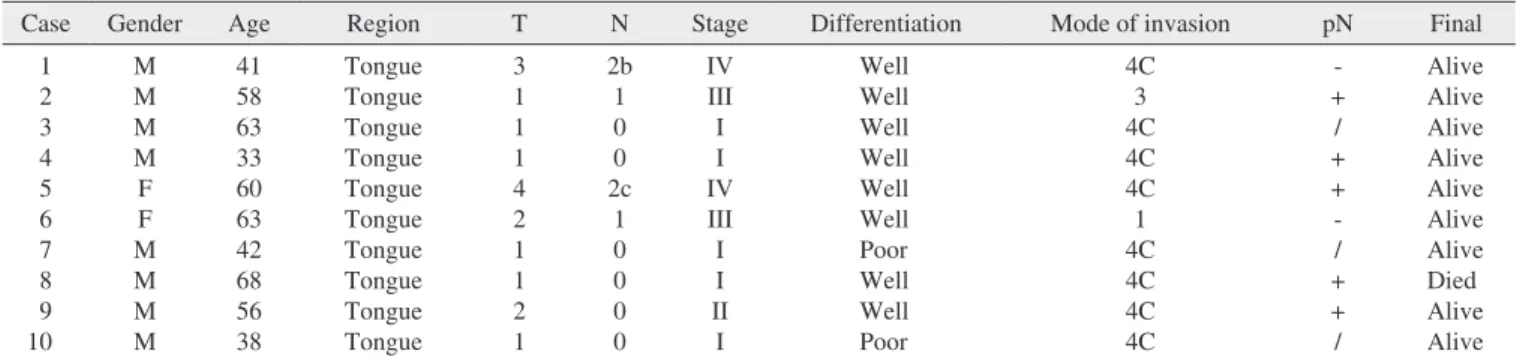
에Table 1. Summary of clinicopathological features in 10 SCCs
Case Gender Age Region T N Stage Differentiation Mode of invasion pN Final
1 2 3 4 5 6 7 8 9 10
M M M M F F M M M M
41 58 63 33 60 63 42 68 56 38
Tongue Tongue Tongue Tongue Tongue Tongue Tongue Tongue Tongue Tongue
3 1 1 1 4 2 1 1 2 1
2b 1 0 0 2c
1 0 0 0 0
IV III I I IV III I I II I
Well Well Well Well Well Well Poor Well Well Poor
4C 3 4C 4C 4C 1 4C 4C 4C 4C
- + / + + - / + + /
Alive Alive Alive Alive Alive Alive Alive Died Alive Alive (M, Male; F, Female; pN, pathological Node; Well, well differentiated; Poor, poorly differentiated)
Kuroiwa Tsukasa et al: Analysis of copy number abnormality (CNA) and loss of heterozygosity (LOH) in the whole genome using single nucleotide polymorphism (SNP) genotyping arrays in tongue squamous cell carcinoma. J Korean Assoc Oral Maxillofac Surg 2011
어레이해석을통해
CNA
로분류된마이크로새틀라이트 영역(Table 2)
을PCR-LOH
해석을이용하여10
증례전체를 검증하였다. PCR
및전기영동,
밴드염색은본강좌가이 전부터실행해온방식에준하여실시하였다4.
LOH
가인정된각증례의결과에재현성이있음을확인하기위해
,
다시한번PCR
마이크로새틀라이트분석을실 시하였다.
3. 헤테로접합성소실의 평가법
획득한마이크로새틀라이트밴드를컴퓨터상에스캔한 후
NIH image (version 1.62)
에의해그강도를수치화하였 다.
종양DNA
의시그널강도를대응되는정상DNA
와비 교하여시그널강도가50%
이하를LOH
로판정하였다.
III. 결 과
1. GeneChip Mapping 10 K Array를 이용한 DNA 매핑 어레이 해석의 평가
설편평상피암환자
10
증례중3
증례(
증례1, 2, 3)
에대 한후, 95
oC, 15
분간의조건으로열실활 처리를실행하였다
. 4%TBE
아가로스겔상에서120 V, 30
분-1
시간의 조 건으로 전기영동한 후,
하이브리다이제이션 믹스190 μ L (0.056 M MES buffer·5.0%DMSO·2.5×Denhardt’s Solution·5.77 mM EDTA·0.115 mg/mL HSDNA·1×origo- nu cleotide control [Affymetrix]·Human Cot-1 [11.5 μg/mL]·
0.0115%Tween-20·2.69 M TMACL)
으로라벨화한타겟DNA
를 혼합하여하이브리다이제이션칵테일을 조제후,
95
oC, 15
분간의조건으로열변성시켜마이크로어레이카트리지에주입
, 45
oC, 16-18
시간의조건으로인큐베이팅하 였다.
마지막으로어레이상에서10 μg/mL
의Streptavidin phycoerythrin
과반응시켜non-stringent wash buffer
로세정 한후, 5 μg/mL
의 비오틴화Streptavidin
항체와인큐베이 팅하여, non-stringent wash buffer
로세정하였다. Gene Chip Mapping 10 K 2.0 Array
에의한스캔이미지는Affymetrix Genotyping Tools software
을이용하여해석하였고23,
복제 수0.8-1.2
를LOH
영역으로판정하였다24,25.
2. PCR 및 마이크로 새틀라이트 해석법
GeneChip Mapping 10 K 2.0 Array
를이용한DNA
매핑Fig. 1. DNA labeling, hybridization, wa- shing and staining of the Affymetrix 10K SNP Mapping Array were performed according to the standard Single Primer GeneChip Mapping 10K Assay protocol (Affymetrix). The array was scanned with a GeneChip Scanner 3000, and the scanned array images were processed with GeneChip Operating software.
Kuroiwa Tsukasa et al: Analysis of copy number abnormality (CNA) and loss of heterozygosity (LOH) in the whole genome using single nucleotide polymorphism (SNP) genotyping arrays in tongue squamous cell carcinoma. J Korean Assoc Oral Maxillofac Surg 2011
Table 2. Microsatellite markers used in our LOH study
Markers Locations Size of PCR products (bp) Sequence of primers
D1S1189 D1S2151 D1S2595
1q31.1 1q31.1 1q31.1
341 260 102
5'-CTGAACTAACACGGAGAAAC-3' 5'-GTAGACTGTTAAAAGAAGAGC-3' 5'-CTGTATAAAGAGCGCTGTGGG-3' 5'-GGATGGGGCAGTGAGAAG-3' 5'-GGGCGTTCCAATACTTAGAGG-3' 5'-AGGACACCACAAGTTCCAGG-3' (LOH: loss of heterozygosity)
Kuroiwa Tsukasa et al: Analysis of copy number abnormality (CNA) and loss of heterozygosity (LOH) in the whole genome using single nucleotide polymorphism (SNP) genotyping arrays in tongue squamous cell carcinoma. J Korean Assoc Oral Maxillofac Surg 2011
PCR-LOH
법을 이용하여10
증례 전체를 검증하였다.
그 결과, 3
마커모두10
증례전체(100%)
에LOH
가인정되었 다. Fig. 3
에LOH
해석결과를표시하였다.
IV. 고 찰
본강좌에서는지금까지구강편평표피암을대상으로한
LOH
해석을실행하여구강편평표피암의 발생및 진전에 관여하는미확인암억제유전자의후보좌위의국재를규명 해왔다26.
그러나,
종래의LOH
법을이용한염색체분석은 숫자와부위의염색체이상영역의동정에한정되어전염 하여GeneChip Mapping 10 K Array
를 이용한전염색체상의
DNA
매핑어레이 해석을 실시, CNA
와LOH
의 유 무에대해 검토하였다.
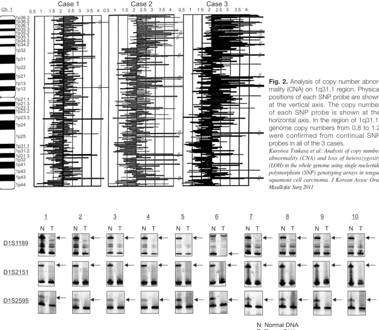
그결과, 3
증례모두1 q31. 1
영역(D1S1189, D1S2151, D1S2595)
에게놈복제수0.8-1.2
의연 속이3
개의SNP
프로브로부터인정되어CNA
와LOH
를나 타내는염색체영역이동정되었다.(Fig. 2)
2. LOH의 평가
DNA
매핑어레이해석에있어CNA
와LOH
가동정된1 q31. 1
영역(D1S1189, D1S2151, D1S2595)
에 대하여Fig. 2. Analysis of copy number abnor- mality (CNA) on 1q31.1 region. Physical positions of each SNP probe are shown at the vertical axis. The copy number of each SNP probe is shown at the horizontal axis. In the region of 1q31.1, genome copy numbers from 0.8 to 1.2 were confirmed from continual SNP probes in all of the 3 cases.
Kuroiwa Tsukasa et al: Analysis of copy number abnormality (CNA) and loss of heterozygosity (LOH) in the whole genome using single nucleotide polymorphism (SNP) genotyping arrays in tongue squamous cell carcinoma. J Korean Assoc Oral Maxillofac Surg 2011
Fig. 3. Analysis of LOH using microsatellite markers (D1S1189, D1S2151, D1S2595). Case numbers are shown at the top and locus symbols on the left. Paired normal (N) and tumor (T) samples for all of 10 patients demonstrating loss of the upper allele (LOH).
Kuroiwa Tsukasa et al: Analysis of copy number abnormality (CNA) and loss of heterozygosity (LOH) in the whole genome using single nucleotide polymorphism (SNP) genotyping arrays in tongue squamous cell carcinoma. J Korean Assoc Oral Maxillofac Surg 2011
실시함으로써설편평상피암의발생
·
진전에관여하는암관 련유전자군을 결정지으려한다.
또한증례수의증가를통해
1 q31. 1
이외영역의복제수와헤테로접합성소실의검색도가능할것으로생각된다
.
V. 결 어
설편평상피암
10
증례를대상으로3
증례(
증례1, 2, 3)
에서전게놈상의
DNA
매핑어레이해석을실시하여, 3
증례모두
1 q31. 1
영역(D1S1189, D1S2151, D1S2595)
에CNA
와LOH
가동정되었다.
실험결과를바탕으로10
증례에대해D1S1189, D1S2151, D1S2595
를PCR-LOH
법을이용하여 검증한결과, 10
증례전체에LOH
가인정되었다.
이번연구의결과
1 q31. 1
영역은설편평상피암의발생에관여하는유전자좌위임이시사되어본영역에는신규미확인암억제 유전자가존재하고있음이시사되었다
.
이논문의요지는제
51
회일본구강외과학회총회(2006
년10
월, 北九州
시)
에발표되었다.
References
1. Ogawa S. Analysis of DNA copy number. Blood Frontier 2006;16:35-42.
2. Yamamoto N, Mizoe JE, Numasawa H, Yokoe H, Uzawa K, Shibahara T, et al. Allelic loss of chromosome 2 in human oral squamous cell carcinoma: correlation with lymph node metastasis.
Oral Oncol 2003;39:64-8.
3. Numasawa H, Yamamoto N, Katakura A, Shibahara T. Loss of heterozygosity and microsatellite instability on chromosome 2q in human oral squamous cell carcinoma. Bull Tokyo Dent Coll 2005;46:17-25.
4. Kakimoto Y, Numasawa H, Yamamoto N, Takeda E, Yamauchi T, Shibahara T. Loss of heterozygosity and microsatellite instability on the long arm of chromosome 2 in human oral squamous cell carcinoma. Jap J Oral Maxillofac Surg 2005;51:374-81.
색체상을망라한해석과유전자의동정은이루어지지않았 다
.
최근개발된
GeneChip Mapping 10 K 2.0 Array
는전염색체상에서단일염기변이다형
(SNP) 10, 204
개소를제노타이핑함으로써각각의
SNP
프로브에 대응하는 게놈복제 수변화의검출이가능하게되었다.
즉,
종양게놈중의유전 자복제수의변화를1
회해석을통해전염색체영역에서검 색할수있는탁월한수단이라하겠다.
통상염색체는부모 로부터1
개씩계승하여복제수는2
가되지만,
염색체혹은 유전자의일부가결실되면복제수의감소가발생한다.
연 속된SNP
프로브에복제수1
이연속하는경우,
이영역에LOH
가존재하게된다.
이번연구는설편평상피암환자
10
증례중3
증례(
증례1, 2, 3)
를대상으로GeneChip Mapping 10 K Array
를이용한 전염색체상의DNA
매핑어레이해석을실시, 3
증례모두 에서1 q31. 1
영역(D1S1189, D1S2151, D1S2595)
에CNA
와LOH
를나타내는염색체영역을동정하였다.
또한10
증례에
PCR-LOH
해석을실시하여밴드를시그널강도로검증한결과
, 3
마커모두10
증례전체에LOH
가인정되는흥 미로운결과를얻게되었다.
기타장기의
GeneChip Mapping 10 K Array
를이용한전 염색체상의DNA
매핑어레이해석에관한지금까지의보 고를Table 3
에제시한다.
이번동정된
1 q31. 1
영역은아직어떤유전자도클로닝 되지않고있어이영역에는신규미확인암억제유전자가 존재하고있을가능성이시사되었다.
이번연구를통해설편평상피암에있어서의고밀도
SNP
Genotyping
어레이를이용한전게놈상의복제수와헤테로접합성의소실에대해해석하였다
.
향후증례수를증가시 켜임상지표와의관련및1 q31. 1
영역의상세한염색체해 석과이영역에존재하는암관련유전자의해석을아울러Table 3. Reports of copy number abnormality (CAN) using single nucleotide polymorphism (SNP)
Reporters Regions
Breast cancer Bladder cancer Prostate cancer
Osteosarcoma Lung cancer
Oral SCC-derived cell lines
Wang, et al.12 Primdahl, et al.14 Hoque, et al.15 Lieberfarb, et al.16 Dumur, et al.17
Wong, et al.18 Lindblad-Toh, et al.19 Jänne, et al.20 Zhou, et al.23
10q23-24, 11q23-24, 13q14, 14q32, 16q23-24, 17p13, 17q11.2-12, 17q21
Ch6, 8, 9, 11, 17 1p, 2q, 12q, 16p
1p33-34, 3q27, 8p21, 10q23, 15q12, 16q23-24, 17p13 1p31.2, 3p12.1-22.1, 6p25.1-25.3, 8p22-23.2, 10q11.21, 10q22.1, 11q22.1-24.2, 12p13.1, 16p23.1-23.2, 17p13.3, 17q21.31, 19q13.12-13.42
6q12-13, 6q14-27
3p, 4q, 5q, 13q, 15q, 16q, 22q
2p24.1, 2p22.2, 2p15, 2p12, 4q28.2, 6q22.31-23.1, 7q31.2, 7q31.31, 7q36.2, 8p12, 11p14.1, 11p12, 15q26.3, 16q11.1-11.2
3p35.3, 11q13.5, 14p14.1-13, 11p, 11q14.3-22.2 (Ch: Chromosome, p: pter, q: qter)
Kuroiwa Tsukasa et al: Analysis of copy number abnormality (CNA) and loss of heterozygosity (LOH) in the whole genome using single nucleotide polymorphism (SNP) genotyping arrays in tongue squamous cell carcinoma. J Korean Assoc Oral Maxillofac Surg 2011
22.
16. Lieberfarb ME, Lin M, Lechpammer M, Li C, Tanenbaum DM, Febbo PG, et al. Genome-wide loss of heterozygosity analysis from laser capture microdissected prostate cancer using single nucleotide polymorphic allele (SNP) arrays and a novel bioinformatics platform dChipSNP. Cancer Res 2003;63:4781-5.
17. Dumur CI, Dechsukhum C, Ware JL, Cofield SS, Best AM, Wilkinson DS, et al. Genome-wide detection of LOH in prostate cancer using human SNP microarray technology. Genomics 2003;81:260-9.
18. Wong KK, Tsang YT, Shen J, Cheng RS, Chang YM, Man TK, et al. Allelic imbalance analysis by high-density single-nucleotide polymorphic allele (SNP) array with whole genome amplified DNA. Nucleic Acids Res 2004;32:e69.
19. Lindblad-Toh K, Tanenbaum DM, Daly MJ, Winchester E, Lui WO, Villapakkam A, et al. Loss-of-heterozygosity analysis of small-cell lung carcinomas using single-nucleotide polymorphism arrays. Nat Biotechnol 2000;18:1001-5.
20. Jänne PA, Li C, Zhao X, Girard L, Chen TH, Minna J, et al. High- resolution single-nucleotide polymorphism array and clustering analysis of loss of heterozygosity in human lung cancer cell lines.
Oncogene 2004;23:2716-26.
21. World Health Organization. International Histological Classi- fication of Tumours. No. 4. Histological Typing of Oral and Oropharyngeal Tumours. Geneva: WHO; 1971:9-28.
22. UICC: TNM Classification of Malignant Tumours. 4th ed. Berlin:
Springer; 1987:16-18.
23. Zhou X, Mok SC, Chen Z, Li Y, Wong DT. Concurrent analysis of loss of heterozygosity (LOH) and copy number abnormality (CNA) for oral premalignancy progression using the Affymetrix 10K SNP mapping array. Hum Genet 2004;115:327-30.
24. Wang J, Meza-Zepeda LA, Kresse SH, Myklebost O.
M-CGH: analysing microarray-based CGH experiments. BMC Bioinformatics 2004;5:74.
25. Lin M, Wei LJ, Sellers WR, Lieberfarb M, Wong WH, Li C.
dChipSNP: significance curve and clustering of SNP-array-based loss-of-heterozygosity data. Bioinformatics 2004;20:1233-40.
26. Yamamoto N, Mizoe J, Numasawa H, Tsujii H, Shibahara T, Noma H. Allelic loss on chromosomes 2q, 3p and 21q: possibly a poor prognostic factor in oral squamous cell carcinoma. Oral Oncol 2003;39:796-805.
5. Arai K, Shibahara T, Yamamoto N, Yakushiji T, Tanaka C, Noma H. Frequent allelic loss/imbalance on the short arm of chromosome 3 in tongue cancer. Bull Tokyo Dent Coll 2001;42:151-7.
6. Arai K, Shibahara T, Yamamoto N, Noma H. The presence of candidate tumor suppressor gene loci at chromosome 3p for oral squamous cell carcinomas. Oral Oncol 2002;38:763-71.
7. Yamamoto N, Uzawa K, Miya T, Watanabe T, Yokoe H, Shibahara T, et al. Frequent allelic loss/imbalance on the long arm of chromosome 21 in oral cancer: evidence for three discrete tumor suppressor gene loci. Oncol Rep 1999;6:1223-7.
8. Yamamoto N, Noma H, Shibahara T. Allelic imbalance on the long arm of chromosome 21 in human oral squamous cell carcinoma:
relationship between allelic imbalances (LOH and MSI) and clinicopathologic features. Bull Tokyo Dent Coll 2001;42:211-23.
9. Huang J, Wei W, Zhang J, Liu G, Bignell GR, Stratton MR, et al. Whole genome DNA copy number changes identified by high density oligonucleotide arrays. Hum Genomics 2004;1:287-99.
10. Zhao X, Li C, Paez JG, Chin K, Jänne PA, Chen TH, et al. An integrated view of copy number and allelic alterations in the cancer genome using single nucleotide polymorphism arrays. Cancer Res 2004;64:3060-71.
11. Schubert EL, Hsu L, Cousens LA, Glogovac J, Self S, Reid BJ, et al. Single nucleotide polymorphism array analysis of flow-sorted epithelial cells from frozen versus fixed tissues for whole genome analysis of allelic loss in breast cancer. Am J Pathol 2002;160:73- 9.
12. Wang ZC, Lin M, Wei LJ, Li C, Miron A, Lodeiro G, et al. oss of heterozygosity and its correlation with expression profiles in subclasses of invasive breast cancers. Cancer Res 2004;64:64-71.
13. Paez JG, Lin M, Beroukhim R, Lee JC, Zhao X, Richter DJ, et al.
Genome coverage and sequence fidelity of phi29 polymerase-based multiple strand displacement whole genome amplification. Nucleic Acids Res 2004;32:e71.
14. Primdahl H, Wikman FP, von der Maase H, Zhou XG, Wolf H, Orntoft TF. Allelic imbalances in human bladder cancer: genome- wide detection with high-density single-nucleotide polymorphism arrays. J Natl Cancer Inst 2002;94:216-23.
15. Hoque MO, Lee CC, Cairns P, Schoenberg M, Sidransky D.
Genome-wide genetic characterization of bladder cancer: a comparison of high-density single-nucleotide polymorphism arrays and PCR-based microsatellite analysis. Cancer Res 2003;63:2216-