대한소화기학회지 2009;53:361-368 DOI: 10.4166/kjg.2009.53.6.361
접수: 2008년 11월 12일, 승인: 2009년 1월 15일 연락처: 윤승규, 137-040, 서울시 서초구 반포동 505번지
가톨릭대학교 강남성모병원 내과 Tel: (02) 590-2622, Fax: (02) 536-9559 E-mail: [email protected]
* 본 논문은 과학기술부에서 추진하는 21세기 프론티어 연 구개발사업인 인간유전체기능연구 사업단(FG08-12-05)의 연구비 지원으로 이루어졌음.
Correspondence to: Seung Kew Yoon, M.D.
Department of Internal Medicine, Kangnam St. Mary's Hos- pital, The Catholic University of Korea, 505, Banpo-dong, Seocho-gu, Seoul 137-040, Korea
Tel: +82-2-590-2622, Fax: +82-2-536-9559 E-mail: [email protected]
차별 발현 역전사중합효소연쇄반응법을 이용한 원발 간세포암종의 유전자 발현 분석
가톨릭대학교 의과대학 공동연구지원센터, WHO 협력 간염바이러스 연구소*, 내과학교실†
이영춘ㆍ허원희*ㆍ최정은*ㆍ박련숙*ㆍ홍성우*ㆍ배시현
†ㆍ최종영
†ㆍ윤승규*
†Analysis of Gene Expression in Primary Hepatocellular Carcinoma Using Differentially Displayed Reverse Transcriptase Polymerase Chain Reaction
Young Chun Lee, Wonhee Hur, Ph.D.*, Jung Eun Choi*, Lian Shu Piao*, Sung Woo Hong*, Si Hyun Bae, M.D.†, Jong Young Choi, M.D.†, and
Seung Kew Yoon, M.D.*†
Integrative Research Support Center, WHO Collaborating Center of Viral Hepatitis*, Department of Internal Medicine, College of Medicine†, The Catholic University of Korea, Seoul, Korea
Background/Aims: The investigation of a specific tumor marker for hepatocellular carcinoma (HCC) is needed to examine the carcinogenesis and to select the patients for treatment options. The aim of this study was to find the genes related to HCC. We also examined the expression level of these genes in cancer cell lines and tissue specimens. Methods: Three pairs of HCC tissue and non-neoplastic hepatic tissue around the HCC were collected from three patients who underwent resection for HCC. Differential display reverse transcriptase-PCR (DD RT-PCR) using GeneFishingTM PCR was used to detect the differences in the gene expression between in HCC tissue and non-neoplatic tissue. Up- or down-regulated genes in HCC tissue were identified through BLAST searches after cloning and sequencing assays. Real-time RT-PCR assay was employed to detect the expression rate in 11 HCC tissues and human cancer cell lines. Results: Differentially expressed 21 genes were identified, and they were classified as genes involved in protein metabolism, ubiquitin-dependent protein catabolism, carbohy- drate metabolism, lipid metabolism, DNA repair, and inflammatory response. Conclusions: We identified differ- entially expressed genes in HCC, and these genes may play an important role in the study of hepatocarcino- genesis, development of biomarker, and target therapy for HCC. (Korean J Gastroenterol 2009;53:361-368) Key Words: Hepatocellular carcinoma; DDRT-PCR; Biomarker
서 론
원발 간세포암종(hepatocellular carcinoma, HCC)은 세계적
으로 5번째로 흔한 암으로 매년 약 100만명의 환자가 발생 하고 있으며 우리나라의 경우 위암, 폐암 다음으로 많이 발 생하는 암으로 40, 50대 남자의 주요 사망원인이 되는 질환
362 대한소화기학회지: 제53권 제6호, 2009
이다. 현재까지 원발 간세포암종의 발생에는 B형 및 C형 간 염 바이러스와 알코올 그리고 환경 요인 및 유전적인 소인 등 다양한 요인이 관여하는 것으로 알려져 있다. 최근 연구 결과에 따르면 우리나라 원발 간세포암종 환자들의 74.2%
가 B형 간염 바이러스 표면 항원 양성, 8.6%가 C형 간염 바 이러스 항체 양성, 6.9%가 장기간의 알코올 음주자 그리고 10.3%가 기타의 원인으로 원발 간세포암종을 유발하는 것 으로 알려져 있다.1 최근 간세포암종에 대한 고주파 열치료 나 다양한 방사선 치료법의 개발에도 불구하고 간세포암종 의 장기 생존율은 매우 낮고 수술 후에도 재발이 많아 예후 가 매우 불량한 암으로 인정되고 있다. 하지만 간세포암종 의 진단 시기에 따라 예후가 달라지기 때문에 간세포암종의 조기 진단 바이오 마커의 개발은 임상적으로 중요한 이슈로 부상하고 있다. 최근 분자생물학적인 기법의 발달에 따라 발암기전이 점차 밝혀지고 있는데 일반적으로 발암유전자 의 활성화와 암 억제유전자의 변이가 중요한 기작으로 작용 한다고 알려져 있다. 현재까지 보고된 바에 따르면 대표적 인 암 억제유전자로는 p53, Rb 및 DLC-1 등이 밝혀졌고, 발 암유전자로는 N-ras, H-ras, K-ras, c-erbA, c-met, c-myc, cyclin-D1 등이 알려져 있으며, 세포주기조절에 관련된 유전 자들로는 p15, p16, p21, p27 등이 있으며 이는 발암의 병태 생리에 기여하는 것으로 알려지고 있다.2-4 그러나 다단계를 거쳐 발생하는 간세포암종은 분자생물학적인 수준에서 다 양한 발병기전들이 보고되고 있어 확실한 정설이 확립되어 있지 않다.5 따라서 원발 간세포암종에서 특이적으로 과발현 되는 특정 유전자를 밝혀내는 것은 간세포암종의 발암 기전 을 규명하는 데 도움을 줄 수 있을 뿐 아니라 간세포암종의 조기발견을 위한 바이오마커의 개발 및 향후 유전자 치료를 위한 기초 자료로 중요한 의의를 가질 것이라 기대된다.
이번 연구에서는 원발 간세포암종에서 차별 발현 역전사 중합효소연쇄반응6(DDRT-PCR)을 이용하여 간세포암종 조 직과 비간세포암종 조직에서 유전자 발현 양상을 비교, 분 석하여 유전자를 탐색함으로써 원발 간세포암종의 발병기 전을 이해하고 진단 및 치료를 위한 계획 수립에 도움을 주 고자 한다.
대상 및 방법
1. 대상조직
2005년 1월부터 2006년 12월까지 가톨릭대학교 의과대학 강남성모병원에 내원하여 원발 간세포암종으로 진단 받고 수술을 받은 환자 3명을 대상으로 원발 간세포암종 조직과 간세포암 조직 주위의 비간세포암종 조직 3쌍을 채취하여 분석에 사용되기 전까지 동결 보관하였다. 상기 조직은 “생
명윤리 및 안전에 관한 법률”과 Helsinki 선언에 따른 환자 의 동의를 받고 가톨릭대학교 생명윤리심의위원회의 허락 을 받아 사용하였다.
2. 차별 발현 유전자의 분리
1) RNA 추출
동결 보관되어 있던 원발 간세포암종 조직과 비간세포암 종 조직 0.1 g을 막자 사발에서 파쇄시킨 다음 trizol reagent (Invitrogen, Carlsbad, CA) 1 mL을 넣고 혼합하여 상온에서 5 분간 방치하였다. 여기에 0.2 mL의 chloroform을 첨가한 다 음 상온에서 2-3분간 방치한 후 4oC, 12,000 g에서 15분간 원심 분리하여 무색의 상층액을 새로운 tube로 옮겼다. 이 상층액에 0.5 mL의 isopropanol을 혼합하고 실온에서 10분간 방치한 다음 4oC, 12,000 g에서 10분간 원심 분리하여 RNA 를 침전시키고 상층액을 제거하였다. 침전된 RNA에 1 mL 의 75% 에탄올을 혼합한 후 4oC, 12,000 g에서 10분간 원심 분리하여 상층액을 제거하고 실온에서 10분간 건조시켰다.
건조된 RNA에 100μL의 증류수를 첨가하여 녹인 후 이 중 1μL를 취하여 spectrometer를 이용하여 흡광도를 측정하고 1% agarose gel에서 전기영동을 수행하여 RNA의 순도를 확 인하였다.
2) 역전사 반응
분리된 total RNA 3μg을 mRNA의 Poly A부위에 특이적 으로 결합하는 dT-ACP를 이용하여 42oC에서 역전사 반응을 통해 first strand cDNA를 합성하였다. 역전사 과정은 3μg의 정제된 RNA, 4μL의 5× reaction buffer (Promega, Madison, WI), 5μL의 dNTPs (각각 2.5 mM), 2μL의 10 uM dT-ACP1 (5'-CGTGAATGCTGCGACTACGATIIIIIT(18)-3'), 0.5μL RNasinⓇ RNase inhibitor (40 U/μL; Promega), 1μL의 Moloney murine leukemia virus reverse transcriptase (200 U/μL; Promega)를 함유한 20μL 용액을 42oC에서 90분 동안 반응시켜 시행하 였다. 합성된 cDNA는 180μL의 증류수로 희석한 뒤 분주하 여 −20oC에 보관하였다.
3) 차별 발현 중합효소연쇄반응
GeneFishing DEG kits (Seegene, Seoul, South Korea)를 사 용하여 ACP-based PCR 방법을 통해 발현된 유전자를 선별 하였다. 먼저 3-5μL (약 50 ng)의 희석된 첫 번째 가닥의 cDNA, 1μL의 dT-ACP2 (10 uM), 1μL의 10 uM arbitary ACP, 10μL의 2× Master Mix가 함유된 20μL 용액을 94oC 에서 1분, 50oC에서 3분, 72oC에서 1분 동안 처리하여 첫 단 계 PCR 과정을 통해 두 번째 가닥의 cDNA를 합성하였다.
두 번째 단계인 PCR 증폭은 94oC에서 40초, 65oC에서 40초, 72oC에서 40초, 72oC에서 5분을 40회 반복하여 시행하였고,
이영춘 외 7인. 원발 간세포암종의 유전자 발현 분석 363
Fig. 1. Annealing control primer (ACP)-based polymerase chain reaction (PCR) for the identification of differentially expressed genes (DEG) from tumor tissue (T) and non tumor tissues (NT).
Total RNA extracted from T and NT were used for the syn- thesis of 1st strand cDNA using dT-ACP1 primer. 2nd strand cDNAs were amplified during second-stage PCR using a combi- nation of dT-ACP2 and one of 120 arbitrary ACP primers (A).
Products were separated on an agarose gel to identify the differ- entially expressed genes that were highly expressed in T and NT tumor liver (B).
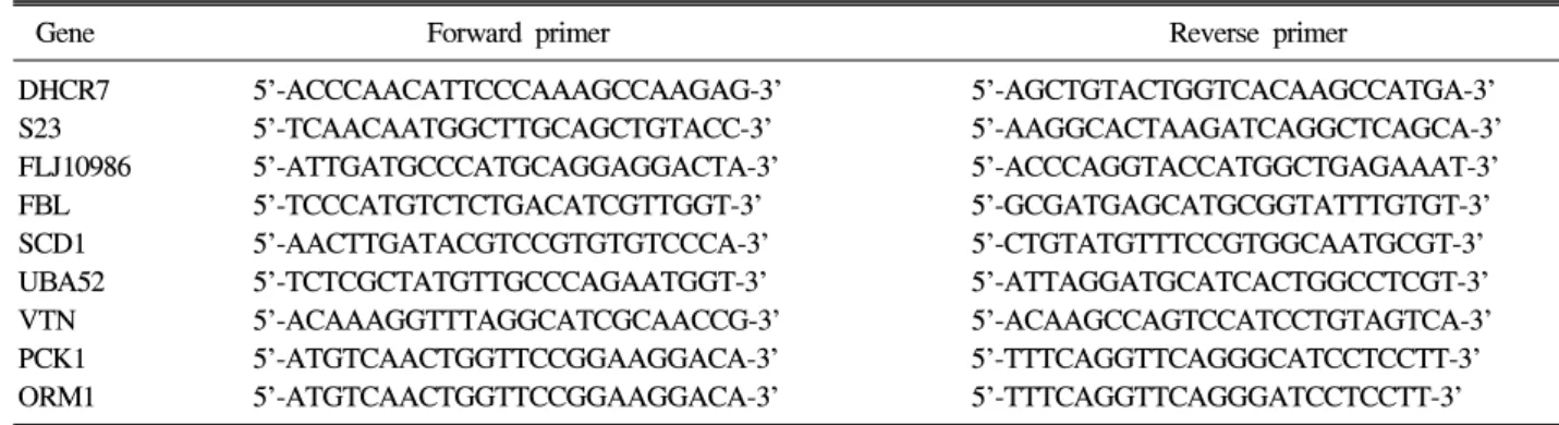
Table 1. The Specific Primer of Genes Identified by Real Time RT-PCR
Gene Forward primer Reverse primer
DHCR7 5'-ACCCAACATTCCCAAAGCCAAGAG-3' 5'-AGCTGTACTGGTCACAAGCCATGA-3' S23 5'-TCAACAATGGCTTGCAGCTGTACC-3' 5'-AAGGCACTAAGATCAGGCTCAGCA-3' FLJ10986 5'-ATTGATGCCCATGCAGGAGGACTA-3' 5'-ACCCAGGTACCATGGCTGAGAAAT-3' FBL 5'-TCCCATGTCTCTGACATCGTTGGT-3' 5'-GCGATGAGCATGCGGTATTTGTGT-3' SCD1 5'-AACTTGATACGTCCGTGTGTCCCA-3' 5'-CTGTATGTTTCCGTGGCAATGCGT-3' UBA52 5'-TCTCGCTATGTTGCCCAGAATGGT-3' 5'-ATTAGGATGCATCACTGGCCTCGT-3' VTN 5'-ACAAAGGTTTAGGCATCGCAACCG-3' 5'-ACAAGCCAGTCCATCCTGTAGTCA-3' PCK1 5'-ATGTCAACTGGTTCCGGAAGGACA-3' 5'-TTTCAGGTTCAGGGCATCCTCCTT-3' ORM1 5'-ATGTCAACTGGTTCCGGAAGGACA-3' 5'-TTTCAGGTTCAGGGATCCTCCTT-3'
증폭된 반응 산물은 2% agarose gel에서 전기영동 하였다.
4) 산물의 클로닝 및 염기서열 분석
증폭된 PCR 산물을 2% agarose gel에 전기 영동하여 확인한 후 대상 조직간 유전자 발현 양상이 뚜렷한 band를 gel extraction kit (Qiagen, Valencia, CA)를 이용하여 분리하고 TOPO TA cloning vector (Invitrogen)를 이용하여 유전자 클로닝을 시 행하였다. 클로닝 후 발현된 유전자들을 plasmid preparation kit 를 이용하여 분리한 후 BigDye 3.1 version kit (Applied Bio- systems, FosterCity, CA)를 이용하여 염기서열을 증폭하고 Blast program을 이용하여 염기서열 분석을 시행하였다.
3. 실시간 중합효소연쇄반응
차별 발현 역전사중합효소반응의 결과로 얻어진 유전자의 발현율을 확인하기 위하여 11쌍의 간세포암종 조직과 비간 세포암 조직 및 간세포암종 세포주[HepG2, Hep3B, Huh7], 자궁암 세포주[HeLa], 대장암 세포주[HT1299], 섬유육종 세 포주[HT1080], 폐암 세포주[A549]에서 RNA를 추출하고 이 중 2μg에 oligo-dT primer를 넣고 역전사 반응을 수행하였 다. 만들어진 cDNA의 1/10을 gene-specific primer set를 이용 하여 유전자 중합효소연쇄반응을 수행하였으며 PCR cycle 은 linear amplification range에 들어가도록 각각의 유전자에 대해서 보정을 해 주었다. 실험에 사용한 primer set은 다음 과 같다(Table 1). Primer set 5’-ACCATGGATGATGATA- TCGC-3'와 5’-ACATGGCTGGGGTGTTGAAG-3'를 이용하여 상대비교를 하였다.
결 과
1. 원발 간세포암종 조직과 비간세포암종 조직에서 차별 발현 유전자 확인
원발 간세포암종 조직 및 주위의 비간세포암종 조직에서
RNA를 추출하고 역전사 반응을 통해 cDNA를 합성한 후 차별발현 역전사중합효소연쇄반응을 시행하여 gel에 전기 영동한 결과 비간세포암종 조직에 비하여 간세포 암조직에 서 새로이 발현되거나 증폭된 유전자와 소실되거나 감소된 유전자의 경우와 같이 다양한 유전자의 발현양상을 확인할 수 있었다(Fig. 1). 이렇게 유전자의 발현도를 분석한 결과 간세포암종 조직에서 발현이 저하되는 유전자의 수가 상대
364 The Korean Journal of Gastroenterology: Vol. 53, No. 6, 2009
Fig. 2. Differentially expressed genes (DEGs) between tumor tis- sue (T) and non-tumor tissue (NT). More than hundreds of DEGs were generated. Among them, 25 DEGs were markedly up-regulated and 10 DEGs were down-regulated in T. These DEG bands were excised from the gel for further cloning and sequencing.
Table 2. Up-regulated Genes in Tumor Tissues Compared with Non-tumor Tissues by DDRT-PCR
No Gene name Accession No Chromosomal location Functions 1 Homo sapiens brain protein 44 (BRP44) NM_015415.1 1q24 Unknown
2 Homo sapiens ribosomal protein S14 (RPS14), NM_001025070.1 5q31-q33 Protein biosynthesis transcript variant2
3 Homo sapiens ribosomal protein S23 (RPS23) NM_001025.4 5q14.2 Protein biosynthesis 4 Homo sapiens aldehyde dehydrogenase 1 family, NM_012190.2 3q21.2 Protein metabolism
member L1 (ALDH1L1)
5 Homo sapiens ubiquitin A-52 residue ribosomal protein NM_001033930.1 19p13.1-p12 Protein metabolism fusion product 1 (UBA52), transcript variant 1
6 Ribosomal protein L36 (RPL36)) NM_033643 19p13.3 Protein metabolism 7 Homosapiensproteasome (prosome, macropain) subunit, NM_002793.2 6q27 Ubiquitin-dependent
betatype, 1 (PSMB1) protein catabolism
8 Homo sapiens hypothetical protein FLJ10986 (FLJ10986) NM_018291.2 1p32.1 Carbohydrate metabolism 9 Homo sapiens stearoyl-CoA desaturase NM_005063.4 10q23-q24 Lipid metabolism
(delta-9-desaturase) (SCD1)
10 Homosapiensacyl-CoenzymeAdehydrogenase, NM_000018.2 17p13-p11 Lipid metabolism verylongchain (ACADVL)
11 Homo sapiens 7-dehydrocholesterol reductase (DHCR7) NM_001360.1 11q13.2-q13.5 Lipid metabolism 12 Homo sapiens poly (ADP-ribose) polymerase family, NM_001618.2 1q41-q42 DNA repair
member 1 (PARP1)
13 Homosapiensdelta-like1homolog (Drosophila) (DLK1), NM_003836.4 14q32 Development transcriptvariant1
14 Homo sapiens fibrillarin (FBL) NM_001436.2 19q13.1 rRNA processing 적으로 많았다. 이 중 원발 간세포암종 조직과 비간세포암
종 조직에서 최소 1.5배 이상의 발현차이가 있는 35개의 밴 드를 영상분석법으로 선택하였다(Fig. 2).
2. 유전자의 염기서열 분석
발현 정도를 확인하고 클로닝 및 유전자 분석을 시행한 결과 조직에서 과발현된 유전자 14종과 저발현된 유전자 7 종을 확인하였다. 원발 간세포암종 조직과 비간세포암종 조 직에서 발현 정도의 뚜렷한 차이를 보이는 유전자는 세포대 사, 이동 전사, 해독, 세포간 상호 작용, 면역 반응, 단백 합 성 등에 관련된 유전자들로 특히 9종의 유전자는 3예에서 모두 동일한 양상으로 변화를 보였으며 그 중 6종의 유전자 는 비간세포암종 조직에 비하여 발현이 증가되었고 3종의 유전자는 발현이 감소되었다. 증가된 유전자는 brain protein 44 (BRP44), ribosomal protein S14 (RPS14), ribosomal protein S23 (RPS23), aldehyde dehydrogenase 1 family member L1 (ALDH1L1), ubiquitin A-52 residue ribosomal protein fusion product 1 (UBA52), ribosomal protein L36 (RPL36), protea- some (prosome, macropain) subunit, beta type, 1 (PSMB1), hypothetical protein FLJ10986 (FLJ10986), stearoyl-CoA desa- turase (SCD1), acyl-Coenzyme A dehydrogenase, very long chain (ACADVL), 7-dehydrocholesterol reductase (DHCR7), poly (ADP-ribose) polymerase family, member 1 (PARP1),
Lee YC, et al. Differentially Expressed Gene in HCC 365
Fig. 4. Relative expression levels measured by semi-quantitative RT-PCR. In order to validate the observed expression on the down-regulated genes (VTN, PCK1, ORM1), we performed quan- titative real-time RT-PCR in 11 HCCs and non-tumor tissue (NT). The obtained mRNA levels for VTN, and ORM1 were on- ly elevated in 3 of 11, and PCK1 was elevated in 4 of 11 in HCCs. All PCRs were conducted in triplicate and normalized for β-actin mRNA expression. Also, data are presented as the level of expression in each HCCs with respect to the corresponding NT.
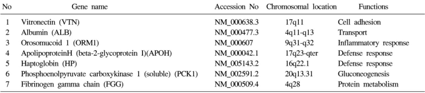
Table 3. Down-regulated Genes in Tumor Tissues Compared to Non-tumor Tissues by DDRT-PCR
No Gene name Accession No Chromosomal location Functions 1 Vitronectin (VTN) NM_000638.3 17q11 Cell adhesion
2 Albumin (ALB) NM_000477.3 4q11-q13 Transport
3 Orosomucoid 1 (ORM1) NM_000607 9q31-q32 Inflammatory response 4 ApolipoproteinH (beta-2-glycoprotein I)(APOH) NM_000042.1 17q23-qter Defense response 5 Haptoglobin (HP) NM_005143.2 16q22.1 Defense response 6 Phosphoenolpyruvate carboxykinase 1 (soluble) (PCK1) NM_002591.2 20q13.31 Gluconeogenesis 7 Fibrinogen gamma chain (FGG) NM_000509.4 4q28 Protein metabolism
Fig. 3. Relative expression levels measured by semi-quantitative RT-PCR. In order to validate the observed expression on the up-regulated genes (DHCR7, S23, FLJ10986, FBL, SCD, UBA52), we performed quantitative real-time RT-PCR in 11 HCCs and non-tumor tissue (NT). The obtained mRNA level for FBL was elevated in 8 of 11 in HCCs, and DHCR7, and UBA52 were elevated in 6 of 11 HCCs, respectively. All PCRs were con- ducted in triplicate and normalized for β-actin mRNA expression. Also, data are presented as the level of expression in each HCCs with respect to the coresponding NT.
delta-like 1 homolog (Drosophila) (DLK1), fibrillarin (FBL)이 며(Table 2), 감소된 유전자는 vitronectin (VTN), albumin (ALB), fibrinogen gamma chain (FGG) orosomucoid 1 (ORM1), apoli- poproteinH (APOH), haptoglobin (HP), phosphoenolpyruvate carboxykinase 1 (PCK1)이 확인되었다(Table 3).
3. 실시간 중합효소연쇄반응의 분석
차별 발현 역전사중합효소연쇄반응을 이용하여 차별 발현 된 유전자의 재현성을 검증하기 위하여 인간 암 세포주와 간세포암종 조직을 이용하여 탐색된 유전자의 실시간 정량 분석을 시행하였다. 이번 연구에서 과발현 또는 저발현되는 것으로 나타난 유전자 중 원발 간세포암종과 관련이 있다고 생각되는 유전자를 선택하여 그 발현 정도를 확인하였다.
간세포암종 조직에서 과발현되는 유전자 6종[RPS23, UBA52, FLJ10986, SCD1, DHCR7, FBL]과 저발현되는 유전 자 3종[VTN, ORM1, PCK1]을 선별하였으며, 11쌍의 간세포 암종 조직과 비간세포암 조직 및 간세포암 세포주[HepG2, Hep3B, Huh7], 자궁암 세포주[HeLa], 대장암 세포주 [HT1299], 섬유육종 세포주[HT1080], 폐암 세포주[A549]에 서 증가된 유전자의 발현 양상을 비교하였다. 11쌍의 간세 포암종 조직과 비간세포암종 조직에서의 발현율을 비교한 결과, DHCR7, UBA 52와 FBL이 50% 이상의 간세포암종 조 직에서 비간세포암종 조직과 비교하여 과발현되는 것으로 나타났으며 VTN과 ORM1은 간세포암종 조직의 약 30%에 서 과발현되는 결과를 얻었다(Fig. 3, 4). 또한 여러 종류의 인간 암세포주와 마우스의 간세포에서의 발현율을 비교한
366 대한소화기학회지: 제53권 제6호, 2009
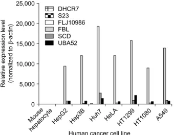
Fig. 5. Relative expression levels measured by semi-quantitative RT-PCR. In order to validate the observed expression on the up-regulated genes (DHCR7, S23, FLJ10986, FBL, SCD, UBA52), we performed quantitative real-time RT-PCR in different human cancer cell lines. The obtained mRNA levels for DHCR7, S23, FLJ10986, FBL, SCD, and UBA52 were elevated in 7 of 7 hu- man cancer cell lines. All PCRs were conducted in triplicate and normalized for β-actin mRNA expression. Also, data are pre- sented as the level of expression in each human cancer cell lines with respect to the corresponding mouse hepatocyte.
Fig. 6. Relative expression levels measured by semi-quantitative RT-PCR. In order to validate the observed expression on the down-regulated genes (VTN, PCK1, ORM1), we performed quan- titative real-time RT-PCR in different human cancer cell lines.
The obtained mRNA levels for VTN, and ORM1 were elevated in 5 of 7 and PCK1 was elevated in 4 of 7 human cancer cell lines. All PCRs were conducted in triplicate and normalized for β-actin mRNA expression. Also, data are presented as the level of expression in each human cancer cell lines with respect to the corresponding mouse hepatocyte.
결과 대부분의 유전자들이 7개의 인간 암세포주에서 높은 발현율을 나타내었으나, VTN, ORM1, PCK1 유전자의 경우 는 암세포주에서 낮은 발현율을 보였다(Fig. 5, 6).
고 찰
원발 간세포암종은 세계적으로 발병률이 높은 치명적인 암으로 간세포암종에서 특이하게 과발현되는 특정 유전자 를 규명하는 것은 간세포암종 발암 기전을 연구하는 데 필 수적이다. 최근 cDNA microarray 기술의 발달로 관련 유전 자 발굴을 통하여 간세포암종 발생에 관여하는 유전자들이 다수 발굴되고 있고7,8 부분적으로 그들의 기능이 분석되어 간세포암종 발생과의 연관성이 서서히 규명되고 있으나,9,10 cDNA microarray의 경우 한 번에 대량의 유전자 분석이 가 능하다는 장점을 가지고 있지만, 교잡 방식을 이용하므로 위양성이 많고, 재현성과 민감도가 낮으며, 비교 가능한 샘 플이 한정되어 있어 각 샘플 간의 발현 차이를 비교하기가 까다롭다는 한계를 가지고 있다. 이에 이번 연구에서는 기 존 방식의 문제점들을 극복하고 발현 차이를 보이는 유전자 를 손쉽고 정확하게 분석할 수 있는 차별 발현 역전사중합 효소연쇄반응법을 이용하여 간세포암종과 연관성이 있는 유전자를 탐색해보고자 하였다.
이번 실험에 사용한 조직은 3쌍의 간세포암종 조직과 비
간세포암종 조직으로 구성되어 있으며 우선 간세포암종 조 직과 비간세포암종 조직만을 따로 분리하여 유사한 조직 안 에서의 유전적 발현 양상이 얼마나 차이가 있는지 조사해 본 결과 간세포암 조직에서 발현이 저하되는 유전자의 수가 상대적으로 많았음을 알 수 있었다. 이번 연구에서는 fibrillarin (FBL), ribosomal protein S23 (RPS23), ribosomal protein S14 (RPS14), ubiquitin A-52 residue ribosomal protein fusion product 1 (UBA52), hypothetical protein 10986 (FLJ10986), poly (ADP- ribose) polymerase-1 (PARP-1) 등과 같이 핵산 및 단백 합성 및 세포사멸에 관여하는 유전자들 이 발현됨으로써 간세포암 조직에서 세포의 성장, 분열, 대 사, 단백 합성 등이 활발하게 일어나는 것을 알 수 있었다.
PARP-1는 DNA 복구 및 증식에 관여하는 유전자로 핵에서 손상된 DNA를 인지하여 활성화된 후 여러 단백들을 poly(ADP- ribosyl)ation이라는 전사과정을 통해 변형시키는 효소로, DNA 손상이 적을 경우에는 핵에서 손상 부위의 복 구를 도와줌으로써 세포의 생존에 기여하는 반면 DNA 손 상이 심해 PARP-1의 활성이 과다하게 높아지면 기질인 NAD를 급격하게 소모하면서 세포 사멸을 유도한다고 보고 되어 있다.11,12 FBL은 핵 내에 위치하며 초기형태의 ribosomal RNA (rRNA)를 성숙한 rRNA 분자로 전사시키는 유전자로 간세포암종 생성과정과의 연관성은 보고된 바 없 으나 실시간 정량 분석 결과 여러 간세포암종 조직에서 발
이영춘 외 7인. 원발 간세포암종의 유전자 발현 분석 367
현율이 높게 나타난 것으로 보아 간세포암종과 관련이 있을 것으로 예상된다.13
간세포암종 조직에서 두드러지게 발현이 감소한 유전자 는 vitronectin (VTN), albumin (ALB), orosomucoid 1 (ORM1), apolipoproteinH (APOH), haptoglobin (HP), phosphoenolpyru- vate carboxykinase 1 (PCK1), fibrinogen gamma chain (FGC) 등으로 당대사 및 단백 합성, 면역반응 그리고 혈액응고에 관여하는 유전자들이다. 사람의 VTN은 fibronectin이나 laminin 등과 마찬가지로 강한 세포접착 활성을 갖는 혈장 당단백으로 Jaskewickz 등14은 말기 간세포암종에서 발현율 이 증가한다고 보고하였으나 이번 연구결과에서는 간세포 암종 조직에서의 발현율이 저하된 것으로 나타났다. 이것은 간세포암종 조직의 다양성에 따른 분화도 및 간경변증과 관 련이 있을 것으로 생각한다. 이 유전자는 VTN 수용체를 보 유하고 있는 세포에 대해 접착 활성을 갖는 것 외에 생리기 능으로는 혈액 응고, 보체의 기능조절 등이 알려져 있다.
PCK1은 피루브산, 아미노산과 글리세롤과 같은 비탄수화물 전구체(noncarbohydrate precursors)로부터 당의 합성에 관여 하는 유전자로, 포도당 신생합성(gluconeogenesis)이 간에서 90% 이상 일어나므로 간세포암종 조직에서 PCK1 유전자의 발현율이 감소한다는 것은 암이 진행될수록 간의 기능이 떨 어진다는 것을 설명해 줄 수 있는 현상이라고 할 수 있을 것이다.
이외에 지질 대사에 관여하는 DHCR7 및 SCD1 유전자는 간세포암종 조직에서 발현율이 저하될 것으로 예상하였으 나 오히려 증가한 것으로 관찰되었다. SCD1는 지방산 형성 에 관여하는 유전자로 전립선암에서 발현이 저하되어 있다 는 보고는 있으나 간세포암종 발생과의 연관성은 알려진 바 가 없다.15,16 그러나 개인의 유전자 발현 양상이 매우 다양 하고 그 기능이 명확하지 않기 때문에 향후에는 이 유전자 들에 대한 기능적인 연구가 추가로 수행되어야 할 것으로 생각한다.17,18
이번 연구를 통해서 비간세포암종 조직에 비하여 간세포 암종에서 발현율이 증가하거나 감소 또는 생성되는 유전자 를 분석하여 간세포암종 발생과정에서 관련 유전자 간의 변 이에 대한 기초 자료를 얻을 수 있었으나 기존에 간세포암 종 조직에서 두드러지게 관찰된다고 알려진 유전자들은 이 번 연구에서는 검출되지 않았다.19-22 이는 이번 연구에 적용 했던 간세포암종 조직의 수가 적었기 때문이라고 생각하며, 연구의 대상 및 결과에 대하여는 간세포암종의 원인에 따른 분류가 같이 진행된다면 좀 더 자세한 유전자 관련성을 확 인할 수 있을 것이라고 생각한다. 이렇게 발현 양상이 다른 것으로 나타난 유전자들은 다양한 단계의 간세포암종 조직 에서 향후 그 발현이 검증되어야 할 것이며 이런 연구 결과 들이 간세포암종 발생 과정과 그의 치료법에 대한 이해도를
높이고 간세포암종의 형성과 억제 기작을 더 자세히 밝힘으 로써 새로운 간세포암종 치료 방법과 치료제의 개발에 유용 하게 이용될 수 있을 것으로 생각한다.
요 약
목적: 원발 간세포암종에서 특이하게 과발현되는 특정 유전자를 규명하는 것은 간세포암종 발암 기전의 연구나, 암의 조기진단 및 암 특이 유전자를 표적으로 한 유전자 치 료에 매우 중요한 정보를 제공한다. 이에 이번 연구에서는 차별 발현 역전사중합효소연쇄반응법을 활용한 유전자 분 석을 통해 간세포암에서 특이하게 과발현되는 특이 유전자 를 밝혀내고 이 유전자의 발현율을 여러 시료에서 확인하여 간세포암 관련 유전자들의 기능을 탐색하고자 하였다. 대상 및 방법: 원발 간세포암종으로 수술을 시행 받았던 3명의 간 세포암 환자의 간 적출물에서 간세포암종 조직과 암주변의 비 간세포암 조직 3쌍을 채취한 후 차별 발현 역전사중합효 소연쇄반응법을 이용하여 간세포암종 조직에서 과발현되거 나 소실된 유전자 혹은 저발현되어 있는 유전자를 겔로부터 DNA를 용출하여 이를 클로닝한 후 염기서열분석을 통하여 나타난 유전자의 서열을 유전자 검색프로그램에 넣어 그 유 전자의 이름이나 특성들을 탐색하였다. 이후 새로 규명된 유 전자의 기능을 검증하기 위하여 11쌍의 간세포암종 및 비간 세포암종 조직과 인간 암세포주인 간세포암종 세포주[HepG2, Hep3B, Huh7], 자궁암 세포주[HeLa], 대장암 세포주[HT1299], 섬유육종 세포주[HT1080], 폐암 세포주[A549]에서 실시간 중합효소연쇄반응을 이용하여 이들 유전자의 발현을 확인 한 후 분석하였다. 결과: 이번 연구를 통해서 단백 대사, 유 비퀴틴 의존성 단백 대사, 탄수화물 대사, 지방대사, 유전자 복구, 염증 반응 등의 다양한 기능을 가지는 21종의 유전자 들을 탐색하였다. 결론: 이번 연구를 통하여 간세포암종 조 직과 비간세포암종 조직에서 발현차이를 보이는 유전자를 탐색하였고, 이러한 유전자의 특성 규명은 간세포암종의 발 암 기전을 규명하거나 조기 간세포암을 진단하는 바이오 마 커의 개발 혹은 분자표적을 대상으로 하는 유전자 치료의 개발에 기초자료로 유용할 것으로 생각한다.
색인단어: 간세포암, 차별 발현 역전사중합효소연쇄반응, 바이오마커
참고문헌
1. Park JW, Kim CM. Epidemiology of hepatocellular carcinoma in Korea. Korean J Hepatol 2005;11:303-310.
2. Kaino M. Alterations in the tumor suppressor genes p53, RB,
368 The Korean Journal of Gastroenterology: Vol. 53, No. 6, 2009
p16/MTS1, and p15/MTS2 in human pancreatic cancer and hepatoma cell lines. J Gastroenterol 1997;32:40-46.
3. Ashida K, Kishimoto Y, Nakamoto K, et al. Loss of hetero- zygosity of the retinoblastoma gene in liver cirrhosis accom- panying hepatocellular carcinoma. J Cancer Res Clin Oncol 1997;123:489-495.
4. Kondoh N, Wakatsuki T, Ryo A, et al. Identification and characterization of genes associated with human hepatocellular carcinogenesis. Cancer Res 1999;59:4990-4996.
5. Song JY, Choi JH, Lee KJ, et al. Identification of the gene associated with hepatocellular carcinoma using differential gene expresion. Korean J Hepatol 2001:7:265-272.
6. Cui XS, Shin MR, Lee KA, Kim NH. Identification of differ- entially expressed genes in murine embryos at the blastocyst stage using annealing control primer system. Mol Reprod Dev 2005;70:278-287.
7. Okabe H, Satoh S, Kato T, et al. Genome-wide analysis of gene expression in human hepatocellular carcinomas using cDNA microarray: identification of genes involved in viral carcinogenesis and tumor progression. Cancer Res 2001;61:
2129-2137.
8. Jia HL, Ye QH, Qin LX, et al. Gene expression profiling re- veals potential biomarkers of human hepatocellular carcinoma.
Clin Cancer Res 2007;13:1133-1139.
9. Villanueva A, Newell P, Chiang DY, Friedman SL, Llovet JM. Genomics and signaling pathways in hepatocellular carcinoma. Semin Liver Dis 2007;27:55-76.
10. Wang ZX, Wang HY, Wu MC. Identification and character- ization of a novel human hepatocellular carcinoma-associated gene. Br J Cancer 2001;85:1162-1167.
11. Nomura F, Yaguchi M, Togawa A, et al. Enhancement of poly-adenosine diphosphate- ribosylation in human hep- atocellular carcinoma. J Gastroenterol Hepatol 2000;15:529- 535.
12. Yu SW, Andrabi SA, Wang H, Kim NS, Poirier GG, Dawson TM, et al. Apoptosis-inducing factor mediates poly(ADP-ri- bose) (PAR) polymer-induced cell death. PNAS 2006;103:
18314-18319.
13. Westra R, Jansen TL, Gouw AS, de Jong KP. Anti-nucleair antibody (ANA) positivity caused by paraneoplastic antibodies due to abundant p53 expression in early hepatic carcinoma.
Neth J Med 2003;61:300-303.
14. Jaskiewicz K. Chasen MR, Robson SC. Differential ex- pression of extracellular matrix proteins and integrins in hep- atocellular carcinoma and chronic liver disease. Anticancer Res 1993;13: 2229-2237.
15. Moore S, Knudsen B, True LD, et al. Loss of stearoyl-CoA desaturase expression is a frequent event in prostate carcinoma. Int J Cancer 2005;114:563-571.
16. Falvella FS, Pascale RM, Gariboldi M, et al. Stearoyl-CoA desaturase 1 (SCD1) gene overexpression is associated with genetic predisposition to hepatocarcinogenesis in mice and rats. Carcinogenesis 2002;23:1933-1936.
17. Jiang J, Nilsson-Ehle P, Xu N. Influence of liver cancer on lipid and lipoprotein metabolism. Lipids Health Dis 2006;5:4.
18. Yahagi N, Shimano H, Hasegawa K, et al. Co-ordinate acti- vation of lipogenic enzymes in hepatocellular carcinoma. Eur J Cancer 2005;41:1316-1322.
19. Huang J, Zhang X, Zhang M, et al. Up-regulation of DLK1 as an imprinted gene could contribute to human hep- atocellular carcinoma. Carcinogenesis 2007;28:1094-1103.
20. Lencioni RA, Allgaier HP, Cioni D, et al. Small hep- atocellular carcinoma in cirrhosis: randomized comparison of radio-frequency thermal ablation versus percutaneous ethanol injection. Radiology 2003;228:235.
21. Liao C, Zhao M, Song H, Uchida K, Yokoyama KK, Li T.
Identification of the gene for a novel liver-related putative tu- mor suppressor at a high-frequency loss of heterozygosity re- gion of chromosome 8p23 in human hepatocellular carci- noma. Hepatology 2000;32:721-727.
22. Shirahashi H, Sakaida I, Terai S, Hironaka K, Kusano N, Okita K. Ubiquitin is a possible new predictive marker for the recurrence of human hepatocellular carcinoma. Liver 2002;22:413-418.