Korean J Gastroenterol Vol. 69 No. 5, 321-324 https://doi.org/10.4166/kjg.2017.69.5.321 pISSN 1598-9992 eISSN 2233-6869
CASE REPORT
Korean J Gastroenterol, Vol. 69 No. 5, May 2017 www.kjg.or.kr
내시경 초음파 유도하 배액술로 치료한 고립성 화농성 췌장 농양 2예
이중엽, 김태현, 전형구
원광대학교 의과대학 내과학교실
Isolated Pyogenic Pancreatic Abscess Successfully Treated via Endoscopic Ultrasound-guided Drainage
Jung Yeop Lee, Tae Hyeon Kim and Hyung Ku Chon
Department of Internal Medicine, Wonkwang University School of Medicine, Iksan, Korea
An isolated pyogenic pancreatic abscess (IPPA) without pancreatitis is extremely rare but can occur in patients with uncontrolled diabetes. This pathologic condition poses a clinical challenge in diagnosis and management because it can be confused easily with a malignancy. Endoscopic ultrasound (EUS) may be a useful diagnostic modality for indeterminate pancreatic lesions and IPPA. Here, we report two cases with elevated carbohydrate antigen 19-9 levels and pancreatic masses on cross sectional imaging. The patients were subsequently diagnosed with IPPA by EUS. EUS-guided drainage was performed successfully and the patients’ clinical symptoms and radiologic findings improved. In our experience, EUS and EUS-guided drainage are crucial steps for the diagnosis and manage- ment of patients with an indeterminate pancreatic lesion. In addition, EUS-guided drainage has excellent technical and clinical out- comes for the treatment of IPPA. (Korean J Gastroenterol 2017;69:321-324)
Key Words: Pancreas; Abscess; Drainage
Received March 27, 2017. Revised May 2, 2017. Accepted May 8, 2017.
CC This is an open access article distributed under the terms of the Creative Commons Attribution Non-Commercial License (http://creativecommons.org/licenses/
by-nc/4.0) which permits unrestricted non-commercial use, distribution, and reproduction in any medium, provided the original work is properly cited.
Copyright © 2017. Korean Society of Gastroenterology.
교신저자: 전형구, 54538, 익산시 익산대로 460, 원광대학교 의과대학 내과학교실
Correspondence to: Hyung Ku Chon, Department of Internal Medicine, Wonkwang University School of Medicine, 460 Iksan-daero, Iksan 54538, Korea.
Tel: +82-63-859-2564, Fax: +82-63-855-2025, E-mail: [email protected]
Financial support: This paper was supported by Wonkwang University in 2017. Conflict of interest: None.
INTRODUCTION
An isolated pyogenic pancreatic abscess (IPPA) without pancreatitis is rare and usually caused by tuberculosis, sal- monellosis, or underlying diabetes.1-3 Surgical debridement or computed tomography (CT)-guided percutaneous drain- age remains the standard treatment for a pancreatic abscess. Recently, fine needle aspiration (FNA) or drainage catheter placement performed by endoscopic ultrasound (EUS) has become a treatment option because they are more effective and less invasive. Herein, we report two cases of IPPA in patients with underlying diabetes mellitus (DM) who
were treated successfully by EUS-guided drainage.
CASE REPORT
Case 1: A 62-year-old man with DM presented with a his- tory of fever, nausea, and pain in the epigastrium for 1 month.
He denied any smoking or alcohol consumption. Laboratory studies revealed an elevation of C-reactive protein 43.52 mg/L, glucose 244 mg/dL, hemoglobin A1c 8.5%, and carbo- hydrate antigen 19-9 (CA 19-9) 93 U/mL, but all other find- ings were unremarkable. An abdominal CT and magnetic res- onance cholangiopancreatography showed a 4.8 cm ill-de-
322 이중엽 등. 내시경 초음파 유도하 배액술로 치료한 화농성 췌장 농양
The Korean Journal of Gastroenterology
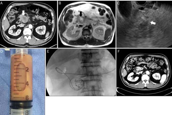
Fig. 1. (A) Transverse abdominal CT scans shows an ill-defined low attenuating mass (white arrow) along the pancreatic head. (B) T-2 weighted images demonstrate high signal intensity (black arrow) in the central portion of the mass. (C) A 20-G FNA needle (white arrow) entering the ill-defined low echoic lesion for aspiration is visible. (D) Yellowish pus discharge from the aspiration fluid collected by EUS-FNA. (E) Fluoroscopic view, demonstrating a naso-abscess catheter (5 Fr) placed into the abscess cavity through the duodenal wall. (F) Follow-up CT shows the reso- lution of an isolated pyogenic pancreatic abscess. CT, computed tomography; FNA, fine needle aspiration; EUS, endoscopic ultrasound.
fined low density mass in the pancreas head with regional lymph node enlargement (Fig. 1A, B). After obtaining informed consent, EUS via a longitudinal echoendoscope (Olympus GF-UCT 260, Tokyo, Japan) revealed a 4 cm anechoic lesion containing echogenic materials. Color Doppler was applied to identify any interposing vessels. EUS-FNA using a 20-G EUS needle (Echotip ProCore® HD Ultrasound biopsy needle;
Wilson-Cook Medical Inc., Bloomington, IN, USA) was per- formed (Fig. 1C) and a thick, opaque, purulent fluid con- sistent with an abscess was aspirated (Fig. 1D). A naso-ab- scess drainage catheter (5 Fr) was also placed (Fig. 1E) and removed 5 days later when the amount of drainage had de- creased significantly. The fluid aspiration tested positive for Klebsiella pneumonia and negative for acid-fast staining.
The patient recovered uneventfully and was discharged after 10 days on antibiotics. A repeated CT scan revealed complete resolution of the abscess (Fig. 1F).
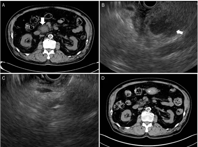
Case 2: A 51-year-old man was admitted to our department complaining of abdominal pain, 5 kg weight loss, fever up to 38oC, and chills. He had a history of DM, hypertension, chron- ic kidney disease, and complete atrioventricular block treat- ed with a pacemaker. The physical examination revealed mild direct tenderness of the epigastrium. A blood test performed after admission revealed the following: white bleed cell count, 6,620/mm3 (69.3% neutrophil); hemoglobin, 11.3 g/dL; aspartate aminotransferase, 23 U/L; alanine amino- transferase, 22 U/L; alkaline phosphatase, 252 IU/L; total bi- lirubin, 0.86 mg/dL; amylase, 54 U/L; lipase, 20 U/L; blood urea nitrogen, 58.8 mg/dL; creatinine, 2.57 mg/dL; glucose, 312 mg/dL; hemoglobin A1c, 8.8%; C-reactive protein, 63.12 mg/L; and CA 19-9, 97 U/mL. Non-contrast enhanced ab- dominal CT showed an irregular exophytic mass in the pan- creas uncinate process (Fig. 2A). EUS revealed an ill-defined, approximately 2.8×2.3 cm hypo-echoic mass-like lesion with
A B C
D E F
Lee JY, et al. Isolated Pyogenic Pancreatic Abscess Successfully Treated Via Endoscopic Ultrasound-guided Drainage 323
Vol. 69 No. 5, May 2017
A B
C D
Fig. 2. (A) Non-contrast enhanced abdominal CT showing an exophytic mass in the uncinate process of the pancreas. (B) On EUS, a heteroge- neous low echoic lesion with floating echogenic materials (white arrow) is visible. (C) EUS image of the 2nd abscess drainage using a 22 gauge needle in the remaining abscess cavity. (D) After 3 months, abdominal CT shows the resolved state of the exophytic pancreatic mass. CT, computed tomography; EUS, endoscopic ultrasound.
floating echogenic materials in the pancreas uncinate process. EUS-FNA was performed using a 22-G EUS needle (Echotip® Ultra Endoscopic Ultrasound needle; Wilson-Cook Medical Inc., Bloomington, IN, USA) (Fig. 2B) and a pus-like discharge was removed. Klebsiella pneumonia was isolated from the pus, which was negative for tuberculosis or a malig- nancy upon the cytopathologic examinations. Seven days after the procedure, the patient developed a fever and leukocytosis.
The follow-up EUS showed a remnant abscess in the uncinate process, which required further intervention. A 2nd EUS-guid- ed drainage was performed successfully (Fig. 2C). Since then, the patient has been asymptomatic. After 3 months, abdomi- nal CT demonstrated successful resolution of the pancreatic abscess (Fig. 2D).
DISCUSSION
In general, a pancreatic abscess is a complication of a pan-
creatic pseudocyst infection that is correlated with high mor- tality due to acute pancreatitis, primary and recurrent chronic pancreatitis, abdominal trauma, or surgery. Tuberculosis, scrub typhus, and salmonellosis are other rare causative fac- tors of pancreatic abscesses.1-3 In the present two cases, there was no definite etiology, but underlying uncontrolled DM. DM is correlated with depressed cellmediated immunity and disorders of humoral immunity. Consequently, DM in- creases the frequency and severity of various types of bacte- rial infections, including abscesses.4,5 Several reports have demonstrated underlying diabetes as a predisposing factor for pancreatic abscesses secondary to Klebsiella pneumo- nia, similar to these two cases.6
On abdominal CT or magnetic resonance imaging, an iso- lated pancreatic solid or cystic lesion with marked elevated CA 19-9 is often confused with various pancreatic pathologic conditions, such as adenocarcinoma, cystadenocarcinoma, neuroendocrine tumor, or pancreatic pseudocysts. EUS can
324 이중엽 등. 내시경 초음파 유도하 배액술로 치료한 화농성 췌장 농양
The Korean Journal of Gastroenterology
provide a detailed ultrasound image, and may help reduce the diagnostic errors and offer a precise diagnosis of an in- determinate pancreatic lesion on cross-sectional abdominal imaging. A pathologic evaluation of the lesion is also essen- tial and EUS-guided FNA may be the preferred diagnostic mo- dality because of its high sensitivity and specificity for identi- fying the etiology of pancreatic masses.7
In the second case, intravenous contrast for CT scans was unavailable due to renal function impairment; thus, the im- age quality was suboptimal. The exophytic solid mass of the pancreas uncinate process on non-contrast enhanced CT with a marked elevated CA 19-9 level mimicked a pancreatic malignancy. On the other hand, on EUS, a low echoic lesion with an irregular margin and floating material was more com- patible with an abscess and the abscess fluid was sub- sequently aspirated via EUS-FNA. Klebsiella pneumonia was found with no evidence of malignancy.
The treatment of pancreatic abscesses is complicated.8 Surgical approaches are associated with significant morbid- ity and mortality.9 The lesion in the pancreas may be difficult to target with CT or ultrasound-guided percutaneous drainage.
Since the advent of linear-array EUS scopes with the evolving EUS techniques and accessories, EUS-guided abscess drain- age of the liver, pelvic cavity, or pancreas has been inves- tigated with a high clinical success rate achieved and a low rate of adverse events.
This report describes the safe and successful EUS-guided drainage of IPPA in two patients. The lesion showed a remark- able resolution following the EUS-guided drainage with nor- malization of CA 19-9. EUS-guided drainage has several ad- vantages: (1) excellent visualization of the lesion on the pan- creas with the surrounding structures; (2) access to the near- est site of the abscess while avoiding puncturing large ves- sels under Doppler ultrasound guidance; (3) direct passage of the needle into the abscess pocket through the gastric or duodenal wall alone; and (4) avoiding transcutaneous infections.
Although more well-designed studies will be required, EUS- guided drainage is a safer and more effective alternative to surgery or percutaneous drainage.10
In conclusion, EUS and EUS-guided drainage should be considered as a crucial step in the diagnosis and treatment of indeterminate pancreatic lesions. In addition, as the imag- ing findings may mimic neoplasms, clinicians should consid- er pancreatic abscesses in a differential diagnosis, partic- ularly in patients with DM.
REFERENCES
1. Liu Q, He Z, Bie P. Solitary pancreatic tuberculous abscess mim- icking pancreatic cystadenocarcinoma: a case report. BMC Gastroenterol 2003;3:1.
2. Arya M, Arya PK. Pancreatic abscess caused by s. typhi. Indian J Med Microbiol 2001;19:18-19.
3. Yi SY, Tae JH. Pancreatic abscess following scrub typhus asso- ciated with multiorgan failure. World J Gastroenterol 2007;13:
3523-3525.
4. Eliashiv A, Olumide F, Norton L, Eiseman B. Depression of cell- mediated immunity in diabetes. Arch Surg 1978;113:1180-1183.
5. Peleg AY, Weerarathna T, McCarthy JS, Davis TM. Common in- fections in diabetes: pathogenesis, management and relation- ship to glycaemic control. Diabetes Metab Res Rev 2007;23:3-13.
6. Chong VH. Isolated pyogenic pancreatic abscess mimicking a neoplasm. JOP 2008;9:309-312.
7. Harewood GC, Wiersema MJ. Endosonography-guided fine nee- dle aspiration biopsy in the evaluation of pancreatic masses. Am J Gastroenterol 2002;97:1386-1391.
8. Seewald S, Groth S, Omar S, et al. Aggressive endoscopic therapy for pancreatic necrosis and pancreatic abscess: a new safe and effective treatment algorithm (videos). Gastrointest Endosc 2005;62:92-100.
9. Fernández-del Castillo C, Rattner DW, Makary MA, Mostafavi A, McGrath D, Warshaw AL. Débridement and closed packing for the treatment of necrotizing pancreatitis. Ann Surg 1998;228:
676-684.
10. Kida M, Itoi T. Current status and future perspective of interven- tional endoscopic ultrasound in Japan. Dig Endosc 2009;21 Suppl 1:S50-S52.