Study on the position of the posterior superior alveolar artery in relation to the performance of the maxillary sinus
bone graft procedure in a Korean population
Woo-Hyun Park, So-Young Choi, Chin-Soo Kim
Department of Oral and Maxillofacial Surgery, School of Dentistry, Kyungpook National University, Daegu, Korea
Abstract(J Korean Assoc Oral Maxillofac Surg 2012;38:71-7)
Objectives: This study sought to investigate the positioning of the posterior superior alveolar artery in relation to the performance of the maxillary sinus bone graft procedure in a Korean population.
Materials and Methods: We identified the position of the posterior superior alveolar artery relative to 93 maxillary sinuses in 58 patients and determined the distance from the inferior border of the artery in the premolar and molar areas to the alveolar ridge and sinus floor.
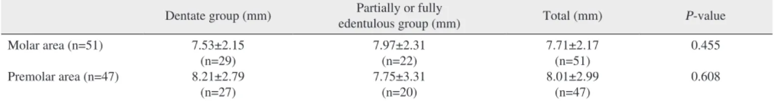
Results: The mean distance from the alveolar ridge to the posterior superior alveolar artery in the dentate group (20.62±3.05 mm in the premolar region, 17.50±2.84 mm in the molar region) was greater than as compared to the edentulous group (18.83±2.79 mm in the premolar region, 15.50±1.64 mm in the molar region), and this difference was statistically significant (P<0.05). In contrast, there was no statistically significant difference (P>0.05) between the mean distance from the sinus floor to the posterior superior alveolar artery in the dentate group (8.21±2.79 mm in the premolar region, 7.52±2.07 mm in the molar region) or in the edentulous group (7.75±3.31 mm in the premolar region, 7.97±2.31 mm in the molar region).
Conclusion: Prior to surgery, it is important to evaluate the position of the posterior superior maxillary artery by using computed tomography scans.
The premolar area is safer than the molar area for performing the maxillary sinus bone graft without bleeding.
Key words: Maxillary sinus, Posterior superior alveolar artery, Sinus floor augmentation
[paper submitted 2011. 11. 11 / revised 2011. 12. 3 / accepted 2012. 3. 15]
extension restoration was suggested as an alternative method, but higher failure rate was recorded with longer distal extension area3,4. The placement of implant in maxillary tuberosity was suggested, but it can damage the maxillary artery. As such, maxillary sinus bone graft was suggested as an alternative method5,6.
Implantation combined with maxillary sinus bone graft has been used widely as a predictable method with high survival rate, and it is useful in overcoming the limit of restoration of the maxillary posterior area with insufficient residual alveolar bone7-10.
Performing maxillary sinus bone graft requires under- standing the anatomy of maxillary sinus first. The pyramid shaped maxillary sinus is the largest of the paranasal sinuses.
The base area consists of the lateral wall of the nasal cavity, and the apex is stretched laterally to the zygomatic process of maxillary bone. The upper wall of the maxillary sinus forms the orbital floor, and the lower wall is the maxillary tuberosity. The anterior region is the facial part of the maxillary bone, with the clinically important sinus floor
I. Introduction
The maxillary posterior area is likely to have tooth loss due to periodontal disease or dental caries1. To implant in the maxillary posterior area, the relationship between the residual alveolar bone and maxillary sinus should be considered. It is difficult to place implants in a defective maxillary posterior area because the alveolar bone becomes constricted and that between the sinus floor and alveolar ridge is decreased by the pneumatization of the maxillary sinus2. As an alternative method, short type has been implanted, but they are likely to fail due to the poor bone quality. Implant-supported distal
Chin-Soo Kim
Department of Oral and Maxillofacial Surgery, Kyungpook National University School of Dentistry, 2177 Dalgubeoldae-ro, Jung-gu, Daegu 700-412, Korea TEL: +82-53-600-7551 FAX: +82-53-426-5365
E-mail: [email protected]
This is an open-access article distributed under the terms of the Creative Commons Attribution Non-Commercial License (http://creativecommons.org/licenses/by-nc/3.0/), which permits unrestricted non-commercial use, distribution, and reproduction in any medium, provided the original work is properly cited.
CC
caused by damaged artery is not life-threatening because only small blood vessel is involved, it may interfere with the elevation of the maxillary sinus membrane and fixation of bone graft material by obstructing the visual field during surgery20. Therefore, understanding of the artery's course and position is an important factor for increasing the success rate of maxillary sinus bone graft.
In previous research, the path of the posterior superior alveolar artery was researched by autopsy or a computed tomography (CT) scan20-23. This study sought to investigate the artery path in relation to the sinus floor and alveolar ridge- which are very important anatomy for implanation on the maxillary posterior area- in the premolar/molar region dependant on the presence or absence of the dentition.
II. Materials and Methods
1. Materials
We took CT scans of 58 patients (19 were male and 39 were female) older than 20 years old and who visited the Local Dental Clinic from September 2008 to March 2010 and obtained the maxillary sinus images of 93 regions (27 regions of male patients and 66 regions of female patients) by excluding images with poor quality and images of patients with maxillary sinus disease or who have undergone related surgery. In addition, to clarify the standards of the measurement region, patients with maxillary anterior teeth were selected. The average age was 41.61±12.95, ranging forming the base area of the alveolar process11. The maxillary
sinus grows rapidly as permanent tooth eruption until 15- 20 years on the average, but the sinus floor can be expanded downward with the loss of the maxillary posterior area12-14.
Maxillary sinus bone graft is largely divided into 2 types: one is the lateral approach introduced in the 1970s by Tatum15, and the other is the alveolar ridge approach introduced in the 1980s by Summers16. Recently, maxillary sinus bone graft using the alveolar ridge approach has been increasingly used with the development of equipment, but it is hard to secure a clear view, and the survival rate is low if the amount of residual bone is insufficient17,18. Therefore, the lateral approach has been used widely as a much stabler, predictable maxillary sinus bone graft.
Anatomical limits that may interfere with maxillary sinus bone graft using the lateral approach include maxillary sinus septum, maxillary sinus disease, and artery invaginated in the side wall of the maxillary sinus19. A lesion in the maxillary sinus and maxillary sinus septum may cause the failure of the maxillary sinus bone graft when related to a perforation of the maxillary sinus membrane during the surgery. On the other hand, the artery running along the side wall of the maxillary sinus is intraosseous artery anastomosing with the infraorbital artery and the posterior superior alveolar artery (hereinafter referred to “posterior superior alveolar artery”). This artery may be damaged in the formation of bony window for bone graft20,21.(Figs. 1, 2) Though bleeding
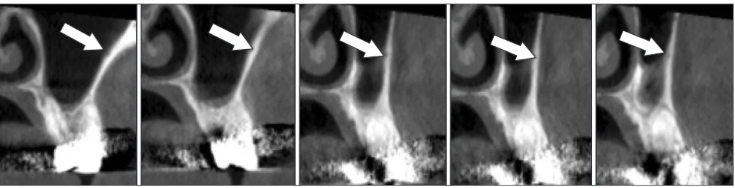
Fig. 1. Schematic representation of the cross-sectional view of the maxillary sinus. The bone canal is intraosseous anastomosis from the posterior superior alveolar artery (arrow indicates intraosseous anastomosis).
Woo-Hyun Park et al: Study on the position of the posterior superior alveolar artery in relation to the performance of the maxillary sinus bone graft procedure in a Korean population. J Korean Assoc Oral Maxillofac Surg 2012
Fig. 2. Schematic representation of sagittal section of the maxillary sinus with blood vessels. (MA: maxillary artery, IA: infra-orbital artery, PSAA: posterior superior alveolar artery, ANS: anterior nasal spine, AR: alveolar ridge)
Woo-Hyun Park et al: Study on the position of the posterior superior alveolar artery in relation to the performance of the maxillary sinus bone graft procedure in a Korean population. J Korean Assoc Oral Maxillofac Surg 2012
view. In coronal views, we examined the existence of artery, the circular or oval radiolucent structure that runs by being fully or partially embedded in the side wall of the maxillary sinus. If there is artery, the distance from the inferior border of the artery to the buccal alveolar ridge in the premolar and molar areas was measured, including the distance from the inferior border of the artery to the sinus floor. The distance between anatomical structures was measured by a “measuring tool” and was gauged up to 2 decimal points.(Fig. 3) We then calculated the detection rate of artery in CT images and measured the distance from the artery to the inferior border of the maxillary sinus and alveolar ridge in the premolar area and molar area. In addition, we measured in the dentate group and the edentulous group (more than one of mission premolar or molar). The measurement standard of premolar was the contact point of the first and second premolars, and the same method was applied for the molar area. If there were many missing teeth, the same standard was applied in the opposite area; the area equidistant from the contact point of the maxillary central incisor became the measurement standard. To distinguish the “artificial defect” generated while reconstructing CT images and actual artery, we compared the axial images and coronal images to reconfirm that the regions contain artery. Afterward, we checked both images for continuity in coronal views close to the posterior superior alveolar artery and measured the distance.(Fig. 4) We compared the detection rate of the posterior superior alveolar artery in male and female groups with chi-square test and conducted t-test to check whether the distance from the posterior superior alveolar artery to the sinus floor and alveolar ridge is different in premolar/molar groups and dentate/partially or fully edentulous groups. Finally, Pearson correlation analysis was performed to determine whether there is correlation between the measured values.
from 20 to 79 years.
2. Methods
We performed imaging of the maxillary sinus using a dental CT (PointCT; Pointnix, Seoul, Korea) and obtained images of 93 regions. To standardize the distortion caused by the change of location of the patient's head, images were taken in Natural Head Position state. We set the guide curve in the axial view of the connected computer program's (Realscan; Pointnix) Dental Volume Reformat window and obtained panoramic view images. Afterward, we examined the coronal images of the maxillary posterior area continuously by moving the sectional plane in panoramic
Fig. 3. Measurement of the distance from the alveolar ridge to the posterior superior alveolar artery on the computed tomography image.
Woo-Hyun Park et al: Study on the position of the posterior superior alveolar artery in relation to the performance of the maxillary sinus bone graft procedure in a Korean population. J Korean Assoc Oral Maxillofac Surg 2012
Fig. 4. Continuity of the posterior superior alveolar artery on the computed tomography T image.
Woo-Hyun Park et al: Study on the position of the posterior superior alveolar artery in relation to the performance of the maxillary sinus bone graft procedure in a Korean population. J Korean Assoc Oral Maxillofac Surg 2012
group, the mean distance from the posterior superior alveolar artery to the alveolar ridge was 20.62±3.05 mm in the premolar region and 17.50±2.84 mm in the molar region. In case of the partially or fully edentulous group, it was 18.83
±2.79 mm in the premolar region and 15.50±1.64 mm in the molar region.(Table 2)
3. Distance from the posterior superior alveolar artery to the sinus floor
Among 51 images containing the posterior superior alveolar artery, measurement in the premolar region was impossible for 4 images because the maxillary sinus was not extended forward. In the case of the dentate group, the mean distance from the posterior superior alveolar artery to the sinus floor was 8.21±2.79 mm in the premolar region and 7.52
±2.07 mm in the molar region. In the case of the partially or fully edentulous group, it was 7.75±3.31 mm in the premolar region and 7.97±2.31 mm in the molar region.(Table 3)
In the correlation analysis of values obtained from CT images, the distance from the alveolar ridge to the posterior superior alveolar artery between the premolar and molar area shows close correlation (P<0.01). Similarly, the distance from the sinus floor to the posterior superior alveolar artery between the premolar and molar areas reveals close correlation (P<0.01). Finally, the distance from the alveolar ridge to the artery shows significant correlation between the dentate group and edentulous group (premolar area: P<0.05,
III. Results
1. Expression ratio of the posterior superior alveolar artery
On the CT scan images, the posterior superior alveolar artery was observed in 18 images among 27 male images and in 33 images among 66 female images. The detection rate of the posterior superior alveolar artery was 66.7% in males and 50% in females, but the difference was not statistically significant (P>0.05).(Table 1)
2. Distance from the posterior superior alveolar artery to the alveolar ridge
Among 51 images containing the posterior superior alveolar artery, there were dentate state in 29 regions and edentulous state in 22 regions. In the case of the dentate
Table 1. Detection rate of the artery by gender
Male Female Total
Detecting site / total site (n) Detection rate (%)
18/27 66.7
33/66 50
51/93 54.8 P=0.351 by chi-square test.
The male and female groups did not show statistically significant difference.
Woo-Hyun Park et al: Study on the position of the posterior superior alveolar artery in relation to the performance of the maxillary sinus bone graft procedure in a Korean population. J Korean Assoc Oral Maxillofac Surg 2012
Table 2. Distance of the alveolar ridge from the artery (mm) Dentate group
(n=29)
Partially or fully
edentulous group (n=22) Total (n=51) P-value
Molar area (mm)
Premolar area (mm) 17.50±2.84
20.62±3.05 15.50±1.64
18.83±2.79 16.64±2.58
19.85±3.04 0.005
0.037 The dentate group and partially or fully edentulous group showed statistically significant difference (P<0.05).
Values are mean±standard deviation.
Woo-Hyun Park et al: Study on the position of the posterior superior alveolar artery in relation to the performance of the maxillary sinus bone graft procedure in a Korean population. J Korean Assoc Oral Maxillofac Surg 2012
Table 3. Distance of the sinus floor from the artery (mm)
Dentate group (mm) Partially or fully
edentulous group (mm) Total (mm) P-value
Molar area (n=51) Premolar area (n=47)
7.53±2.15 (n=29) 8.21±2.79
(n=27)
7.97±2.31 (n=22) 7.75±3.31
(n=20)
7.71±2.17 (n=51) 8.01±2.99
(n=47)
0.455 0.608
The dentate group and partially or fully edentulous group did not show statistically significant difference (P>0.05).
Values are mean±standard deviation.
Woo-Hyun Park et al: Study on the position of the posterior superior alveolar artery in relation to the performance of the maxillary sinus bone graft procedure in a Korean population. J Korean Assoc Oral Maxillofac Surg 2012
amount of sample, and verification is necessary with ample population in the future.
In the group without missing teeth in the posterior area, the distance from the alveolar ridge to the posterior superior alveolar artery was 20.62±3.05 mm in the premolar region and 17.50±2.84 mm in the molar region. In the group with missing teeth, however, it was 18.83±2.79 mm in the premolar region and 15.50±1.64 mm in the molar region.
These results are similar to those of existing research20-24. In the correlation analysis of the distance from the alveolar ridge to the posterior superior alveolar artery in the dentate group and edentulous group, significant correlation was noted (P<0.05 in the premolar region, P<0.01 in the molar region).
It reflects the result of the alveolar bone before and after tooth defect.
In the group without missing teeth in the posterior area, the distance from the sinus floor to the posterior superior alveolar artery was 8.21±2.79 mm in the premolar region and 7.52±2.07 mm in the molar region. In the group with missing teeth, it was 7.75±3.31 mm in the premolar region and 7.97±2.31 in the molar region. In 4 images, measurement in the premolar region was impossible because the maxillary sinus was not extended forward. In the premolar region, the mean distance decreased in the edentulous group than in the dentate group because of the generally less pneumatization of the maxillary sinus in the premolar region than in the molar region and minimal effect of the extraction of premolar on the pneumatization of the maxillary sinus25. In addition, this research used fewer samples and included the extracted premolar in missing teeth. For the distance from the sinus floor to the posterior superior alveolar artery in the dentate group and edentulous group, the difference was not statistically significant (P>0.05 in the premolar region and P>0.05 in the molar region). It did not reflect the pneumatization of the maxillary sinus after the extraction of a tooth in the maxillary posterior area. According to Sharan molar area: P<0.01). Other values did not show correlation.
(Table 4)
IV. Discussion
When damaged in the formation of bony window, the artery in the side wall of the maxillary sinus may affect the process and result of maxillary sinus bone graft surgery because excessive bleeding obstructs the visual field or interferes with the fixation of bone graft material20,21.
Elian et al.22 studied CT images to determine the detection rate of the posterior superior alveolar artery and the distance from the inferior border of the posterior superior alveolar artery to the alveolar ridge. In 50 CT images, the detection rate of the posterior superior alveolar artery was 52.9%, and the distance from the inferior border of the artery to the alveolar ridge was 16.4±3.5 mm. In addition, Mardinger et al.23 examined the existence of the posterior superior alveolar artery in 180 maxillary sinuses using CT scan. The detection rate was 55%, and the mean distance from the residual alveolar ridge to the artery in the first molar area was 16.9
±4.46 mm. Kim et al.24 conducted a research on the Korean population and found that, among 87 CT scan images, the detection rate of artery was 67.5% and the distance from the buccal residual alveolar ridge to the artery was 17.7±4.2 mm in the first premolar area, 14.0±3.4 mm in the first molar area, and 15.9±3.7 mm in the second molar area.
In this study, the detection rate of the posterior superior alveolar artery in the CT scan images of 93 maxillary sinuses was 54.8%. This result is very similar to the expression ratio of the posterior superior alveolar artery in previous studies22-24. Moreover, this study investigated the detection rate of the posterior superior alveolar artery in male and female groups. Though each group's result was different (male: 66.7%, female: 50%), statistical significance was not established. This may be attributed to the insufficient
Table 4. Pearson correlation analysis Pearson correlation
coefficient (r) Edentulous state Distance in P Distance in M
C-A S-A C-A S-A
Edentulous state Distance in P Distance in M
C-A S-A C-A S-A
- -0.293*
-0.083 -0.388**
0.105
- 0.079 0.588**
-0.105
- -0.014
0.434** -
0.147 -
(P : premolar area, M : molar area, C : alveolar ridge, S : sinus floor, A : posterior superior alveolar artery) (*P<0.05, **P<0.01)
Woo-Hyun Park et al: Study on the position of the posterior superior alveolar artery in relation to the performance of the maxillary sinus bone graft procedure in a Korean population. J Korean Assoc Oral Maxillofac Surg 2012
tooth loss in the permanent dentition of adults: United States, 1988- 1991. J Dent Res 1996;75:684-95.
2. Pietrokovski J. The bony residual ridge in man. J Prosthet Dent 1975;34:456-62.
3. Jaffin RA, Berman CL. The excessive loss of Branemark fixtures in type IV bone: a 5-year analysis. J Periodontol 1991;62:2-4.
4. Shackleton JL, Carr L, Slabbert JC, Becker PJ. Survival of fixed implant-supported prostheses related to cantilever lengths. J Prosthet Dent 1994;71:23-6.
5. Choi J, Park HS. The clinical anatomy of the maxillary artery in the pterygopalatine fossa. J Oral Maxillofac Surg 2003;61:72-8.
6. Smiler DG, Johnson PW, Lozada JL, Misch C, Rosenlicht JL, Tatum OH Jr, et al. Sinus lift grafts and endosseous implants.
Treatment of the atrophic posterior maxilla. Dent Clin North Am 1992;36:151-86.
7. Jensen OT, Shulman LB, Block MS, Iacono VJ. Report of the Sinus Consensus Conference of 1996. Int J Oral Maxillofac Implants 1998;13 Suppl:11-45.
8. Del Fabbro M, Testori T, Francetti L, Weinstein R. Systematic review of survival rates for implants placed in the grafted maxillary sinus. Int J Periodontics Restorative Dent 2004;24:565-77.
9. Olson JW, Dent CD, Morris HF, Ochi S. Long-term assessment (5 to 71 months) of endosseous dental implants placed in the augmented maxillary sinus. Ann Periodontol 2000;5:152-6.
10. Tong DC, Rioux K, Drangsholt M, Beirne OR. A review of survival rates for implants placed in grafted maxillary sinuses using meta-analysis. Int J Oral Maxillofac Implants 1998;13:175-82.
11. The Korean Association of Oral and Maxillofacial Surgeons.
Textbook of oral maxillofacial surgery. 2nd ed. Seoul: Uichihaksa;
2005. p. 195.
12. Ikeda A, Ikeda M, Komatsuzaki A. A CT study of the course of growth of the maxillary sinus: normal subjects and subjects with chronic sinusitis. ORL J Otorhinolaryngol Relat Spec 1998;60:147- 52.
13. Uchida Y, Goto M, Katsuki T, Soejima Y. Measurement of maxillary sinus volume using computerized tomographic images.
Int J Oral Maxillofac Implants 1998;13:811-8.
14. Barone A, Santini S, Sbordone L, Crespi R, Covani U. A clinical study of the outcomes and complications associated with maxillary sinus augmentation. Int J Oral Maxillofac Implants 2006;21:81-5.
15. Tatum H Jr. Maxillary and sinus implant reconstructions. Dent Clin North Am 1986;30:207-29.
16. Summers RB. A new concept in maxillary implant surgery: the osteotome technique. Compendium 1994;15:152, 154-6, 158 passim; quiz 162.
17. Rosen PS, Summers R, Mellado JR, Salkin LM, Shanaman RH, Marks MH, et al. The bone-added osteotome sinus floor elevation technique: multicenter retrospective report of consecutively treated patients. Int J Oral Maxillofac Implants 1999;14:853-8.
18. Toffler M. Osteotome-mediated sinus floor elevation: a clinical report. Int J Oral Maxillofac Implants 2004;19:266-73.
19. van den Bergh JP, ten Bruggenkate CM, Disch FJ, Tuinzing DB.
Anatomical aspects of sinus floor elevations. Clin Oral Implants Res 2000;11:256-65.
20. Traxler H, Windisch A, Geyerhofer U, Surd R, Solar P, Firbas W. Arterial blood supply of the maxillary sinus. Clin Anat 1999;12:417-21.
21. Solar P, Geyerhofer U, Traxler H, Windisch A, Ulm C, Watzek G.
Blood supply to the maxillary sinus relevant to sinus floor elevation procedures. Clin Oral Implants Res 1999;10:34-44.
22. Elian N, Wallace S, Cho SC, Jalbout ZN, Froum S. Distribution of the maxillary artery as it relates to sinus floor augmentation. Int J Oral Maxillofac Implants 2005;20:784-7.
23. Mardinger O, Abba M, Hirshberg A, Schwartz-Arad D. Prevalence, diameter and course of the maxillary intraosseous vascular canal with relation to sinus augmentation procedure: a radiographic study. Int J Oral Maxillofac Surg 2007;36:735-8.
and Madjar26, however, the sinus floor was pneumatized 2.18
±2.89 mm after the extraction of a tooth in the maxillary posterior area. Likewise, Jung et al.25 reported that the maxillary sinus was pneumatized 1.52±2.15 mm after the extraction of a molar, and that the extraction of premolar did not affect the pneumatization of the maxillary sinus in Korean population. Therefore, even though the result of this study is different from that of previous studies on maxillary sinus pneumatization, it can possibly change if we use more samples and divide the population into the dentate group and edentulous group.
In addition, the distance from the alveolar ridge to the posterior superior alveolar artery in the premolar and molar region shows statistically significant difference (P<0.01). It means the path of the posterior superior alveolar artery in the side wall of the maxillary sinus has a certain pattern based on the alveolar ridge. In addition, the distance from the sinus floor to the posterior superior alveolar artery in the premolar region and molar region has high statistical significance (P<0.01). This suggests that the path of the posterior superior alveolar artery also has a certain pattern based on the sinus floor.
V. Conclusion
This study took the CT scans of 93 maxillary sinuses and analyzed the location of the posterior superior alveolar artery which runs along the inner wall of the maxillary sinus in 51 sites where it is detectable. As a result, the location of the posterior superior alveolar artery in the alveolar ridge was found to be shorter in edentulous group than dentate group.
In both groups, the distance from the alveolar ridge to the posterior superior alveolar artery is shorter in the molar region than the premolar region.
Therefore, by considering the test results, the posterior superior alveolar artery should be checked using CT scans prior to surgery because the probability of identifying it with CT is 55%. If it is not identified, setting the upper limit of bony window available in the premolar region- not the molar region- as the lower limit is recommended to increase the success rate of the maxillary sinus bone graft by minimizing damage to posterior superior alveolar artery, getting clear field while not disturbing the angiogenesis of bone graft material.
References
1. Marcus SE, Drury TF, Brown LJ, Zion GR. Tooth retention and
maxillary molar extraction assessed with cone beam computed tomography. Korean J Oral Maxillofac Radiol 2009;39:109-13.
26. Sharan A, Madjar D. Maxillary sinus pneumatization following extractions: a radiographic study. Int J Oral Maxillofac Implants 2008;23:48-56.
24. Kim KY, Kim SG, Seo HS, Song YJ, Kim MJ, Hong SM, et al. Arterial arcade of the maxillary sinus related to sinus bone graft in korean population ; A preliminary study using computed topographies. J Korean Assoc Oral Maxillofac Surg 2008;34:475- 9.
25. Jung YH, Nah KS, Cho BH. Maxillary sinus pneumatization after