95
불소를 첨가한 10% Carbamide Peroxide의 법랑질표면 변화와 미백효과
이혜진
†·김현대·김민영·권태엽
1·김교한
1동부산대학 치위생과, 1경북대학교 치과대학 치과생체재료학 교실 및 생체재료연구소
Effect of Fluoridated 10% Carbamide Peroxide on Enamel Surface Change and Whitening
Hye-Jin Lee
†, Hyun-Dae Kim, Min-Young Kim, Tae-Yub Kwon
1and Kyo-Han Kim
1Dept of Dental Hygiene, Dongpusan College University, Busan 612-715, Republic of Korea
1
Dept of Dental Biomaterials, School of Dentistry and Institute for Biomaterials Research and Development, Kyungpook National University, Daegu 700-412, Republic of Korea
Abstract
The purposes of this study were to examine the effect of different fluoridated bleaching solution on the changes in physical and chemical characteristics of tooth. Forty-eight bovine incisors were divided into four groups to receive bleaching treatments, over a 14days period, as follows: no treatment; 10% carbamide peroxide (CP) bleaching;
10% CP containing 0.05% fluoride; and 10% CP containing 0.1% fluoride. All the specimens were highly polished and discolored with commercial COCK.Color and enamel changes were determined with colorimeter, microhardness tester, scanning electron microscope, atomic force microscopy. All the collected data were analyzed with one-way ANOVA.
After the bleaching, bleached groups showed the color change(E
*)
.Microhardness of 10% CP group decreased after tooth bleaching. But microhardness of containing fluoride bleached groups increased after tooth bleaching. Enamel surface of 10% CP bleached group showed any apparent morphology and roughness changes compared to the enamel which was stored in distilled water only. These results demonstrated that Fluoridated 10% Carbamide Peroxide have appreciable bleaching effect on bovine teeth and were not adversely affects enamel. Supporting influence of fluoride-containing bleaching solution on remineralization could be observed and further research must be carried out in various active environments to confirm these results clinically.
Key words
Color, Enamel surface, 10% Carbamide peroxide, Microhardness, Roughness
서 론
산업사회가 발전하고경제활동인구가증가하면서외모 에 대한 관심이 증가되고 있다
.
아름다운 외모를만드는과정의 일환으로 치학 분야에서는 치아교정술
,
심미보철 등과같은 치아의 심미성증가에큰 관심을 가지게되었 다1).
또한사회적으로 미백에대한환자의요구증가에따라치과임상에서는치아에대한심미성의증진을위해치 아의 색조를 밝게 변화시켜주는 치아미백술이 도입되었 으며
,
그에 따른 미백제품의 발달과 미백기술의 발전이 서로 맞물려 치아미백술의 활용이 급진적으로 증가하고 있다2).
치아미백이란
,
강한산화제인과산화수소의oxygen releasing,
mechanical cleansing
작용기전으로치질내착색부분을산화시 켜 착색을 제거하고 치아를원래의 색으로 회복시켜주는 술식을말한다3).
이렇게함으로써자신감있는 미소는첫 인상을 긍정적이고 적극적으로 보이게하는 기능을 부여 한다.
치아미백술의종류로는병원에내원하지않고가정에서
1-2
주간 스스로 시행하는 자가미백술과 병원에 내원하여치과의사의 지도하에 이루어지는 전문가 미백술이 있으 며4)
,
자가미백제로는10%, 16%, 20%
등의Carbamide
peroxide
가 주로 사용되며 전문가미백제로는 주로30-
35%
의Hydrogen peroxide
나35% Carbamide peroxide
가주로사용되고있다
.
자가미백술은
1989
년Haywood
와Hey mann
4)에 의해“Nightgurard vital bleaching”
이라는 제목으로 처음 발표되었고
, 10% Carbamide peroxide
를tray
에 담아환자자 신이 직접시행할 수 있는방법으로,
홈블리칭,
마우스가 드 미백으로도 불려져왔다.
이러한CP
를 이용한치아미†
Corresponding author Tel: 051-540-3877 Fax: 051-540-3823
E-mail: [email protected]
백술이임상에서 성공적으로 사용된경우는
52%
에 달하고 있으며
,
술식의 유용성과 전반적인 임상적 성공에 의 해빠르게받아들여지고있다.
치아미백제처리가치아의색조를변화시킨다는보고가있는데
,
특히치아의명도를 유의하게 증가시킨다는 많은 연구가 있다5-7).
또한Leonard
등8)은 저농도미백제를 장기간적용시 결과적으로고농도미백제를적용한것과비슷한미백효과를얻을 수 있다고 하였다
.
그러나 치아 미백시에는 치아의 색조변화뿐만 아니라 구강연조직 손상이나 법랑질경도 감 소
,
치아과민성증가와같은여러 가지부작용이일어날수있다고하였다9-10)
. McCracken
과Haywood
10), Attin
등12), Lewinstein
등13)은10% Carbamide peroxide
처치 후 법랑질경도가감소한다고보고하였다
.
Bitter
14)는 미백제에 장기간노출된경우법랑질표면의부식으로인하여과민성의증가현상을지적하였다
.
이에반해 미백제가 치아의구조와 구성성분에 영향을미치지 않는다는연구들도다수있어아직의견이분분한실정이
다15-18)
.
미백을통해일어나는경도의감소나치아표면의변화 등에관한 문제들은 불소를 적용함으로써 완화시킬
수 있다는 연구가 진행되고 있다
. Armenio
등19)은 미백후불소를도포함으로써지각과민증의감소를가져왔다고 보고하였으며
, Gladwell
등20)은 불소가 함유된Carbamide
peroxide
는 미백에 영향을 미치지 않으면서 재광화를 촉진시킬수있다고보고하였다21-22)
.
이에 본 연구에서는 자가미백제로 가장 많이 사용되고 있는
10% Carbamide peroxide
를 사용하여,
콜라를이용하여 인공적으로 착색시킨 우치의 법랑질에 미백처리를 한 후 법랑질 표면의 색조변화량
(
∆E*)
과 미세경도,
주사전자현미경에의한표면의미세구조변화및표면의거칠 기를관찰하여미백이법랑질표면에미치는영향과불소 양치용액으로 매일 사용시 권장되는농도인
0.05% NaF
와
0.1%NaF
를 첨가하여 미백 시 불소가 법랑질 표면에미치는영향과미백효과에대해서알아보고자하였다
. 재료 및 방법
1. 연구재료
1)
시편제작우식이나 결함이 없는 건전한치질의 우치의 전치부를 백악법랑경계부에서 치근단측으로
5 mm
정도 되는 지점 에서절단 후 발수하여 증류수로깨끗이 세척한후 실험 전까지0.1%
티몰 용액(Sigma, U.S.A)
에 넣어 냉장보관하였다
.
우치시료 협면의1×1 cm
가량의 평활면을 제외한 다른부위를 모두아크릭 레진으로 포매하여 경화 후 자동연마기
(Automatic Polisher; Labopol-1, Struers,
Denmark)
를 사용하여240
번부터2000
번까지의 사포로단계적으로연마한후
1
µm
의다이아몬드 페이스트로최 종 연마하였다.
색 변화를 측정할 부위는 상아질이 노출되지않도록법랑질에한하여평활하게연마하였으며
,
모든 시편은초음파세척기를 사용하여
30
분간충분히 세척 한후사용하였다.
2)
시편착색시편착색을위해준비된콜라를
24
시간정도충분히교반하여탄산가스를 완전히방출시킨다음사용하였다
.
탄 산가스를 방출시킨콜라에 시편을24
시간 동안담가두었으며
,
침적24
시간 후 꺼내어3
차 증류수로 세척하였다.
착색된 시편들은 초기 색과 경도를 측정한 후
,
순차적으로
12
개씩4
그룹으로분류하였다(Table 1).
2. 연구방법
1)
미백시행제작된시편은하루에
8
시간씩10% Carbamide peroxide (Sigma, USA)
와0.05%NaF, 0.1%NaF
를 첨가하여 미백 시행후 나머지 시간동안은3
차 증류수에 보관하였으며,
이러한 과정을
2
주 동안 반복하였다.
분석전 모든 시편 은3
차증류수를 사용하여충분히세척하여분석에 사용하였다
.
2)
색조변화측정법랑질 색조변화 측정은 색조색차계
(Colorimeter; TC- 8600A, Nippon Denshoku, Japan)
를 사용하여미백 전과후의 색상을 측정하였다
.
색 측정 전 색차계의 감지부에 흡광통을 밀착시켜 영점 조정을 하고,
표준 백색판과흑색판으로
L*, a*, b*
의표시치에대해표준조정을시행한후
,
각시편의색상을측정하였다.
색조변화량은국제조명Table 1.
According to time of bleaching group
Group Classification pH Application period 1 Control group 6.9 Distilled water(D.W.) 2 Experimental
group 5.0 10% Carbamide peroxide 8hours/day
3 Experimental
group 5.02 10% Carbamide peroxide + 0.05% NaF 8hours/day 4 Experimental
group 5.23 10% Carbamide peroxide + 0.1% NaF 8hours/day
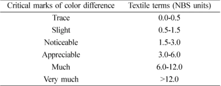
Table 2.
Critical marks of color change according to the national bureau of standards
Critical marks of color difference Textile terms (NBS units)
Trace 0.0-0.5
Slight 0.5-1.5
Noticeable 1.5-3.0
Appreciable 3.0-6.0
Much 6.0-12.0
Very much >12.0
위원회에서 규정한
CIE L*a*b*
측정체계를 사용하였으며
, L*
는0(
검정)
에서100(
백색)
까지의 범위로물체에대 한 명도를 나타내고, a*
와b*
는 채도를 나타낸다. a*
는적색채도
(Red-green chromaticity)
의값을, b*
는 황색채도(Yellow-blue chromaticity)
의값을나타내고, a*
와b*
는0
값에 근접 할수록 무채색에 가깝다
.
색조변화량(
∆E*)
은아래의식에 의해계산되었으며 ∆
E*
은 임상적 색변화량 의 지표가 되는NBS
기준에 의하여 환산한 뒤 비교하였다
(Table 2).
∆
E*={(
∆L*)
2+(
∆a*)
2+(
∆b*)
2}
½NBS unit=
∆E*×0.92
3)
법랑질표면의미세경도측정(VHN)
Vickers diamond indenter
가 부착된 미세경도측정기(MVK-H100, Hardness Testing Machine, Akashi Corporation,
Japan)
를 사용하여 법랑질 표면의 비커스경도(Vickers
Hardness Number)
를 측정하였다.
시편의 법랑질 표면이표면경도계의 압인 방향에 직각이 되도록 위치한 다음
200 gm
의하중으로10
초간압인하고계측하여법랑질표면경도를 측정하였으며
,
한 시편당3
회씩 측정하여 평균을구하였다
.
4)
주사전자현미경관찰시편을 진공상태에서 금이온증착장치
(IB-3, Eiko Co.,
Japan)
로180~200Å
두께의 금으로 피복시킨 후, EDX
(Energy Dispersive X-Ray Spectrophotometer)
가 장착된 주사전자현미경(Scanning Electron Microscope; S-4200, Hitachi Co., Japan)
으로20 kV
상태에서10,000
배의배율 로시편의표면형태변화를분석하였다.
5)
표면거칠기측정시편 표면의 미세 거칠기 변화는 원자현미경
(Atomic Force Microscopy; Nanoscope III Multimode, Digital Instruments, USA)
을 사용하여 표면거칠기값(Ra)
과 양상을분석하였다
.
원자현미경의cantilever
길이450
µm,
탄 성계수0.02-0.1 N/m
인etched sililcon cantilever
를 사용하여
tapping mode
로시행하였다.
3. 자료분석
군별색조변화량과경도변화차이는일변량분산분석법
(one-way ANOVA)
을이용하였고,
유의수준5%
에서Tukey
의 다중비교법으로 사후검정하였다
.
수합된 자료는 통계 분석용소프트웨어인SPSS (SPSS 13.0 KO for Windows, SPSS Inc, Chicago, USA)
를이용하여분석하였다.
결 과
1. 색조 변화 분석
미백 전 인공착색 후 색조변화량은
34.01~34.29
였으며 미백후 색조변화량을 비교해본결과 미백처리군 모두시 간의경과에따라많은색조변화를보였으며통계적으로도 유의한차이가있었다
.
또한NBS unit
로환산해본결과 모든 미백처리군이
very much
에 해당되어 임상적인의미를 지니는것으로 나타나불소 첨가시 치아의 색조
변화에 방해를 하지 않는 것으로 나타났다
(p<0.001,
Table 3,4).
2. 법랑질의 표면경도 분석(VHN)
법랑질의 표면경도 변화
(VHN)
는 대조군에서는14
일동안 거의 변화를보이지 않았으며 통계적으로도 유의한
차이를 나타내지않았다
(P>0.05). 10% CP
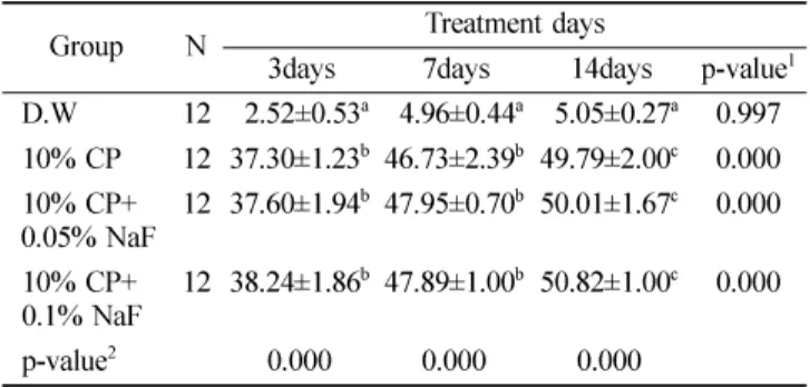
미백군에서는Table 3.
The calculated color change of enamel during the process ( ∆ E*)
Group N Treatment days
3days 7days 14days p-value
1D.W 12 02.52±0.53
a04.96±0.44
a05.05±0.27
a0.997 10% CP 12 37.30±1.23
b46.73±2.39
b49.79±2.00
c0.000 10% CP+
0.05% NaF 12 37.60±1.94
b47.95±0.70
b50.01±1.67
c0.000 10% CP+
0.1% NaF 12 38.24±1.86
b47.89±1.00
b50.82±1.00
c0.000
p-value
20.000 0.000 0.000
Values are mean±S.D.
p-values
1were significantly different during among experimental time by repearted measures ANOVA procedure.
p-values
2were significantly different among the group at each time by one-way ANOVA procedure.
a,b,c
Same letters indicate no significant difference by Tukey's multiple com- parison at
α=0.05 in each group.
Table 4.
National bureau of standards (NBS) units of each group
Group N Treatment weeks
3days 7days 14days
D.W 12 02.31±0.53 (Noticeable) 04.56±0.44 (Appreciable) 04.64±0.27 (Appreciable)
10% CP 12 34.31±1.23 (Very much) 42.99±2.39 (Very much) 45.80±2.02 (Very much)
10% CP + 0.05% NaF 12 34.59±1.94 (Very much) 44.11±0.72 (Very much) 46.00±1.67 (Very much)
10% CP + 0.1% NaF 12 35.18±1.86 (Very much) 44.05±1.01 (Very much) 46.75±1.08 (Very much)
Values are mean±S.D.
법랑질의 표면경도 값이 감소하였고 통계적으로도 유의 한차이를나타냈다
(P<0.05).
반면10% CP
에NaF
를첨 가한 불소미백군에서는 표면경도 값이 통계적으로 유의 한차이가없었다(P>0.05, Table 5).
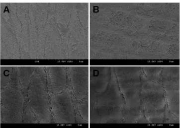
3. 법랑질 표면의 미세구조 분석
Fig. 1
에서A
는 미백처리를 하지 않은법랑질 표면, B
는
10% CP
미백제로 처치한 법랑질 표면, C
는10%
CP+0.05% NaF
로처치한법랑질표면, D
는10% CP+0.1%
NaF
로 처치한법랑질표면을보여주고 있다. B
는미백처리에 의해법랑질 표면이불규칙하고 부분적으로 붕괴양 상을보이며
. C
와D
에서는법랑질표면이불소첨가미백제의사용에의해재광화가되어불소미포함미백군과비 교하여상대적으로평활한면을관찰할수있었다
.
4. 표면 거칠기 변화
미백처리하지 않은시편과비교해보았을 때
,
미백을시행한모든군에서표면이더 거칠어진양상을보였다
.
특 히 미백처리군중 불소미포함 미백군에서 불소첨가 미백군보다 더 거칠어진모습을나타냈다
.
각각의Ra
값은미 백 미처리 군에서는4.090 nm, 10% CP
처리군34.581 nm, 10% CP
에0.05% NaF
를 첨가한 군이27.569 nm, 10%
CP
에0.1% NaF
를 첨가한 군이21.744 nm
로 표면 거칠기값에있어서도정성적분석결과와동일하였다
(Fig. 2).
고 찰
최근 치과에 내원하는 환자들 중 치과 치료를 통해서 구강영역의 기능뿐 아니라심미적인 면까지개선되는 것 을 희망하는환자가 많아지고 심미적인 개선만을 목적으 로내원하는경우도크게늘고있으며사회·문화적발전 과 생활수준이 높아지고 환자들의 심미적요구도가 늘어 나면서 종전의 치아를 삭제하는 도재 수복 등의 보철적 방법보다는 치질 삭제량이 없으면서 간편한 치아미백술 이부각되고있다3)
.
그러나 치아미백술로 인하여발생 가능한 여러 문제점 들이 제기되고 있다
.
현재 상용화되고 있는 미백제는미Table 5.
VHN values of enamel during the process
Group N Baseline Treatment days
3days 7days 14days p-value
1D.W 12 244.63±5.30
a243.36±4.79
a241.73±5.16
a240.18±10.10
a0.708
10% CP 12 243.90±17.27
a223.93±21.80
b217.14±14.93
c216.84±17.23
c0.002
10% CP + 0.05% NaF 12 244.05±15.23
a237.84±20.21
ab242.34±9.27
a242.10±10.37
a0.752 10% CP + 0.1% NaF 12 240.71±16.79
a241.60±8.24
a243.22±6.98
a249.11±12.00
a0.299
p-value
20.910 0.011 0.000 0.000
Values are mean±S.D.
p-values
1were significantly different during among experimental time by repearted measures ANOVA procedure.
p-values
2were significantly different among the group at each time by one-way ANOVA procedure.
a,b,c
Same letters indicate no significant difference by Tukey's multiple comparison at
α=0.05 in each group.
Fig. 1.
SEM images of enamel surfaces.
A: Distilled water 8hours for 14days, B: 10% Carbamide peroxide 8hours for 14days, C: 10% Carbamide peroxide + 0.05% NaF 8hours for 14days , D: 10% Carbamide peroxide + 0.1% NaF 8hours for 14 days (×10,000)
Fig. 2. Atomic force microscope surface topography of enamel surface after bleaching.
(A: Distilled water, B: 10% Carbamide peroxide, C: 10%
Carbamide peroxide + 0.05% NaF, D:10% Carbamide peroxide +
0.1% NaF)
백의효과는탁월하나미백제의사용으로인해발생가능 한 지각과민증
,
치아의 형태학적 구조변화와 그로 인한 색소침착의 용이성 증가로 야기될 수 있는 변색,
치주조직의염증반응등이발생할수있다14)
.
본 연구에서는사람의법랑질성분과비슷한무기질함 량을가지며과거의연구에서 사람의치아와비교하여실 험하였을 때 비슷한경도 및 미세구조를 가진우치를 이 용하여 자가미백제로 널리 사용되는
10% Carbamide peroxide
의 안정성을확인하고자하였다23-24).
또한재광화 효과가확인된NaF
를 첨가하여미백효과와 법랑질 표면에미치는영향에대해서알아본결과색조변화량은모든 미백 처리군에서 치아의 색이 밝아짐을 알 수 있었다
. Gross
와Moser
25)는 ∆E*
가0-2
이면 색 차이를 육안으로인지할 수 없으나
, 2-3
이면 거의 인지할 수 있는 정도이고
, 3-8
이면 보통 인지할수 있으며, 8
이상이면현저하게인지할수있다고보고한결과와비교해보면본연구에서
∆
E*
및NBS
값으로환산한결과30
보다높은수치로미백전·후현저한색조변화량을보여주고있다
.
모든미백 처리군에서very much
로 나타나 임상적으로 의미 있는결과를보였으며
,
주에6
시간씩4
주간미백처리한Andreas
등의연구26)에서와비슷한결과를보였다
.
또한Haywood
와
Heymann
등4)의Carbamide peroxide
를이용한가정용치아미백제의 사용에따른 색조변화량에 관한보고와 같 은경향을나타냈다
.
또한 법랑질 표면의 변화를 관찰하기 위하여 미세경도 측정
,
주사전자현미경,
원자현미경을 이용하였으며 법랑질 표면경도의 변화는미세경도 측정기를이용하여측정 한 결과미백 처리군에서 표면경도가 낮아진것을 알 수 있었으며
,
이는McCracken
과Haywood
10), Attain
등12)의10% Carbamide peroxide
처치 후 법랑질경도가감소한다는 보고와 일치한 결과였다
.
반면 불소를 첨가한 미백처리군에서는 통계적인 유의성은 확인할 수 없었으나 경 도 값이 높아짐을 알 수 있었다
.
이는 미백제에 불소를첨가하여 도포함으로써 미백에의해 발생 가능한치아의 형태학적구조변화를미백처리전과유사한정도로재광 화를시킬수있다고보고한결과와유사하였다21,27-28)
.
그 러나 같은 농도의 미백제를pH
를 달리하여 사용하였던연구들을 살펴보면
pH
가5.2
아래인경우표면 경도값이감소한반면
pH
가 중성에 가까울수록 표면 경도값에 변화가없는것으로나타났다29-30)
.
이러한결과를미루어볼때
pH
는 미백시 중요한 요소가 되므로 중성의pH
와 적 정농도의 불소첨가 미백제를 사용하게 되면 미백으로 인 해표면경도감소에영향을미치지않을것으로사료되었 다.
원자 현미경과 주사전자현미경을사용하여 정량 및 정 성적으로 표면변화양상을 확인한 결과
,
미백미처리군과미백처리군을 비교했을 때 미백처리군에서
Ra
값이 상대 적으로높게 나타났으며 특히 불소미포함 미백군에서 원자현미경의 정량적인 결과와 일치하는 거칠어진 양상을 확인할 수있었다
. Basting
등31)과Turkun
등32)의 연구에서 미백후법랑질표면의부분적인탈회를보였던결과와일 치하는것을알수있었다.
이상의결과들을종합해볼때
, 10% Carbamide peroxide
를 이용한자가미백술 시 법랑질 표면에미세 구조적변 화를야기할수 있으므로
,
미백시 불소를포함한미백제 를 사용하거나 미백 후 불소도포를 시행함으로써 자가미 백술 시 야기될수 있는 지각과민증이나법랑질 미세구 조변화 등 여러문제점을감소시킬 수있을 것으로 사료 된다.
본연구의한계점으로는임상적으로 사용되는 미백
gel
을 사용하지 않고 불소의 재광화 효과를 관찰하기위해
CP
용액을 사용하였으며,
구강 내 환경을 완벽히 재현할 수 없는 in vitro실험으로 진행되었기 때문에 외인성 및 내인성 치아착색들을 완벽하게 구현하지 못하는 제한성 을 가진다.
뿐만아니라 구강환경에서는타액에의한 탈회와 재광화가 서로 상호 보완되고
,
불소,
칼슘,
인산 등에 의해 치아의형태학적 구조와미세경도의 변화가 완화될 수있으나,
in vitro실험여건상구강환경을반영할수없 다는 문제점을 지적할수있다.
따라서향후 in vitro실험의 한계점을 보완한 in vivo
,
in situ연구를 통하여 보다세밀하고종합적인분석이필요할것으로사료된다
.
요 약
본 연구는 자가 미백제로 많이 사용되고 있는
10%
Carbamide Peroxide
에NaF
를 첨가하여사용하였으며 미백 후인공적으로 변색시킨법랑질의색
,
경도및 표면변 화에미치는영향을관찰하여다음과같은결론을얻었다.
1. 10% CP
미백처리군과10% CP+NaF
미백처리군의색조변화량은미백 후색조 변화를보였으며 통계적 으로도유의한 차이를나타냈으며
NBS unit
로 환산해본 결과 미백처리군 모두
very much
에 해당되어임상적인의미를지니는것으로나타났다
(p<0.001).
2. 10% CP
미백처리군은미백처리 후의법랑질표면경도 값이미백처리전에비해 감소하였으며통계적으 로도유의한차이를나타냈다
.
불소첨가미백군은미 백처리 전과 비교하여 표면경도 값이 거의 변하지 않았다.
3.
미백처리 전·후의 법랑질표면에 대한 미세구조를 분석하기 위해주사전자현미경과원자현미경으로관 찰한 결과 미백처리 후 탈회현상에 의해 법랑질 표 면이 거칠어짐을 관찰 할 수 있었으며 미백제와 함 께 불소를 적용한 처리 군에서는 미백 처리에 의해 거칠어진 법랑질표면이불소에의해 재광화가됨을 관찰할수있었다.
이상의결과를미루어볼때
10% CP
치아미백은법랑질 표면에영향을 미쳐미백 후 치아의시림이나표면의 형태변화