https://doi.org/10.4174/astr.2019.96.2.86 Annals of Surgical Treatment and Research
Oncologic evaluation of obesity as a factor in patients with rectal cancer undergoing laparoscopic surgery: a propensity-matched analysis using body mass index
Il Tae Son1,2, Duck-Woo Kim1, Eun Kyung Choe3, Young Hoon Kim4, Kyoung Ho Lee4, Soyeon Ahn5, Sung Il Kang1, Myung Jo Kim1, Heung-Kwon Oh1, Jae-Sung Kim6, Sung-Bum Kang1
1Department of Surgery, Seoul National University Bundang Hospital, Seongnam, Korea
2Department of Surgery, The Catholic University of Korea, Uijeongbu St. Mary's Hospital, Uijeongbu, Korea
3Seoul National University Hospital Gangnam Center, Seoul, Korea
4Department of Radiology, Seoul National University Bundang Hospital, Seoul National University College of Medicine, Seongnam, Korea
5Medical Research Collaborating Center, Seoul National University Bundang Hospital, Seongnam, Korea
6Department of Radiation Oncology, Seoul National University Bundang Hospital, Seoul National University College of Medicine, Seongnam, Korea
Received July 18, 2018, Revised October 5, 2018, Accepted October 16, 2018
Corresponding Author: Duck-Woo Kim
Department of Surgery, Seoul National University Bundang Hospital, 82 Gumi-ro 173beon-gil, Bundang-gu, Seongnam 13620, Korea
Tel: +82-31-787-7101, Fax: +82-31-787-4078 E-mail: [email protected]
ORCID code: https://orcid.org/0000-0001-9218-4676
• The abstract of this article was presented at the 50th Annual Meeting of the Korean Society of Coloproctology in Seoul, Korea, on April 1–2, 2017.
Copyright ⓒ 2019, the Korean Surgical Society
cc Annals of Surgical Treatment and Research is an Open Access Journal. All articles are distributed under the terms of the Creative Commons Attribution Non- Commercial License (http://creativecommons.org/licenses/by-nc/4.0/) which permits unrestricted non-commercial use, distribution, and reproduction in any medium, provided the original work is properly cited.
Purpose: This study evaluated the oncologic impact of obesity, as determined by body mass index (BMI), in patients who underwent laparoscopic surgery for rectal cancer.
Methods: The records of 483 patients with stage I–III rectal cancer who underwent laparoscopic surgery between June 2003 and December 2011 were reviewed. A matching model based on BMI was constructed to balance obese and nonobese patients. Cox hazard regression models for overall survival (OS) and disease-free survival (DFS) were used for multivariate analyses. Additional analysis using visceral fat area (VFA) measurement was performed for matched patients.
The threshold for obesity was BMI ≥ 25 kg/m2 or VFA ≥ 130 cm2.
Results: The score matching model yielded 119 patients with a BMI ≥ 25 kg/m2 (the obese group) and 119 patients with a BMI < 25 kg/m2 (the nonobese group). Surgical outcomes including operation time, estimated blood loss, nil per os periods, and length of hospital stay did not differ between the obese and the nonobese group. The retrieved lymph node numbers and pathologic CRM positive rate were also similar in between the 2 groups. After a median follow-up of 48 months (range, 3–126 months), OS and DFS rates were similar between the 2 groups. A tumor location-adjusted model for overall surgical complications showed that a BMI ≥ 25 kg/m2 were not risk factors. Multivariable analyses for OS and DFS showed no significant association with a BMI ≥ 25 kg/m2.
Conclusion: Obesity was not associated with long-term oncologic outcomes in patients undergoing laparoscopic surgery for rectal cancer in the Asian population.
[Ann Surg Treat Res 2019;96(2):86-94]
Key Words: Body mass index, Rectal neoplasms, Laparoscopy
INTRODUCTION
Obesity is an uncontrollable host factor, and a predictor of surgical outcomes, including technical difficulties, post- operative complications, and anthropometric events in patients undergoing gastrointestinal surgery [1]. Body mass index (BMI) and visceral fat area (VFA) have been widely used to define threshold values for obesity, although these markers do not always reflect the degree of intra-abdominal or intrapelvic fat, which may be associated with technical difficulties during surgical procedures [2]. In colorectal cancer, obesity is not only an etiologic risk factor, but a predictive marker for morbidity and mortality [3]. Moreover, obesity, as evaluated by BMI and VFA, has been reported to affect outcomes in patients with colorectal cancer who underwent open or laparoscopic surgery [4-9].
The impact of obesity in rectal cancer may differ from that in colon cancer because visceral fat and the volume of the pelvis may have greater effects on surgical procedures for rectal cancer. Studies assessing the effects of obesity as determined by BMI on outcomes in rectal surgery patients have yielded conflicting results [10-12]. Previous studies using VFA have also suggested a strong consensus that measures of visceral adiposity are more accurate than BMI, and that these measures of visceral adiposity predict more difficult resections and a higher incidence of postoperative complications, although these studies reached disparate conclusions [9,13].
However, in clinical practice, it may not be feasible for surgeon to take the approach of measurement of VFA for the prediction of surgical outcomes. Furthermore, the long-term oncologic effect of obesity, as determined by BMI in laparoscopic surgery for rectal cancer, remains still unclear. This study therefore evaluated surgical complications and the oncologic impact of obesity, based on BMI, in patients who underwent laparoscopic surgery for rectal cancer, using a matching model that balanced clinicopathologic factors in obese and non-obese patients. Additionally, we investigated distribution of VFA in the matched patients to evaluate a relationship with the BMI.
METHODS
Patients who underwent laparoscopic surgery for stage I–
III rectal cancer at the Department of Surgery, Seoul National University Bundang Hospital, between June 2003 and December 2011 were retrospectively analyzed. Rectal cancer was defined as an adenocarcinoma located within 15 cm of the anal verge.
Patients with stage IV disease, synchronous colorectal cancer, multiple malignancies or a previous history of abdominal surgery were excluded, as were patients who underwent noncurative resection or trans-anal excision. Patients were divided into 2 groups based on BMI, the BMI cutoff for obesity
based on classification by the Asia Cohort Consortium of the World Health Organization (WHO). Patients with a BMI ≥ 25 kg/m2 were defined as obese and those with a BMI < 25 kg/m2 as nonobese [14].
VFA was preoperatively measured by Fat Scan software, Rapidia version 2.8, on cross-sectional CT scans, obtained at the middle of L4. Adipose tissue was determined by setting the attenuation level within a range of -190 to -30 Hounsfield units [15]. VFA was defined by manual contour tracing and calculated automatically by Fat Scan. The VFA cutoff for obesity was based on the classification in a previous study with VFA < 130 cm2 defined as nonobese and VFA ≥ 130 cm2 as obese [16].
Differences between nonobese and obese groups, based on BMI and VFA, were investigated. Factors evaluated included baseline characteristics, pathologic parameters (retrieved lymph nodes and circumferential resection margin status), short-term surgical outcomes (conversion, technical difficulty, operation time, estimated blood loss, nil per os [NPO] period, and days hospitalized), postoperative surgical complications, and long-term oncologic outcomes, including recurrence pattern, overall survival (OS) and disease-free survival (DFS).
The technical difficulty of surgical procedures was defined as a significant deviation from 3 surgical procedure categories as described; step 1, visualization and localization of the tumor after trocar insertion; step 2, lymphovascular dissection and bowel mobilization; step 3, transection and anastomosis [17].
Preoperative chemoradiotherapy was performed for locally advanced rectal cancer as recommended by the National Comprehensive Cancer Network guideline, which was described in our previous study [18]. Long-course radiotherapy was given over 5.5 weeks at a dose of 50.4 Gy, of which 45 Gy was applied in 25 fractions to the pelvis, and a 5.4 Gy boost was applied in 3 fractions to the primary tumor. The chemotherapeutic regimens consisted of 2 cycles of an intravenous bolus of fluorouracil (400 mg/m2 per day) and racemic D, L-leucovorin (20 mg/m2 per day) for 3 days in weeks 1 and 5 of radiotherapy, or continuous oral administration of capecitabine (825 mg/m2 twice daily) during radiotherapy.
All operations were performed by 2 experienced surgeons, each of whom had performed > 50 laparoscopic rectal operations. Tumor stage was classified using the 7th edition of the American Joint Committee on Cancer staging system.
Patterns of recurrence were classified as local (tumor recurrence around the anastomosis or the region of the primary operation), systemic, or combined. In the pathological assessment of the completeness of total mesorectal excision (TME) and the involvement of a pathological circumferential resection margin (pCRM) defined as the shortest distance from a tumor of ≤ 1 mm, we examined the quality of the TME specimens from selected patients via a multidisciplinary team approach, using the method of Nagtegaal and Quirke [19]. The quality of the
TME specimens and the pCRM assessment were validated in our previous study [18].
Patients in the obese and nonobese groups, as defined by the BMI cutoff of 25 kg/m2, were matched 1:1 to adjust for potential biases that may influence surgical and oncologic outcomes as in previous studies [6,7,20]. Covariates for matching included preoperative factors (age, sex, tumor height, American Society of Anesthesiologists (ASA) physical status classification, and preoperative treatment) and postoperative factors (differentiation type, T-stage, N-stage) (Supplementary Fig. 1). All variables including surgical complications, and long- term oncologic outcomes were compared between the obese and nonobese groups. In addition, the radiologic measurement of VFA was performed in only matched patients with the exclusion of 59 patients because the quality of their CT scans was poor. The correlation between BMI and VFA was also assessed by scatter plot analysis.
Categorical variables were analyzed using the chi-square test or Fisher exact test. Continuous variables expressed as means ± standard deviation were compared using Student t-test. OS and DFS were analyzed by the Kaplan-Meier method and compared by the log-rank test. A logistic regression model for overall surgical complications and a Cox proportional hazards regression model for OS and DFS were performed for risk stratification. Risk of overall surgical complications was stratified using tumor location-adjusted multivariate analysis.
Multivariable analyses for OS and DFS were performed using the tumor location. Data were analyzed using IBM SPSS Statistics ver. 20.0 (IBM Co., Armonk, NY, USA) and R statistical software (R Foundation for Statistical Computing, Vienna, Austria; www.r-project.org). Two-tailed statistical significance was set at P < 0.050. This study was approved by the Institutional Review Board (IRB) of the Seoul National University Bundang Hospital, Korea (approval number: B-1504-
296-109). And the IRB authority did waive the requirement to obtain informed consent because this retrospective study did not include the personal information.
RESULTS
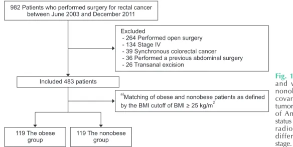
Of the total 982 patients who had undergone surgery for rectal cancer during the study period, 483 patients who underwent laparoscopic surgery for stage I–III rectal cancer were included after 499 patients were excluded (Fig. 1). Based on a BMI cutoff of 25 kg/m2, there were 119 patient pairs matched by age, sex, tumor height, ASA physical status classification, preoperative radiotherapy, chemotherapy, differentiation type, and T–N stage. Included patients had a mean BMI of 24.61 kg/
982 Patients who performed surgery for rectal cancer between June 2003 and December 2011
Excluded
- 264 Performed open surgery - Stage IV
- Synchronous colorectal cancer
- Performed a previous abdominal surgery - Transanal excision
134 39 36 26
a)
2
Matching of obese and nonobese patients as defined by the BMI cutoff of BMI > 25 kg/m
Included 483 patients
119 The obese group
119 The nonobese group
Fig. 1. Flow chart for matching and validation of obese and nonobese patients. a)Matched covariates including age, sex, tumor height, American Society of Anesthesiologists physical status classification, preoperative radiotherapy, chemotherapy, differentiation type and T–N stage. BMI, body mass index.
15 30
VFA(cm)2
BMI (kg/m )2 0
300
200
100
35
20 25
Fig. 2. The relationship of body mass index (BMI) with visceral fat area (VFA). Coefficient of correlation, R2 = 0.436, P < 0.001.
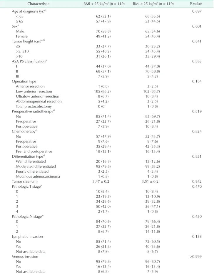
Table 1. Baseline characteristics and pathologic parameters
Characteristic BMI < 25 kg/m2 (n = 119) BMI ≥ 25 kg/m2 (n = 119) P-value
Age at diagnosis (yr)a) 0.697
< 65 62 (52.1) 66 (55.5)
≥ 65 57 (47.9) 53 (44.5)
Sexa) 0.601
Male 70 (58.8) 65 (54.6)
Female 49 (41.2) 54 (45.4)
Tumor height (cm)a,b) 0.841
≤5 33 (27.7) 30 (25.2)
>5, ≤10 55 (46.2) 54 (45.4)
>10 31 (26.1) 35 (29.4)
ASA PS classificationa) 0.883
I 44 (37.0) 44 (37.0)
II 68 (57.1) 70 (58.8)
III 7 (5.9) 5 (4.2)
Operation type 0.184
Anterior resection 1 (0.8) 3 (2.5)
Low anterior resection 105 (88.2) 102 (85.7)
Ultralow anterior resection 8 (6.7) 10 (8.4)
Abdominoperineal resection 5 (4.2) 3 (2.5)
Total proctocolectomy 0 (0) 1 (0.8)
Preoperative radiotherapya) 0.819
No 85 (71.4) 83 (69.7)
Preoperative 27 (22.7) 26 (21.8)
Postoperative 7 (5.9) 10 (8.4)
Chemotherapya) 0.824
No 57 (47.9) 52 (43.7)
Preoperative 9 (7.6) 9 (7.6)
Postoperative 35 (29.4) 42 (35.3)
Pre- and postoperative 18 (15.1) 16 (13.4)
Differentiation typea) 0.851
Well differentiated 20 (16.8) 15 (12.6)
Moderated differentiated 95 (79.8) 99 (83.2)
Poorly differentiated 3 (2.5) 4 (3.4)
Mucinous adenocarcinoma 1 (0.8) 1 (0.8)
Tumor size (cm) 3.47 ± 0.2 3.51 ± 0.2 0.942
Pathologic T stagea) 0.470
0 10 (8.4) 10 (8.4)
1 23 (19.3) 13 (10.9)
2 34 (28.6) 39 (32.8)
3 50 (42.0) 56 (47.1)
4 2 (1.7) 1 (0.8)
Pathologic N stagea) 0.430
0 84 (70.6) 79 (66.4)
1 27 (22.7) 26 (21.8)
2 8 (6.7) 14 (11.8)
Lymphatic invasion 0.138
No 85 (71.4) 72 (60.5)
Yes 26 (21.8) 40 (33.6)
Not available data 8 (7.8) 8 (6.7)
Venous invasion >0.999
No 95 (79.8) 96 (80.7)
Yes 16 (13.4) 16 (13.4)
Not available data 8 (6.8) 7 (5.9)
Table 1. Continued
Characteristic BMI < 25 kg/m2 (n = 119) BMI ≥ 25 kg/m2 (n = 119) P-value
Perineural invasion 0.347
No 93 (78.2) 85 (71.4)
Yes 18 (15.1) 27 (22.7)
Not available data 8 (6.7) 7 (5.9)
Values are presented as mean ± standard deviation or number (%).
BMI, body mass index; ASA PS, American Society of Anesthesiologists physical status.
a)Covariates for matching preoperative and postoperative factors in included patients. b)Tumor height was classified according the distance from anal verge.
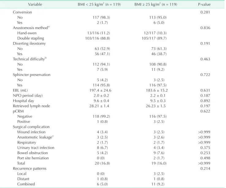
Table 2. Surgical outcome including perioperative parameters, short-term complication and, recurrence
Variable BMI < 25 kg/m2 (n = 119) BMI ≥ 25 kg/m2 (n = 119) P-value
Conversion 0.281
No 117 (98.3) 113 (95.0)
Yes 2 (1.7) 6 (5.0)
Anastomosis methoda) 0.836
Hand-swen 13/116 (11.2) 12/117 (10.3)
Double stapling 103/116 (88.8) 105/117 (89.7)
Diverting ileostomy 0.191
No 63 (52.9) 73 (61.3)
Yes 56 (47.1) 46 (38.7)
Technical difficultyb) 0.463
No 112 (94.1) 108 (90.8)
Yes 7 (5.9) 11 (9.2)
Sphincter preservation 0.722
No 5 (4.2) 3 (2.5)
Yes 114 (95.8) 116 (97.5)
EBL (mL) 197.4 ± 24.6 183.6 ± 15.2 0.631
NPO period (day) 2.0 ± 0.2 2.2 ± 0.1 0.187
Hospital day 9.6 ± 0.4 9.5 ± 0.3 0.892
Retrieved lymph node 28.21 ± 1.4 26.23 ± 1.5 0.197
pCRM 0.622
Negative 118 (99.2) 116 (97.5)
Positive 1 (0.8) 3 (2.5)
Surgical complication
Wound infection 4 (3.4) 3 (2.5) >0.999
Anastomotic leakagec) 3 (2.5) 3 (2.6) >0.999
Respiratory 2 (1.7) 2 (1.7) >0.999
Urinary tract infection 8 (6.7) 4 (3.4) 0.375
Bowel obstruction 5 (4.2) 9 (7.6) 0.253
Port site herniation 0 (0) 2 (1.7) 0.498
Total 20 (16.8) 19 (16.0) >0.999
Recurrence patterns 0.214
Local 0 (0) 3 (2.5)
Distant 1 (0.8) 1 (0.8)
Combined 6 (5.0) 11 (9.2)
Values are presented as number (%) or mean ± standard deviation.
BMI, body mass index; EBL, estimated blood loss; NPO, nil per os; pCRM, pathological circumferential resection margin.
a)Not available data (n = 5). b)Defined as a significant deviation from the ordinary surgical procedure [25]. c)Excluded patients who did not perform a sphincter preservation surgery.
15 30
Operationtime(min)
BMI (kg/m )2 0
700 600 500
400 300 200 100
35
20 25
A
Coefficient of correlation for BMI, R = 0.149, P < 0.0212 Tumor location (cm)
: <5 : >5, <10 : >10
B
Operation time (min) BMI < 25 kg/m (n = 119)
2BMI >
(n = 119) 25 kg/m2
P-value
Tumor location
<5 from AV
>5, <10 from AV
>10 from AV Total
249.3 + 12.0 215.9 + 10.1 185.2 + 9.8 217.4 + 6.5
277.8 + 14.0 240.1 + 13.2 195.7 + 11.7 236.5 + 8.2
0.137 0.150 0.491 0.070
Fig. 3. (A) Scatter plots showing the relationships of body mass index with operation time according to tumor location. (B) Operation time was compared between obese and nonobese patients, based on a body mass index (BMI) cutoff of 25 kg/m2. AV, anal verge.
0 96
Overallsurvival(probability)
After surgery (mo) 0.0
1.0
0.8
0.6
0.4
0.2
12 24 36 48 60 72 84 0 96
Disease-freesurvival(probability)
After surgery (mo) 0.0
1.0
0.8
0.6
0.4
0.2
12 24 36 48 60 72 84
BMI < 25 kg/m : 87.4%
BMI > 25 kg/m : 91.6%
Log rank P = 0.239
2 2
BMI < 25 kg/m : 94.1%
BMI > 25 kg/m : 87.4%
Log rank P = 0.076
2 2
A B
Fig. 4. Kaplan-Meier analyses of overall survival (A) and disease-free survival (B) in the 119 matched pairs of obese and nonobese patients, based on the World Health Organization cutoff of body mass index (BMI) of 25 kg/m2. Patients were matched 1:1 based on age, sex, tumor height, American Society of Anesthesiologists physical status classification, preoperative treatment, differentiation type, and T–N stage.
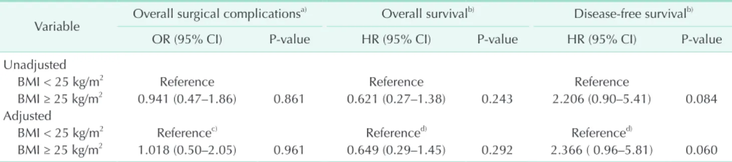
Table 3. The risk stratification for surgical complication and survival according to the body mass index
Variable Overall surgical complicationsa) Overall survivalb) Disease-free survivalb)
OR (95% CI) P-value HR (95% CI) P-value HR (95% CI) P-value
Unadjusted
BMI < 25 kg/m2 Reference Reference Reference
BMI ≥ 25 kg/m2 0.941 (0.47–1.86) 0.861 0.621 (0.27–1.38) 0.243 2.206 (0.90–5.41) 0.084 Adjusted
BMI < 25 kg/m2 Referencec) Referenced) Referenced)
BMI ≥ 25 kg/m2 1.018 (0.50–2.05) 0.961 0.649 (0.29–1.45) 0.292 2.366 ( 0.96–5.81) 0.060 OR, odd ratio; CI, confidence interval; HR, hazard ratio; BMI, body mass index.
a)Logistic regression model. b)Cox proportional hazards regression model. c)Tumor location – adjusted logistic regression analysis.
d)Tumor location – adjusted Cox proportional hazards regression model.
m2 (range, 16.90–33.15 kg/m2), and a mean VFA of 114.65 cm2 (range, 23.14–265.90 cm2). The median of obesity between the obese and nonobese groups was as follows; BMI, 22.31 vs.
26.59 kg/m2; VFA, 70.75 vs. 138.4 cm2. BMI was significantly associated with VFA (coefficient of correlation, R2 = 0.436, P <
0.001) (Fig. 2). Furthermore, the BMI-matched model showed that the characteristics and pathologic parameters of the non- obese and obese patients did not differ significantly (Table 1).
Perioperative parameters, including technical difficulties, conversion rate, anastomosis method and sphincter pre- servation, did not differ significantly between the nonobese and obese groups (Table 2). Surgical outcomes, including operation time, estimated blood loss, NPO period, and length of hospital stay also did not differ, nor did the number of retrieved lymph nodes and resection margin status. Postoperative surgical complications did not differ in the obese and nonobese groups.
Operation time correlated significantly with BMI (coefficient of correlation, R2 = 0.149, P = 0.021) (Fig. 3A). Operation time according to tumor location did not differ significantly between the obese and nonobese groups (Fig. 3B).
Kaplan-Meier analysis showed that OS and DFS did not differ significantly in the obese and nonobese groups based on a BMI cutoff of 25 kg/m2 (Fig. 4A, B). Rates of local (2.5% vs. 0%), distant (0.8% vs. 0.8%), and combined (9.2% vs. 5.0%) recurrence did not differ significantly.
Univariate analyses showed that BMI ≥ 25 kg/m2 was not associated with overall surgical complications, OS or DFS (Table 3). Multivariate analyses, using a tumor location-adjusted model for overall surgical complications, showed that a BMI ≥ 25 kg/
m2 was a not risk factor for overall surgical complications (odds ratio [OR], 1.018; 95% confidence interval [CI], 0.50–2.05; P = 0.961). Multivariable analyses for OS (hazard ratio [HR], 0.649;
95% CI, 0.29–1.45; P = 0.292) and DFS (HR, 2.366; 95% CI, 0.96–
5.81; P = 0.060) showed no significant associations with a BMI
≥ 25 kg/m2.
DISCUSSION
This study evaluated the oncologic impact of obesity, based on BMI, in patients with rectal cancer who underwent laparoscopic surgery. This study found that obesity did not add to technical challenges or oncologic hazards. These outcomes were further supported by both BMI-matched and VFA- measured models. Similar to our findings, previous studies reported similar oncologic outcomes in obese and nonobese patients, suggesting that obesity did not increase postoperative complications in patients who underwent laparoscopic surgery for colorectal cancer [6,7], although other studies have reported that obesity was useful in predicting surgical complications [5,8,9]. In rectal cancer needing TME procedure related directly to surgical quality and prognosis, visceral adiposity was
associated with postoperative, oncologic, and survival outcomes [13].
However, conflicting outcomes were observed in some studies [21] similar to our findings, giving some reasons including a lower rate of positive CRM, the benefit of neoadjuvant therapy, and resections done at a specialty cancer center with dedicated oncologic colorectal surgeons. Even, obesity has been found to have a positive oncologic effect in patients with rectal cancer.
Rectal cancer patients with a BMI ≥ 25 kg/m2 had a higher DFS rate and a lower distant metastasis rate than patients with a BMI < 25 kg/m2 [12,22]. In this study, only 4 patients had a positive CRM after the completion of TME. Furthermore, we considered that any potential adverse effects of obesity may have been masked in the setting of a high-volume specialized colorectal unit.
Obesity may be oncologically relevant in patients with rectal cancer. Obesity can reveal the underlying nutritional status of patients undergoing major intra-abdominal cancer surgery [23].
Furthermore, cachexia, one of the most life-threatening factors in cancer, can induce alterations in intermediary metabolism through mechanisms that include the release of cytokines, lipid-mobilizing. and proteolysis-inducing factors [24]. During adjuvant treatment, obese patients are less likely to develop chemotherapy-related toxicities than nonobese patients, likely because obesity facilitates the administration of appropriate doses and the continuation of chemotherapy [25].
In this study, the correlation coefficients between BMI and VFA was low but significant, although BMI could not reflect the distribution of intraabdominal adipose tissue in Asian populations having lower BMI but higher proportion of intraabdominal adipose tissue [15,26]. This study also attempted to assess the relationship of obesity with operation time according to tumor location. Determinations of obesity by VFA and BMI may differ in predicting the technical difficulty of laparoscopic rectal cancer surgery. The correlation coefficients between obesity and operation time were very low, showing that BMI was more closely correlated than VFA, in disagreement with previous studies suggesting that VFA could be a better predictor for surgical outcomes than general obesity measured by the BMI [8,9,13]. A possible explanation for these correlations might be that the degree of obesity in our study was not severe without patients excessing BMI > 35 or 40 kg/m2. Therefore, obesity may seem not to influence rectal cancer surgery in this study. Furthermore, we consider that the VFA measurement may be limited in terms of potential interobserver differences and a difficulty of being generalized due to needing Fat Scan software, compared to BMI simply calculated as weight in kilograms divided by height in meters squared (kg/m2).
This study had several limitations. First, its retrospective design has resulted in an inherent bias in spite of adoption of propensity score matching to reduce selection bias. Second, our
criterion for obesity, BMI ≥ 25 kg/m2, differs from that typically used for Western populations (BMI ≥ 30 kg/m2) [27]. The prevalence of obesity is much lower in Asian than in Western populations, whereas Asians have a higher rate of body fat compared to Caucasians of the same BMI [14]. A prospective cohort study based on a larger population showed that the average BMI of Koreans was 23.2 kg/m2, with patients having a BMI between 23.0–24.9 kg/m2 being at lowest risk of death [28]. In this study, only 10 patients, or 2.1%, had a BMI > 30 kg/m2. Furthermore, because the WHO recommended a lower threshold for obesity in Asian versus Western populations [29], our criteria for obesity may have been reasonable.
In conclusion, the present study showed that obesity was
not associated with long-term oncologic outcomes in patients undergoing laparoscopic rectal cancer surgery in Asian populations.
CONFLICTS OF INTEREST
No potential conflict of interest relevant to this article was reported.
SUPPLEMENTARY MATERIAL
Supplementary Fig. 1 can be found via https://www.astr.or.kr/
src/sm/astr-96-86-s002.pdf.
REFERENCES
1. STARSurg Collaborative. Multicentre pro- spective cohort study of body mass index and postoperative complications fol low- ing gastrointestinal surgery. Br J Surg 2016;103:1157-72.
2. Bouchard C, Despres JP, Mauriege P. Ge- ne tic and nongenetic determinants of regional fat distribution. Endocr Rev 1993;
14:72-93.
3. Calle EE, Rodriguez C, Walker-Thurmond K, Thun MJ. Overweight, obesity, and mortality from cancer in a prospectively studied cohort of U.S. adults. N Engl J Med 2003;348:1625-38.
4. Watanabe J, Tatsumi K, Ota M, Suwa Y, Suzuki S, Watanabe A, et al. The impact of visceral obesity on surgical outcomes of laparoscopic surgery for colon cancer. Int J Colorectal Dis 2014;29:343-51.
5. Moon HG, Ju YT, Jeong CY, Jung EJ, Lee YJ, Hong SC, et al. Visceral obesity may affect oncologic outcome in patients with colorectal cancer. Ann Surg Oncol 2008;15:1918-22.
6. Nakamura T, Miura H, Ikeda A, Sato T, Naito M, Ogura N, et al. Laparoscopic sur- gery for obese patients with colon cancer:
a case-matched control study. Surg Today 2013;43:763-8.
7. Makino T, Trencheva K, Shukla PJ, Rubino F, Zhuo C, Pavoor RS, et al. The influence of obesity on short- and long-term out-
comes after laparoscopic surgery for colon cancer: a case-matched study of 152 pa- tients. Surgery 2014;156:661-8.
8. Ishii Y, Hasegawa H, Nishibori H, Watanabe M, Kitajima M. Impact of vis- ceral obesity on surgical outcome after lapa ro scopic surgery for rectal cancer. Br J Surg 2005;92:1261-2.
9. Seki Y, Ohue M, Sekimoto M, Takiguchi S, Takemasa I, Ikeda M, et al. Evaluation of the technical difficulty performing lapa ro scopic resection of a rectosigmoid carcinoma: visceral fat reflects technical difficulty more accurately than body mass index. Surg Endosc 2007;21:929-34.
10. Ballian N, Yamane B, Leverson G, Harms B, Heise CP, Foley EF, et al. Body mass index does not affect postoperative mor bidity and oncologic outcomes of total meso- rectal excision for rectal adeno carcinoma.
Ann Surg Oncol 2010;17:1606-13.
11. Aytac E, Lavery IC, Kalady MF, Kiran RP.
Impact of obesity on operation perfor- med, complications, and long-term out- comes in terms of restoration of in testi- nal continuity for patients with mid and low rectal cancer. Dis Colon Rectum 2013;
56:689-97.
12. Seishima R, Okabayashi K, Hasegawa H, Sugiyama D, Ishii Y, Tsuruta M, et al.
Obesity was associated with a decreased postoperative recurrence of rectal cancer
in a Japanese population. Surg Today 2014;44:2324-31.
13. Chen B, Zhang Y, Zhao S, Yang T, Wu Q, Jin C, et al. The impact of general/visceral obesity on completion of mesorectum and perioperative outcomes of laparoscopic TME for rectal cancer: a STARD-com pli- ant article. Medicine (Baltimore) 2016;95:
e4462.
14. Zheng W, McLerran DF, Rolland B, Zhang X, Inoue M, Matsuo K, et al. Association between body-mass index and risk of death in more than 1 million Asians. N Engl J Med 2011;364:719-29.
15. Examination Committee of Criteria for
‘Obesity Disease’ in Japan; Japan Society for the Study of Obesity. New criteria for
‘obesity disease’ in Japan. Circ J 2002;66:
987-92.
16. Kang J, Baek SE, Kim T, Hur H, Min BS, Lim JS, et al. Impact of fat obesity on laparoscopic total mesorectal excision:
more reliable indicator than body mass index. Int J Colorectal Dis 2012;27:497- 505.
17. Kang SB, Park JS, Kim DW, Lee TG. Intra- operative technical difficulty during lapa- ro scopy-assisted surgery as a prog nostic factor for colorectal cancer. Dis Colon Rectum 2010;53:1400-8.
18. Kang SB, Park JW, Jeong SY, Nam BH, Choi HS, Kim DW, et al. Open versus lapa ro-
scopic surgery for mid or low rectal cancer after neoadjuvant chemo radiotherapy (COREAN trial): short-term outcomes of an open-label rando mised controlled trial.
Lancet Oncol 2010;11:637-45.
19. Nagtegaal ID, Quirke P. What is the role for the circumferential margin in the mo d ern treatment of rectal cancer? J Clin Oncol 2008;26:303-12.
20. Delaney CP, Pokala N, Senagore AJ, Casillas S, Kiran RP, Brady KM, et al. Is lapa ro scopic colectomy applicable to patients with body mass index >30? A case-matched comparative study with open colectomy. Dis Colon Rectum 2005;
v48:975-81.
21. Chern H, Chou J, Donkor C, Shia J, Guil- lem JG, Nash GM, et al. Effects of obe sity in rectal cancer surgery. J Am Coll Surg 2010;211:55-60.
22. Min Y W, K im SA, Lee JH, K im JY, Chang DK, Rhee PL, et al. Overweight
is associated with a favorable survival in patients with colorectal cancer: a pro- spective cohort study in an Asian popula- tion. Ann Surg Oncol 2012;19:3460-4.
23. Mullen JT, Davenport DL, Hutter MM, Hosokawa PW, Henderson WG, Khuri SF, et al. Impact of body mass index on perioperative outcomes in patients under going major intra-abdominal cancer surgery. Ann Surg Oncol 2008;15:2164-72.
24. Inui A. Cancer anorexia-cachexia syn- drome: current issues in research and manage ment. CA Cancer J Clin 2002;52:
72-91.
25. Meyerhardt JA, Tepper JE, Niedzwiecki D, Hollis DR, McCollum AD, Brady D, et al.
Impact of body mass index on outcomes and treatment-related toxicity in patients with stage II and III rectal cancer: findings from Intergroup Trial 0114. J Clin Oncol 2004;22:648-57.
26. WHO Expert Consultation. Appropriate
body-mass index for Asian populations and its implications for policy and inter- vention strategies. Lancet 2004;363:157- 63.
27. Jensen MD, Ryan DH, Apovian CM, Ard JD, Comuzzie AG, Donato KA, et al. 2013 AHA/ACC/TOS guideline for the manage- ment of overweight and obesity in adults:
a report of the American College of Car- dio logy/American Heart Association Task Force on Practice Guidelines and The Obe- si ty Society. J Am Coll Cardiol 2014;63(25 Pt B):2985-3023.
28. Jee SH, Sull JW, Park J, Lee SY, Ohrr H, Guallar E, et al. Body-mass index and mor tality in Korean men and women. N Engl J Med 2006;355:779-87.
29. Kim MK, Lee WY, Kang JH, Kang JH, Kim BT, Kim SM, et al. 2014 clinical practice guidelines for overweight and obesity in Korea. Endocrinol Metab (Seoul) 2014;29:
405-9.
Supplementary Fig. 1. The difference for the matched covariates before and after a propensity score matching. PM scores, propensity matching scores; BMI, body mass index.
Before matching
1. BMI < 25 kg/m2 (n = 361) 2. BMI ≥ 25 kg/m2 (n = 122)
After matching
1. BMI < 25 kg/m2 (n = 119) 2. BMI ≥ 25 kg/m2 (n = 119)
No. of patients
PM score No. of patients
PM score