Robot resection of a choledochal cyst with Roux-en-y hepaticojejunostomy in adults: Initial experiences with 22 cases and a comparison with laparoscopic approaches
Jang Hun Han, Jae Hoon Lee, Dae Wook Hwang, Ki Byung Song, Sang Hyun Shin, Jae Woo Kwon, Young Joo Lee, Song Cheol Kim, and Kwang Min Park
Division of Hepatobiliary and Pancreatic Surgery, Department of Surgery, Asan Medical Center, University of Ulsan College of Medicine, Seoul, Korea
Backgrounds/Aims: In adult choledochal cysts, complete excision of cyst with Roux-en-Y hepaticojejunostomy by lapa- roscopy is typically been performed, but there is now a trend towards adopting robot-assisted resection. Methods:
From January 2014 to December 2017, 22 patients who underwent robotic procedure were classified as Group 1, and 34 patients who underwent the same laparoscopic procedure as Group 2. In addition, from September 2009 to July 2011, 13 patients who underwent laparoscopic procedure were classified as Group 3. The perioperative outcomes and short-term postoperative morbidity levels were evaluated in three groups. Results: In all groups, there were more women than men, and the mean age and BMI did not differ significantly. Since 2014, jejunojejunostomy was performed extracorporeally and the mean operation time was shorter in Group 1 (258.5±52.9 min) and Group 2 (236.2±62.9 min) than Group 3 (395.2±85.9 min). [p=0.00 (1 vs 3), 0.00 (2 vs 3)] The median hospital stay was 7 days in Group 1 and 2, and shorter than 9 days in Group 3. [p=0.00 (1 vs 3), 0.011 (2 vs 3)] In Group 1, there were three postoperative complications, which included cholangitis, bile leakage and umbilical herniation, respectively). In Group 2, there were seven of postoperative complications, which included choledochojejunostomy site stricture & intrahepatic duct stone, choledochojejunostomy site stone, jejunal branch bleeding, portal vein thrombus, acute pancreatitis, adhesive ileus, and A-loop syndrome. In Group 3, there were three of postoperative complications, which included 2 hep- aticojejunostomy site stricture and 1 paralytic ileus. Conclusions: Robot-assisted resection of a choledochal cyst with Roux-en-y hepaticojejunostomy is a safe and feasible approach with short-term results that are comparable to those of laparoscopic surgery. (Ann Hepatobiliary Pancreat Surg 2018;22:359-366)
Key Words: Choledochal cyst; Robotic surgery; Laparoscopic surgery
Received: October 25, 2018; Revised: October 27, 2018; Accepted: October 30, 2018 Corresponding author: Jae Hoon Lee
Division of Hepatobiliary and Pancreatic Surgery, Department of Surgery, Asan Medical Center, University of Ulsan College of Medicine, 88 Olympic-ro 43-gil, Songpa-gu, Seoul 05505, Korea
Tel: +82-2-3010-1521, Fax: +82-2-3010-6701, E-mail: [email protected]
Copyright Ⓒ 2018 by The Korean Association of Hepato-Biliary-Pancreatic Surgery
This is an Open Access article distributed under the terms of the Creative Commons Attribution Non-Commercial License (http://creativecommons.org/
licenses/by-nc/4.0) which permits unrestricted non-commercial use, distribution, and reproduction in any medium, provided the original work is properly cited.
Annals of Hepato-Biliary-Pancreatic Surgery ∙ pISSN: 2508-5778ㆍeISSN: 2508-5859
INTRODUCTION
A choledochal cyst is a rare benign disease of the bili- ary tract.1 Almost 80% of cases are diagnosed in the first decade of life, and this disease is four times more com- mon in women. This disease is also known to be more prevalent in Asian countries.2-4 The choledochal cyst was first described by Vater and Ezler in 1723 and classified by Todani in 1977 in accordance with its anatomical loca- tion and morphology.5
Although choledochal cysts are classified as benign, malignant transformation, cholangitis, pancreatitis and
other complications may occur if they are not treated.6 Surgical resection is the standard treatment method be- cause a malignant transformation rate of 10-30% is possible.7-10 The standard surgical procedure is a complete excision of the cyst with Roux-en-Y hepaticojejunostomy anastomosis and laparoscopic surgery has been increas- ingly used since it was first described by Farello et al.11 in 1995. This is still a challenging way to perform a hep- aticojejunostomy due to the small diameter of the bile duct and the possibility of bile leak or stricture.
As robotic surgery has continued to develop, it has be- come possible to perform complex minimal access proce-
overcome the shortcomings of laparoscopic surgery. By providing a three-dimensional view, magnification of the area in question, articulated instrumentation and an in- creased freedom of movement of the surgical instru- ments.12,13 Moreover, robotic systems are ergonomically better for the surgeon. Also, since choledochal cysts are common in young women,14,15 the improved cosmetic out- comes when using robotic equipment are desirable and have led to these surgeries becoming more widespread to treat this disease.
Despite many reports on robotic procedures for hep- atectomy and pancreatic surgery, there have not been many previous studies of the use of robotic-assisted ex- cision for choledochal cysts in adults. We have used lapa- roscopic surgery at our institute since 2009 to treat these cysts but started to perform robotic surgery for these cases from January 2014. The aims of our current study were to evaluate the safety and feasibility of using robotic pro- cedures to treat a choledochal cyst, and to compare the robotic approach with our early and late experiences with the laparoscopic procedure.
MATERIALS AND METHODS
Patient selection
We try in principle at our institute to apply a minimally invasive approach to treating a choledochal cyst if there is no suspicion of biliary tract malignancy. We here per- formed a retrospective analysis of adult patients (≥18 years) who underwent a robotic and laparoscopic proce- dure for cyst excision and Roux-en-Y hepaticojejuno- stomy at our hospital, Asan Medical center, Seoul, Korea, between January 2014 and December 2017. We received appropriate informed consent from all patients and we re- ceived IRB approval.
We previously reported in 2012 our initial experiences with the laparoscopic approach to treating a choledochal cyst in adults.16 Since that time, a large number of lapa- roscopically-based pancreaticoduodenectomy and chol- edochal cyst operations have been conducted in our center and the technical skill and knowledge of these procedures has therefore developed and evolved. In our study, we divided our choledochal cyst patients into 3 treatment groups: Robotic (Group 1), late laparoscopic (Group 2),
hepaticojejunostomy.
According to surgeon’s preference and status of pa- tient’s private insurance, the approach choice of robotic or laparoscopic procedure was determined. There were no hybrid procedures, which mix the laparoscopic and ro- botic procedure, among the Group 1 cases. The extent of cystic involvement, anomalous pancreatobiliary ductal un- ion (APBDU), biliary stone or malignancy was evaluated through CT, MRCP or ERCP, and classified according to the Todani classification.
Robotic surgical technique
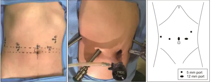
The surgical techniques used at out hospital for chol- edochal cyst excision are similar between the robotic and laparoscopic approaches, and our laparoscopic procedure has been fully described in a previous report that de- scribed early experience of laparoscopic surgery for chol- edochal cyst.16 Briefly, the patients were placed in a re- verse Trendelenburg position with the table tilted towards the left side, so that right side was elevated by about 15-20 degrees. In the robotic procedure, the da Vinci Robotic Surgical System (Si and Xi model, Intuitive Surgical, Sunnyvale, CA) was used. Five ports (4 robotic trocars including a 12-mm camera port , and 12-mm ac- cessory port) were used and the port site is shown in the Fig. 1. The abdominal cavity was inflated with CO2 gas to a pressure of 10-12 mmHg to generate a pneumo- peritoneum.
After docking was completed, the dilated cyst was care- fully separated from the hepatic artery and portal vein us- ing a bipolar Maryland dissector on robotic arm 2 and monopolar hook or scissor and harmony scalpel on arm 1. Cadiere forceps on robotic arm 3 helped to create a better visual field to accomplish liver or duodenum traction. After circumferential dissection of the dilated choledochal cyst, the cyst was dissected down from the hepatic hilum to the intrapancreatic portion of the com- mon bile duct until the transitional area with downward traction of duodenum (Fig. 2A). The distal portion of the cyst was safely ligated and divided using hemo-loc or en- do-stapler (Fig. 2B). Using the distal stump of the cyst as a retractor, further upward dissection was performed until the hepatic ducts were sufficiently observed with identification of the right hepatic artery and main portal
Fig. 1. Site and size of the trocars in robotic procedure.
Fig. 2. Operative photographs. (A) Robotic view of choledochal cyst. (B) Distal portion of the choledochal cyst being ligated and resected. (C) A hepaticojejunostomy was performed intracorporeally.
vein. The bile duct was then transected just below the hi- lum and the cyst and gallbladder were placed into a LapBag (Sejong Co., Paju, Korea). After dedocking, the specimen including the bile duct and gallbladder, was re- moved through extension of the periumbilical port site (about 2.5-3 cm). The jejunum at 40 cm distal to the Trieitz ligament was then exteriorized through the peri- umbilical port site and was divided using an EndoGIA lin- ear stapler.
Jejunojejunostomy anastomosis was performed ex- tracorporeally by manual suturing or an EndoGIA stapler.
The da Vinci system continued to be used after the bowel was repositioned into the peritoneal cavity. A hep- aticojejunostomy was performed intracorporeally using a needle holder on robotic arms 1 and 2 (Fig. 2C). Interrupt- ed sutures were made with 2 or 3 mm intervals using 5-0 PDS (Ethicon, Somerville, NJ) at the posterior wall. For the anterior wall, 5-0 Prolene (Ethicon) was used in the
same manner. We preferred interrupted suturing at the posterior wall as this helps to for prevent anastomosis stricture, but we applied a continuous suture using 4-0 V-Loc (Covidien, Norwalk, CT) when the diameter of bile duct was greater than 10 mm or when it was decided that running suture was appropriate. After the hepaticojejuno- stomy was completed, a Jackson-Pratt drain was inserted through the port on robotic 3th arm and placed posteriorly to the hepaticojejunostomy.
Postoperative management and follow-up Patients were commenced on water on postoperative day 1, followed by a soft diet on day 2. The Jackson- Pratt drain was removed on postoperative day 3 or 4 if no bile leak or intraabdominal complicated fluid was seen.
At 2 weeks after discharge, patients visited the outpatients department and checked a liver function test and an evalu- ation for any complications. If there were no abnormal
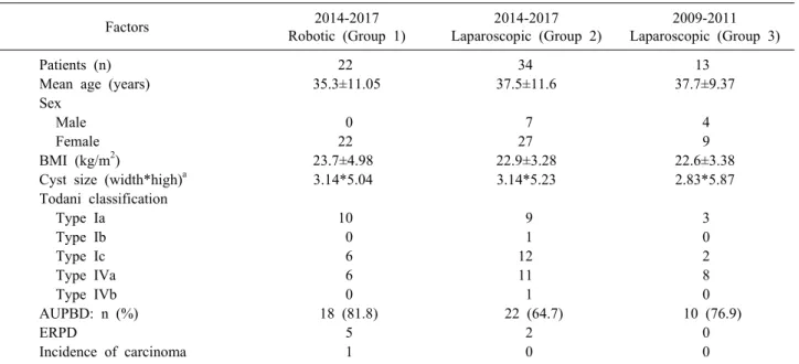
Table 1. Patient demographics
Factors 2014-2017
Robotic (Group 1)
2014-2017 Laparoscopic (Group 2)
2009-2011 Laparoscopic (Group 3)
Patients (n) 22 34 13
Mean age (years) 35.3±11.05 37.5±11.6 37.7±9.37
Sex
Male 0 7 4
Female 22 27 9
BMI (kg/m2) 23.7±4.98 22.9±3.28 22.6±3.38
Cyst size (width*high)a 3.14*5.04 3.14*5.23 2.83*5.87
Todani classification
Type Ia 10 9 3
Type Ib 0 1 0
Type Ic 6 12 2
Type IVa 6 11 8
Type IVb 0 1 0
AUPBD: n (%) 18 (81.8) 22 (64.7) 10 (76.9)
ERPD 5 2 0
Incidence of carcinoma 1 0 0
BMI, body mass index; AUPBD, anomalous union of the pancreatobiliary duct; ERPD, endoscopic retrograde pancreatic drainage Cyst size - measured size by pre-operative MRCP
Group 1 - Underwent robotic surgery between January 2014 and December 2017 Group 2 - Underwent laparoscopic surgery between January 2014 and December 2017 Group 3 - Underwent laparoscopic surgery between September 2009 and July 2011 months at the discretion of the surgeon. In this study, the
overall follow-up period ranged from 5 months to a max- imum of 100 months.
Statistic analysis
The Kolmogorov-Smirnov and Shapiro-Wilk tests were used to test the normality of the data in the three groups.
One way ANOVA was applied to compare the differences between the three groups for factors that satisfy the normality. And the post-hoc analysis was perfomed through Tukey's HSD(honest significant difference) test for the operation time, which is the difference between groups. p-value of <0.05 was considered statistically significant.
On the other hand, the Kruskal-Wallis test was applied to compare the differences between the three groups for factors that don’t satisfy the normality. Mann-Whitney test was used for the post-hoc analysis and statistical sig- nificance was considered when the p-value was less than 0.017 (=0.05/3) by Bonferroni’s method. The data were analyzed using SPSS statistics (Version 21, IBM, USA).
Clinical characteristics of patients
We compared 22 patients who underwent robotic sur- gery (Group 1) and 34 who underwent laparoscopic sur- gery (Group 2) from January 2014 to December 2017 for a choledochal cyst, in addition to 13 patients who under- went laparoscopic surgery from September 2009 to July 2011 for this same condition (Group 3). The character- istics of the patients included in the three groups are shown Table 1.
The mean age was 35.3±11.05 years in Group 1, 37.5±11.6 years in Group 2, and 37.7±9.37 years in Group 3. The ratio of males to females in the three groups was 0:22, 7:27, and 4:9, respectively. In all groups, there were more women than men.
The mean body mass index (BMI) was 23.7±4.98 kg/m2 in Group 1, 22.9±3.28 kg/m2 in Group 2 and 22.6±3.38 kg/m2 in Group 3.
The mean cyst size (width*high) was 3.14*5.04 cm2 in Group 1, 3.14*5.23 cm2 in Group2 and 2.83*5.87 cm2 in Group 3.
According to the Todani classification, the Group 1 ro- botic surgery patients included 10 type Ia cases, 6 type
Table 2. Comparison of clinical outcomes by treatment group Factors
2014-2017 Robotic (Group 1)
(n=22)
2014-2017 Laparoscopic (Group 2)
(n=34)
2009-2011 Laparoscopic (Group 3)
(n=13)
Mean operation time (min) 258.5±52.9 236.2±62.9 395.2±85.9
Anastomosis
Hepaticojejunostomy
Intracorporeal 22 34 13
Jejunojejunostomy
Intracorporeal 0 1 11
Extracorporeal 22 33 2
Hepaticojejunostomy suture type Anterior
Interrupt 20 20 13
Continuous 2 14 0
Posterior
Interrupt 10 2 2
Continuous 12 32 11
Length of stay (days) 7±3.8 7±3.5 9.3±7.6
Open conversion 0 0 7§
Complication 3 7 3
§7 cases of the 20 cases that were the first to attempt laparoscopic surgery in 2009.9 - 2011.7 Ic cases, and 6 type IVa cases. Group 2 patients, which
underwent laparoscopic surgery over the same period as the Group 1 robotic cases, included 9 type Ia cases, 1 type Ib case, 12 type Ic cases, 11 type IVa cases, and 1 type IVb case. The Group 3 patients that underwent laparo- scopic surgery from September 2009 to July 2011 in- cluded 3 type Ia cases, 2 type Ic cases, and 8 type IVa cases.
As shown in Table 1, more than 64% of the patients had AUPBD in all groups.
To prevent pancreatitis or pancreatic duct injury after surgery, the number of patients with a preoperative in- sertion of ERPD was 5 in Group 1 and 2 in Group 2 and none in Group 3.
Postoperatively, biopsy specimens showing GB cancer (stage I, pT1NxM0) were found only in the robotic group with no malignancies observed in the other groups.
Clinical findings
The mean operative time were 258.5±52.9 min for ro- botic surgery (Group 1), 236.2±62.9 min for late laparo- scopic surgery (Group 2), and 395.2±85.9 min for the ini- tial laparoscopic surgery group (Group 3).
In all cases, a hepaticojejunostomy was performed intracorporeally. A jejunojejunostomy was performed in-
tracorporeally in 11 of 13 cases and extracorporeally in only 2 cases in the initial laparoscopic surgery (Group 3).
However, for the procedures undertaken at out hospital over the last 4 years, extracorporeal anastomosis was per- formed in all cases in Group 1 and all but one case in Group 2.
The anterior wall suturing of hepaticojejunostomy was performed more interrupted suture than continuous suture in three groups, respectively. On the other hand, the poste- rior wall suturing was performed more continuous suture than interrupt suture in all groups.
The median hospital stay was 7 days for both Group 1 and 2, and 9.3 days for the initial laparoscopic surgery cases in Group 3. The mean of hospital stay was thus lon- ger in the early group [p=0.000 (Group 1 vs 3), 0.011 (Group 2 vs 3)]. There was no open conversion case in Group 1 and Group 2. In Group 3, 20 cases of laparo- scopic procedure were performed originally, but open conversion was performed in 7 cases (Table 2).
Postoperative complications
Postoperative complications occurred in three patients who underwent robotic surgery (Group 1). This included a case of cholangitis which was resolved after con- servative treatment, and bile leakage which was treated
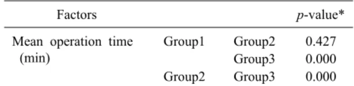
Table 3. Statistical comparison of mean operation time in the three groups
A. One-way ANOVA
Factors p-value*
Mean operation time (min)
Group1 Group2 0.427 Group3 0.000 Group2 Group3 0.000
*p<0.05, considered statistically significant in Tukey’s HSD test
B. Kruskal-Wallis test
Factors p-value*
Jejunojejunostomy Group1 Group2 0.421 Group3 0.000 Group2 Group3 0.000 Length of stay (days) Group1 Group2 0.119 Group3 0.000 Group2 Group3 0.011
*p<0.017=0.05/3, considered statistically significant by Bonferroni’s method in Mann-whitney U test
The third case of an adverse event in Group 1 involved an umbilical herniation which was corrected by laparo- scopic herniorrhaphy.
There were 7 cases of complications in the recent lapa- roscopic surgery patients in Group 2. These included a choledochojejunostomy site stricture & intrahepatic duct stone, choledochojejunostomy site stone due to the suture materials, jejunal branch bleeding, portal vein thrombus, acute pancreatitis, adhesive ileus and A-loop syndrome.
The early laparoscopic surgery patients (Group 3) had complications in 3 cases. A hepaticojejunostomy site stric- ture occurred in 2 patients and a revision operation and biliary stent insertion were performed. Paralytic ileus was also observed in one case and these symptoms improved after conservative care. There were no mortality case in any of the three groups.
DISCUSSION
A choledochal cyst is a rare disease that is usually found in children and is often observed in young women when diagnosed in adults.5 The symptoms of this disease manifest as abdominal pain, a right upper quadrant mass and obstructive jaundice in the pediatric population, and abdominal pain and biliary or pancreatic symptoms in adults.4 Although this disease is typically benign, surgical treatment should be considered as a priority because there is a low probability of malignancy. Long-term follow-up is therefore also needed after surgery.17
Laparoscopic surgery to treat a choledochal cyst was first described by Farello et al.11 in 1995, but more re- cently has been widely adopted and is now preferred over open surgery. Because most patients with a choledochal cyst are young women, laparoscopic surgery has become preferred to open surgery in terms of cosmetic outcomes as well as reduced pain and earlier recovery.14,15 It is gen- erally advantageous to shorten the hospital stay after sur- gery and to have less postoperative pain. However, the adaptation of laparoscopic approaches to perform hep- atobiliary surgery has been slow due to the technical com- plexities of these procedures which have led to consid- erable learning curve, even in experienced centers.
Laparoscopic procedures to treat choledochal cyst com- menced in our hospital in 2009, and this method has un-
Initially, an intracorporeal jejunojejunostomy was per- formed in 11 out of 13 cases at our center and a total laparoscopic procedure without manual procedures was conducted in most cases. In our more recent laparoscopic surgeries for a choledochal cyst, extracorporeal jejunojeju- nostomy was performed in 33 out of 34 cases, which dra- matically reduced the operation time from 395.2±85.9 mi- nutes to 236.2±62.9 minutes [p=0.000]. Table 3 show that there was a statistically difference in jejunojejunostomy anastomosis method between Group 1, 2 and Group 3 [p=0.000 (Group 1 vs 3), 0.000 (Group 2 vs 3)]. Also, Group 1 (258.5±52.9 min) and Group 2 (236.2±62.9 min) were significantly shorter than Group 3 (395.2±85.9 min) in mean operation time [p=0.000 (Group 1 vs 3), 0.000 (Group 2 vs 3)]. The hospital days were also reduced from 9.3 days to 7 days [p=0.000 (Group 1 vs 3), 0.011 (Group 2 vs 3)].
There were two cases of anastomosis stricture among the early 13 cases, and also among the more recent 34 cases who underwent laparoscopic surgery for a chol- edochal cyst. Although there is no statistical significance until now, the problem of anastomosis was relatively less in the recent laparoscopic surgery patients (5.9%) com- pared with the early cases (15.4%). More than 180 laparo- scopic pancreaticoduodenectomy procedures were per-
formed in our center from 2009 to 2014 which has greatly enhanced the experience and skill levels among our sur- geons with the dissection and anastomosis of the bile duct.
There are some noteworthy limitations with the use of laparoscopic instruments which include their rigidity and consequent reduction in the degree freedom. This is cou- pled with the fulcrum effect of laparoscopy and 2-dimen- sional view only.12,13 These drawbacks may contribute to the lack of popularity of this approach. The introduction of robotic surgical systems has overcome some of these hurdles. The principle advantages of robotic surgery are the magnified 3D imaging and the enhanced instrument control and dexterity.18 The camera is mainly controlled by the console surgeon and has very stable vision. The robotic arms can provide intuitive manipulations with tremor filtration, motion scaling, and articulated wrists that enable a high freedom of movement.12,13 Improved er- gonomics for the surgeons are also an important advant- age of robotic systems which allow the surgeon to sit comfortably at the console and perform intricate dis- section, and precise suturing techniques. These advantages of robotic procedures are particularly pertinent when the diameter of hilum duct is small or the level of the cyst is close upward to hilum, and intracorporeal suturing such as hepaticojejunostomy anastomosis is needed.
In our robotic procedure, Cardier forceps on the robotic 3rd arm are used to maintain traction of the liver and ante- rior bile duct wall during the hepaticojejunostomy, which facilitates stable movement and a better visual field. We have not experienced any open conversions or a need for reoperation when using this system. The surgical out- comes of the robotic series of choledochal cyst cases in our present study, in spite of the initial experience, were favorable in comparison with the early and late laparo- scopic patients with this condition. The biliary tract-spe- cific complication rates were similar between patients who underwent laparoscopic surgery and those who underwent robotic surgery. There were 2 cases of postoperative bili- ary leakage or stricture after laparoscopic surgery (5.8%) while only one case of this complication was observed af- ter robotic surgery (4.5%). But, postoperative biliary leak- age after a robotic procedure was mild and transient only, and no biliary strictures were observed after robotic ex- cision of a choledochal cyst.
The operation time is an important practical issue for
robotic procedures. The robot system and more recent lap- aroscopic procedure during the last 4 years for a chol- edochal cyst had operation times of 258.5±52.9 and 236.2±62.9 minutes, respectively. However, this extra 20 minutes required for the robotic procedure was not un- favorable considering the preparation time required for this approach such as docking.
Also, when the learning curve is taken into consid- eration, our initial series of robotic surgery patients have shown quite favorable results. We can expect that the op- eration times will be shortened for the robotic procedures in our hospital as more experience is gained with these surgeries.
This study had some limitations of note. First, it was retrospective investigation with inherent biases. Notably however, a large prospective study is not easy for diseases such as a choledochal cyst which are rare. Second, the number of robotic cases was relatively small. However, this was an initial series however and we would point to the fact that we have to our knowledge described the larg- est number of robotic cases in biliary and pancreatic sur- gery such as pancreaticoduodenectomy. Future multicenter studies with large patient populations and longer-term fol- low-ups are warranted to further evaluate the safety and feasibility of robotic surgery. Currently, robotic procedure is considerably more expensive than laparoscopic procedure. So cost-effectiveness of using a robotic system also should be evaluated.
A robot-assisted resection of a choledochal cyst with a Roux-en-y hepaticojejunostomy is a safe and feasible option with short-term results that are comparable to lapa- roscopic surgery. Compared to laparoscopic surgery, ro- bot-assisted techniques also have distinct advantages such as better intracorporeal suturing and provision of a good 3D visual field.
It is likely that robotic surgery will soon become more widely adopted to complement the current limitations of conventional laparoscopic surgery.
REFERENCES
1. Dumitrascu T, Lupescu I, Ionescu M. The Todani classification for bile duct cysts: an overview. Acta Chir Belg 2012;112:340- 345.
2. Kim OH, Chung HJ, Choi BG. Imaging of the choledochal cyst.
Radiographics 1995;15:69-88.
3. Wiseman K, Buczkowski AK, Chung SW, Francoeur J,
environment. Am J Surg 2005;189:527-531.
4. Soares KC, Arnaoutakis DJ, Kamel I, Rastegar N, Anders R, Maithel S, et al. Choledochal cysts: presentation, clinical differ- entiation, and management. J Am Coll Surg 2014;219:1167- 1180.
5. Todani T, Watanabe Y, Narusue M, Tabuchi K, Okajima K.
Congenital bile duct cysts: classification, operative procedures, and review of thirty-seven cases including cancer arising from choledochal cyst. Am J Surg 1977;134:263-269.
6. Huang CS, Huang CC, Chen DF. Choledochal cysts: differences between pediatric and adult patients. J Gastrointest Surg 2010;
14:1105-1110.
7. Stain SC, Guthrie CR, Yellin AE, Donovan AJ. Choledochal cyst in the adult. Ann Surg 1995;222:128-133.
8. Song HK, Kim MH, Myung SJ, Lee SK, Kim HJ, Yoo KS, et al. Choledochal cyst associated the with anomalous union of pancreaticobiliary duct (AUPBD) has a more grave clinical course than choledochal cyst alone. Korean J Intern Med 1999;
14:1-8.
9. Jan YY, Chen HM, Chen MF. Malignancy in choledochal cysts.
Hepatogastroenterology 2002;49:100-103.
10. Tsai MS, Lin WH, Hsu WM, Lai HS, Lee PH, Chen WJ.
Clinicopathological feature and surgical outcome of choledochal cyst in different age groups: the implication of surgical timing.
C, Chiappetta A. Congenital choledochal cyst: video-guided lap- aroscopic treatment. Surg Laparosc Endosc 1995;5:354-358.
12. Ballantyne GH, Moll F. The da Vinci telerobotic surgical sys- tem: the virtual operative field and telepresence surgery. Surg Clin North Am 2003;83:1293-1304, vii.
13. Lanfranco AR, Castellanos AE, Desai JP, Meyers WC. Robotic surgery: a current perspective. Ann Surg 2004;239:14-21.
14. Okada A, Hasegawa T, Oguchi Y, Nakamura T. Recent advances in pathophysiology and surgical treatment of congenital dilata- tion of the bile duct. J Hepatobiliary Pancreat Surg 2002;9:342- 351.
15. Kimura W. Congenital dilatation of the common bile duct and pancreaticobiliary maljunction: clinical implications. Langen- becks Arch Surg 2009;394:209-213.
16. Hwang DW, Lee JH, Lee SY, Song DK, Hwang JW, Park KM, et al. Early experience of laparoscopic complete en bloc excision for choledochal cysts in adults. Surg Endosc 2012;26:3324-3329.
17. Jang JY, Yoon YS, Kang MJ, Kwon W, Park JW, Chang YR, et al. Laparoscopic excision of a choledochal cyst in 82 consec- utive patients. Surg Endosc 2013;27:1648-1652.
18. Zureikat AH, Nguyen KT, Bartlett DL, Zeh HJ, Moser AJ.
Robotic-assisted major pancreatic resection and reconstruction.
Arch Surg 2011;146:256-261.