ISSN 2234-3806 • eISSN 2234-3814
384 www.annlabmed.org http://dx.doi.org/10.3343/alm.2015.35.3.384 Ann Lab Med 2015;35:384-386
http://dx.doi.org/10.3343/alm.2015.35.3.384
Letter to the Editor
Clinical Microbiology
Flavobacterium ceti From Blood Samples of a Korean Patient With Alcoholic Liver Cirrhosis
Ji Yeon Sung, M.D.1,2, Taek Soo Kim, M.D.1,2, Sue Shin, M.D.2,3, Eun Youn Roh, M.D.2,3, Jong Hyun Yoon, M.D.2,3, and Eui-Chong Kim, M.D.1,2
Department of Laboratory Medicine1, Seoul National University Hospital; Department of Laboratory Medicine2, Seoul National University College of Medicine;
Department of Laboratory Medicine3, Seoul Metropolitan Government-Seoul National University Boramae Hospital, Seoul, Korea
Dear Editor
Flavobacterium species are nonfastidious, oxidase-positive, glu- cose nonfermenting, gram-negative rods. Few Flavobacterium species have been extensively studied [1]. Flavobacterium ceti was first isolated from the lung and liver of two beaked whales, as described in 2007 by Vela et al. [2]. Flavobacterium species are mainly found in aquatic animals and are rarely isolated from human clinical specimens. We present a case in which F. ceti was identified in the blood of a patient admitted to Seoul Metro- politan Government-Seoul National University Boramae Medical Center, Seoul, Korea.
A 43-yr-old man with alcoholic liver cirrhosis presented with mild fever and abdominal distension. Laboratory investigation re- vealed mild leukocytosis with neutrophilia (10.34×10⁹/L; neutro- phils, 83%) and hepatic dysfunction. Ascitic fluid showed leuko- cyte counts >1×10⁹ cells/L (neutrophils, 62%), whereas the se- rum ascites albumin gradient was >1.1 g/dL, total protein was 0.8 g/dL, glucose was 107 mg/dL, and lactate dehydrogenase was 97 U/L. Blood culture was drawn from two separate sites on the day of admission, but ascites cultures were not performed. Intrave- nous antibiotics were administered from the day of admission:
one day of cefotaxime followed by ceftriaxone for the next 20 days. Follow-up blood cultures on day 4, 11, and 14 were all
negative. Ascitic cultures performed after 14 days of antibiotics revealed no organisms. Signs of inflammation were gradually re- solved, and the patient was discharged 25 days after admission.
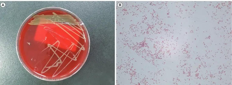
After two days of incubation, bacterial growth was detected in aerobic culture bottles of two separate blood cultures. The next day, circular, orange-colored, non-hemolytic colonies were ob- served on the blood agar plates (Fig. 1A), and gram stain smear revealed gram-negative rods (Fig. 1B). No colonies were ob- served on MacConkey agar plates. Vitek2 GN system (bioMéri- eux, Marcy l’Etoile, France) identified the isolated organism as Brevundimonas diminuta/vesicularis with 95% probability. The contraindicating typical biopattern was phosphatase negative, enzyme activity that is almost always observed in Brevundimo- nas [3]. Matrix-assisted laser desorption/ionization time-of-flight mass spectrometry (MALDI-TOF MS; BrukerDaltonik GmbH, Bremen, Germany) revealed the most matched pattern as Aci- netobacter lwoffii, but showed no reliability with a score of 1.191 (Biotyper Version 3.1, Bremen, Germany). To confirm the identi- fication, 16S rRNA sequence was analyzed by using polymerase chain reaction with 27F and 1492R primers and sequencing with 518F and 801R primers in order to span all bacteria [4].
Sequencing reactions were performed by using a DNA Engine Tetrad 2 Peltier Thermal Cycler (BioRad, Hercules, CA, USA)
Received: November 4, 2014 Revision received: December 1, 2014 Accepted: February 13, 2015 Corresponding author: Sue Shin
Department of Laboratory Medicine, Seoul Metropolitan Government-Seoul National University Boramae Medical Center, 20 Boramae-ro 5-gil, Dongjak-gu, Seoul 156-707, Korea
Tel: +82-2-870-2602, Fax: +82-2-870-2620, E-mail: jeannie@snu.ac.kr
© The Korean Society for Laboratory Medicine.
This is an Open Access article distributed under the terms of the Creative Commons Attribution Non-Commercial License (http://creativecommons.org/licenses/by-nc/3.0) which permits unrestricted non-commercial use, distribution, and reproduction in any medium, provided the original work is properly cited.
Sung JY, et al.
Flavobacterium ceti bacteremia
http://dx.doi.org/10.3343/alm.2015.35.3.384 www.annlabmed.org 385
with a ABI BigDye(R) Terminator v3.1 Cycle Sequencing Kit (Ap- plied Biosystems, Foster City, CA, USA) and were analyzed by using electrophoresis in an ABI 3730xl DNA Analyzer (Applied Biosystems) with ChromasPro 1.7.6 (Technelysium Pty Ltd, South Brisbane, Australia). The 1,367 bp of the 16S rRNA gene sequence shared 99% identity with the GenBank sequence NR_042540 (F. ceti strain 454-2).
Flavobacterium species are well-known pathogens of bacterial cold-water disease, with some causing opportunistic infections.
There have been some reports of human infections due to pathogens that were previously known as Flavobacterium but are currently classified under other genera such as Chryseobacte- rium [5] and Myroides [6]. Thus, known human infections from current Flavobacterium are limited, with only a few reports of dif- ferent species [7, 8]. Mosayebi et al. [9] reported a Flavobacte- rium sepsis outbreak due to contaminated distilled water in a neonatal intensive care unit. To our knowledge, our report dis- cusses the first case of F. ceti infection in human. Physical ex- amination and ascites lab findings were consistent with sponta- neous bacterial peritonitis. F. ceti was considered the causative pathogen because it was isolated from two separate blood cul- tures collected on the day of admission and the cause of bacte- remia was presumed to be peritonitis although ascitic culture was not performed. Clinical manifestations were relatively mild, with good outcomes. Identification of gram-negative non-fer- menting rods is quite challenging, and commercial systems often provide unreliable results. Molecular sequencing is one of the most accurate ways characterizing these bacteria to the species
level. In this case, conventional methods and MALDI-TOF MS were insufficient in identifying Flavobacterium. The MALDI data- base contained a limited number of Flavobacterium species, and F. ceti was not included. 16S rRNA sequence analysis success- fully confirmed the isolate as F. ceti.
Here, we report the first probable human case of F. ceti de- tected in the blood of a patient with liver cirrhosis. Flavobacte- rium may cause an opportunistic infection in humans, although its significance remains unclear owing to the limited number of human infections reported. Molecular sequencing is currently the most reliable method for identifying this unusual pathogen.
Authors’ Disclosures of Potential Conflicts of Interest
No potential conflicts of interest relevant to this article were re- ported.
REFERENCES
1. Bernardet JF and Bowman JP. The genus Flavobacterium. In: Dworkin M, Falkow S, Rosenberg E, Schleifer KH, Stackebrandt E, eds. The prokary- otes: a handbook on the biology of bacteria. 3rd ed. New York: Springer, 2006:455-80.
2. Vela AI, Fernandez A, Sánchez-Porro C, Sierra E, Mendez M, Arbelo M, et al. Flavobacterium ceti sp. nov., isolated from beaked whales (Ziphius cavirostris). Int J Syst Evol Microbiol 2007;57:2604-8.
3. Laffineur K, Janssens M, Charlier J, Avesani V, Wauters G, Delmée M.
Biochemical and susceptibility tests useful for identification of nonfer- menting gram-negative rods. J Clin Microbiol 2002;40:1085-7.
Fig. 1. Colony morphology and microscopic characteristics of Flavobacterium ceti. (A) Orange-colored, non-hemolytic colonies were ob- served after two days of aerobic culture on a blood agar plate. (B) Gram-negative rods from positive aerobic blood culture smear prepara- tions (Gram stain, ×1,000).
A B
Sung JY, et al.
Flavobacterium ceti bacteremia
386 www.annlabmed.org http://dx.doi.org/10.3343/alm.2015.35.3.384 4. CLSI. Interpretive criteria for identification of bacteria and fungi by DNA
target sequencing; Approved guideline, MM18-A. Wayne, PA: Clinical and Laboratory Standard Institute, 2008.
5. Hsueh PR, Hsiue TR, Wu JJ, Teng LJ, Ho SW, Hsieh WC, et al. Flavo- bacterium indologenes bacteremia: clinical and microbiological charac- teristics. Clin Infect Dis 1996;23:550-5.
6. Crum-Cianflone NF, Matson RW, Ballon-Landa G. Fatal case of necro- tizing fasciitis due to Myroides odoratus. Infection2014;42:931-5.
7. Tian GZ, Piao DR, Zhao HY, Jiang H, Cui BY, Li JY. A Flavobacterium lin-
danitolerans strain isolated from the ascites sample of a Chinese patient with EV71 virus infection. Biomed Environ Sci 2011;24:694-6.
8. Kuai S, Huang L, Pei H, Chen Y, Liu J. Imipenem resistance due to class A carbapenemase KPC-2 in a Flavobacterium odoratum isolate. J Med Microbiol 2011;60:1408-9.
9. Mosayebi Z, Movahedian AH, Soori T. Flavobacterium sepsis outbreak due to contaminated distilled water in a neonatal intensive care unit. J Hosp Infect 2011;78:214-5.