J Korean Soc Surg Hand 2015;20(3):96-102.
http://dx.doi.org/10.12790/jkssh.2015.20.3.96
THE HAND
J Korean Soc Surg Hand 2015;20(3):96-103.
http://dx.doi.org/10.12790/jkssh.2015.20.3.96
서론
원위 요골 골절은 전 세계적으로 정형외과 의사가 접하는 가장 흔한 상지의 골절이다1,2. 대퇴 경부 골절, 근위 상완골 골절, 척추 골절과 함께 골다공증과 관련이 있는 골절로 알려 져 있으며, 폐경 후 여성에게서 그 빈도가 높은 것으로 알려 져 있다3.
만성 신부전에서는 신기능 저하로 인하여 저칼슘혈증, 저인 산혈증과 함께 칼시트리올(1,25-dihydroxyvitamin D)이 감 소하며4, 이로 인하여 이차적으로 부갑상선 기능이 항진되어5, 골대사의 이상이 발생한다. 골밀도의 감소뿐 아니라, 피질골 에도 변화가 생기는 것으로 알려져 있으며6, 투석으로 인한 아 밀로이드증(amyloidosis)으로 골내낭종을 형성하며 골파괴를 유발하기도 한다7. 만성 신부전 환자에게서 발생할 수 있는 취
Volar Locking Plate Fixation for Distal Radius Fractures in Hemodialysis Patients
Jin-Hyung Im
1, Sung-Woo Huh
2, Min-Kyu Park
1, Joo-Yup Lee
11Department of Orthopedic Surgery, College of Medicine, The Catholic University of Korea, Seoul, Korea
2Department of Orthopedic Surgery, Hong-Ik Hospital, Seoul, Korea
Received:July 26, 2015 Revised:[1] August 25, 2015
[2] September 1, 2015 Accepted:September 3, 2015 Correspondence to:Joo-Yup Lee Department of Orthopedic Surgery, St.
Vincent’s Hospital, College of Medicine, The Catholic University of Korea, 93 Jungbu- daero, Paldal-gu, Suwon 16247, Korea TEL:+82-31-249-8301
FAX:+82-31-254-7186 E-mail:[email protected]
Purpose: Although the possibility of distal radius fractures is strong in hemodial- ysis patients, there are many difficult problems such as the bleeding tendency, hypervascularity and injury to arteriovenous fistula. We studied the outcomes of open reduction and volar locking plate fixation of the distal radius fractures in hemodyalisis patients with ipsilateral arteriovenous fistula.
Methods:From 2007 to 2009, a retrospective chart review was performed of eleven hemodialysis patients who underwent volar locking plate fixation for treat- ment of distal radius fractures. Eight of them were female and three were male and mean age was 68 years (range, 57-81 years). Mean follow-up period was 19 months (range, 12-28 months). All patients had osteoporosis with mean T-score of -2.7. All operations were performed on the next day of hemodialysis.
Scheduled hemodialysis was possible on the next day of surgery without splint.
We analyzed radiographic results, the wrist range of motion, Mayo wrist score and disabilities of the arm, shoulder and hand (DASH) score at the last follow-up.
Results:All fractures achieved anatomical reduction and united at final follow- up. Complications such as hematoma or occlusion of arteriovenous fistula were not observed. Mean Mayo wrist score was 78 and mean DASH score was 22.
Conclusion:Volar locking plate fixation for distal radius fractures provides immediate support for continuing hemodialysis and exercise. Open reduction of the fractures and the use of tourniquet don’t seem to increase the vascular complications like hematoma and fistula occlusion.
Keywords:Distal radius, Hemodyalisis, Arteriovenous fistula, Fracture, Volar locking plate
This is an Open Access article distributed under the terms of the Creative Commons Attribution Non-Commercial License (http://creativecommons.org/licenses/by- nc/3.0/) which permits unrestricted noncommercial use, distribution, and reproduction in any medium, provided the original work is properly cited.
약 골절(fragility fracture)을 예방하고자 골의 강도 변화를 진단하기 위한 방사선학적 검사나, 혈청학적 검사에 대해 많 은 연구가 이루어져 있다8-10. 만성 신부전 환자에게서 이러한 이차적인 골의 변화에 더하여 폐경이나 고령에 의한 골감소나 골다공증으로 인하여 원위 요골 골절의 발생 가능성이 높지 만, 그 치료에 대해서는 많은 연구가 부족한 실정이다. 대부분 의 만성 신부전 환자들이 혈액투석을 위하여 상완, 전완부에 동정맥루를 가지고 있어 동정맥루의 손상에 대한 위험성과 출 혈성 경향 때문에11, 동정맥루의 동측에 원위 요골 골절이 발생 한 경우 수술을 결정하기 어려우며, 수술을 한다고 하더라도 침습이 적은 경피적 핀이나12, 외고정기를 고려할 수 있었다.
경피적 핀과 외고정기의 원위 요골 골절의 수술적 고정 치료 결과에 대해 다양한 보고가 있었으며, 정복 소실을 보고하는 일부 연구들이 있었다13-15. 전방 잠김 금속판(volar locking plate)은 정복 소실 없이 전방 피질골만으로도 좋은 결과들이
보고되어16-20최근 원위 요골 골절 치료에 널리 사용되고 있다.
본 연구에서는 만성 신부전 환자에게서 동정맥루의 동측에 발생한 원위 요골 골절에 대해서 전방 잠김 금속판을 이용한 수술 방법과 임상적 결과를 알아보고자 하였다.
대상 및 방법
2007년 3월부터 2009년 11월까지 만성 신부전으로 혈액투 석을 위한 동정맥루를 가지고 동측에 발생한 불안정성 원위 요골 골절에 대하여, 전방 잠김 금속판을 이용한 수술을 시행 하고 1년 이상 추시가 가능하였던 환자를 대상으로 하였다. 수 술은 Lafontaine이 정의한 불안정성 원위 요골 골절에 해당 하는 환자에게서 모두 시행하였으며21, 총 11명 중 남자가 3명, 여자가 8명이었고, 평균 연령은 68세이었다. 평균 추시 기간 은 19개월(범위, 12-28개월)이었다. 골밀도 검사에서 평균 T 점수는 -2.7이었다. 수술 시행 후 운동 범위, disabilities of the arm, shoulder and hand (DASH) 점수, Mayo wrist 점 수를 측정하여 임상적 결과를 평가하였으며, 수술 후 방사선 검사를 통하여 유합 시기와 요측 경사, 전방 경사, 요측 길이 등의 방사선학적 지표를 측정하였고, 도플러 초음파 검사를 이용하여 동정맥루의 협착 여부를 확인하였다.
수술은 혈액투석한 다음날 전신마취하에 시행하였으며, 모 든 환자에게 압박대를 사용하였고, 액와부의 바로 원위부에 위치시킨 후 상지를 거상하고 있다가 압착(squeezing)없이 피 부 절개 직전 250 mm Hg의 압력으로 팽창시켰다. 전방 요수 굴근 도달법을 이용하여, 요골 동맥은 요수근굴근막에 포함하 여 요측으로 박리하여 보호하였다. 방형회내근을 상완요골근
옆 요측 경계부위에서 박리하여 골절부위를 노출시키고 견인 없이 올림기(freer elevator)를 이용하여 지렛대(leverage)로 전방 피질골을 정복한 후 전방 잠김 금속판을 이용하여 고정 하였다. 고정 후 압박대의 압력을 제거하고 국소적인 출혈부 위를 지혈한 뒤, 요골의 요측 경계부터 박리하였던 방형회내 근을 상완요골근에 봉합하여 전방 잠김 금속판을 감싸고 보호 한 뒤, 배액관을 삽입하고 피부를 봉합하였다. 수술 다음날 동 정맥루를 이용한 혈액투석을 시행하였으며, 수술 후 2일째에 배액관을 제거한 뒤, 수술 후 3일째부터 관절 운동을 시작하 고, 착탈이 가능한 부목을 6주간 착용하며 관절운동을 지속하 였다(Fig. 1).
결과
11명 모두 1 m 이하에서 발생한 저에너지에 의한 수상이었 으며, 수상일로부터 평균 4.3일에 수술을 시행하였다. 평균 압 박대 사용 시간은 36분이었다. 방사선학적 골절의 유형은 AO 유형 A2: 2명, A3: 4명, C1: 2명, C3: 3명이었으며, 방사선학 적 검사에서 모든 환자에게서 평균 3.2개월에 골유합을 얻었 다. 수술 후 방사선학적 지표들은 각각 평균 요측 경사 25�, 전 방 경사: 13�, 요측 길이: 11 mm로 해부학적 정복을 얻었다 (Fig. 2). 최종 추시에서 평균 관절 운동 범위는 수근관절 신전 62�, 굴곡 43�, 회외전 65�, 회내전 55�이었다(Fig. 3). 평균 Mayo wrist 점수는 78점, 평균 DASH 점수는 22점이었다.
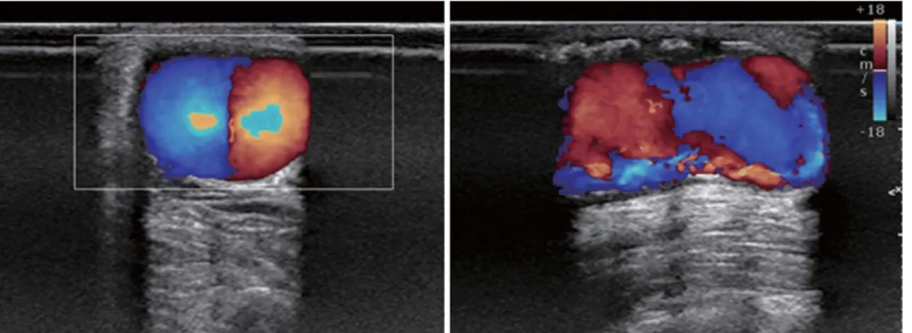
도플러 초음파 검사상에서의 동정맥루의 협착이나 혈종 등 수 술로 인한 합병증은 관찰되지 않았다(Fig. 4).
고찰
수명의 연장, 식생활과 생활패턴의 변화, 운동량 감소 등으 로 당뇨와 고혈압 환자가 증가하는 가운데, 이로 인한 합병증 으로 만성 신부전 환자가 증가하고 있다. 2012년 the U.S Renal Data System Annual data Report에 따르면 이미 약 59만 명이 만성 신부전 환자로 등록이 되어 있으며, 이들 중 약 41만명 이상이 혈액투석을 받고 있고22, 투석 치료의 발전 으로 인하여 2000년도 이후 20% 정도 증가되었다23. 만성 신 부전으로 인한 칼슘-인대사의 변화와 칼시트리올의 감소로 이차적인 부갑상선항진증이 발생하여 부갑상선호르몬이 증가 하고, 골교체(bone turn over)가 감소되어 골연화증(osteo- malacia), 골감소증(osteopenia), 무력성골질환(adynamic bone disease)의 발생이 불가피하여, 이로 인한 골강도의 변 화는 노화에 따른 변화와 함께 그 영향이 클 수 있으며4-7,24, 골
강도의 변화로 인한 골절의 발생률도 증가하고 있기 때문에
7,25,26, 골밀도나 골강도의 감소를 진단하기 위한 연구들이 많이
있었다6,8-10. 골강도가 감소한 만성 신부전 환자들은 동정맥루
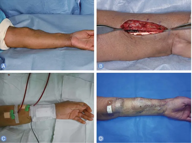
부위의 부자연스러운 사용으로 원위 요골 골절이 발생한 가능 성이 높지만, 아직 이에 대한 뚜렷한 치료방침도 없으며, 일반 적인 방법을 따른다 하더라도 혈액투석을 하고 있는 상황이라 Fig. 1. (A)
The upper arm which have distal radius fracture with ipsilateral arteriovenous fistula is on hand table.
(B)Standard
flexor carpi radialis (FCR) approach is performed. The FCR sheath including the radial artery is retracted to radial side.
(C)Scheduled hemodyalisis was conducted on the first day after operation.
(D)Wrist exercise was started without drain on the third day after operation.
Fig. 2.
Radiographic images of 64 aged female with distal radius fracture and ipsilateral arteriovenous fistula show normal radi-
ologic parameters and union in last follow-up.
면 동정맥루로 인해 지혈대 및 수술절개창의 위치를 결정하기 어려워 수술의 적응증에 해당하더라도 수술적 치료에 어려움 이 발생할 수 있다. 전위가 적은 안정 골절인 경우 석고고정을 시행하면 되겠으나 유합까지 석고고정으로 인한 압박성 궤양, 피부염과 같은 피부합병증, 구획증후군, 관절강직 등이 발생 하거나27, 석고고정으로 인하여 차단된 동정맥루로 인해 혈액
투석 자체가 중단될 수 밖에 없는 상황을 초래한다12. 불안정 골절인 경우 수술 적응증에 해당되지만, 보편적으로 동정맥루 가 있는 경우 지혈대를 사용하는 것뿐만 아니라 혈관손상을 줄 수 있는 압박이나, 침습적 술기 등을 금하고 있는 상황에서 관혈적으로 골절부위에 접근하기가 어렵기 때문에 많은 술자 들이 관혈적 정복을 기피하거나 망설이고 있을 것이다.
Fig. 4.
Doppler sonography shows normal blood flow and no occlusion of fistula.
Fig. 3.
The patients showed good range of motion in last follow-up.
동정맥루가 있을 경우 지혈대의 사용에 대한 연구가 많지 않으나, 만성 신부전 환자에게 발생한 동정맥루 동측의 수근 관 증후군에 대하여 압박대를 사용하고 성공적인 수술 결과를 보고한 연구가 있으며28, 동정맥루를 형성한 환자들에게 지혈 대를 사용한 운동을 이용하여 동정맥루의 성숙(maturation) 을 성공적으로 유도한 연구도 있는 것으로 보아29, 짧은 시간의 지혈대의 사용이 절대적 금기에 해당하지는 않는다고 여겨진 다. 지혈대의 적용에 있어 Green 등30은 상완두동정맥루를 가 진 경우 동정맥루보다 원위부에 위치하는 전완부 압박대의 사 용을 권고하였으나, 저자들은 수술 시 지혈대를 동측의 액와 부 바로 원위부에 적용시켜, 상완두동정맥루, 요두동정맥루 모든 경우에서 동정맥루보다 근위부에 위치하였다. 압력은 250 mm Hg으로 하였으며, 실제 적용시간도 피부절개 직전 부터 금속판 고정까지 평균 36분으로 그 사용을 최소화하였 다. Sugiyama 등31은 동정맥루와 동측의 원위 요골 골절 3예 에 대한 치료 결과를 발표하며, 수술 시 압박대를 사용하지 않 았다고 하였으나, 증례가 적으며, 평균 환자 연령 등에서 본 연구와 차이가 있어 더 많은 연구가 필요할 것으로 보인다.
본 연구에서 모든 환자는 수술 전일과 수술 다음날 혈액투 석 시 헤파린을 사용하였으며, 혈액투석 시작 시 1,500 IU를 일시주사(bolus)로 사용하고 4시간의 투석 동안 시간당 500 IU을 투여하였다. 동정맥루의 혈전 형성 방지를 위하여 사용 하는 헤파린은 수술 전, 후 적용에 대해 고민할 수 있으나, 문 헌에 따라 투석 후 2-4시간이면 헤파린은 체외로 배설되어 항 응고 작용을 하지 않는다고 보고하고 있어32,33, 투석을 예정대 로 진행하고 투석 다음날 수술을 하는 것에 큰 문제가 없을 것 으로 생각하였으며, 수술 후 혈종이나 동정맥루의 합병증 등 이 발생하지 않았다.
수근관증후군은 혈액투석과 관련된 아밀로이드증으로 인한 가장 흔한 합병증으로 문헌마다 차이는 있으나 약 10% 정도의 발생률이 보고되고 있다34. 본 연구에서는 수상 전과 수술 전, 수술 후 수근관증후군의 증상을 보인 환자는 없었으며, 수술 시 수근관 유리술을 시행하지 않았다. 이는 증례 수가 적어 그 러할 것으로 예상되며 증례 수가 많아지면 발생을 예상할 수 있는 합병증으로 생각된다.
원위 요골 골절 수술 시 고정 방법 선택을 논함에 있어 외고 정기기나, 경피적 핀고정이 불안정 골절치료에 효과적인지에 대해서는 아직까지 논란이 있다. 많은 저자들이 두 가지의 치 료 방법에 따른 결과와 전방 잠김 금속판과의 결과를 비교하 였으며, 발표한 결과는 저자들마다 다양하였다16-19,35, 그러나 많은 연구들의 환자군과 본 연구의 만성 신부전 환자군의 골 강도가 다르기 때문에 여러 가지 고려해야 할 점들이 있을 것
으로 생각된다. 두 방법 모두 핀 주위 감염의 가능성을 가지고 있으며, 외고정기기는 고정기간에 따른 관절강직 발생이 가능 하며, 경피적 핀고정은 핀의 이동이나 신경자극 등이 발생할 수 있다36. 가장 중요한 점은 외고정기기, 경피적 핀 고정 모두 고정물을 제거한 후에 정복의 소실에 대해 보고가 있었으며13-
15, 만성 신부전 환자군의 골강도가 더욱 약하기 때문에 전방 피질골 만으로도 견고한 고정이 가능한 전방 잠김 금속판이 적합할 것으로 생각된다. 본 연구에서는 수술 시 수장측 피질 골을 정확히 맞추어 전방 경사를 회복하고, 전방 잠김 금속판 으로 정복의 소실을 예방하여 이로 인해 골유합 후 충분한 파 악력을 유지할 수 있도록 하는 것을 중요하게 생각하였다. 전 방 잠김 금속판 내고정을 통하여 외고정 기기나 경피적 핀 고 정에 비해서 유합까지 정복 소실 없이 견고하게 고정하고 조 기에 운동을 시작하며, 쉽게 착용과 제거가 가능한 부목을 사 용하여 고정 기간을 단축하였다.
본 연구는 몇 가지 제한점을 가지고 있다. 첫째, 의무 기록 평가를 이용한 후향적 연구이며, 대조군 없이 전방 잠김 금속 판 이용한 환자군 만의 평가의 결과로, 다른 고정 방법으로 수 술한 환자와의 방사선학적, 임상적 결과의 비교가 어려운 제 한점이 있다. 둘째, 증례 수가 많지 않아 일반화하기에 어려운 점이 있다. 따라서, 본 연구에서는 합병증 없이 만족할 만한 방사선학적, 임상적 결과를 보이고 있으나, 압박대의 사용이 나 관혈적 정복술에 의한 동측의 동정맥루에 손상 가능성을 염두하여 수술 시 주의해야 할 것으로 생각된다. 여러 가지 제 한점에도 혈액투석 환자의 불안정성 원위 요골 골절 시 동정 맥루에 발생할 수 있는 손상을 주의하여 전방 잠김 금속판을 이용한 관혈적 정복술을 고려해 볼 수 있을 것으로 생각된다.
결론
만성 신부전 환자에게 동정맥루 동측에 발생한 불안정 원위 요골 골절에 대하여, 조기 운동을 가능하게 하고 즉각적인 혈 액투석 재개를 위하여 전방 잠김 금속판을 이용한 수술적 치 료를 고려할 수 있다.
REFERENCES
1. Karl JW, Olson PR, Rosenwasser MP. The epidemiology of upper extremity fractures in the United States, 2009.
J Orthop Trauma. 2015;29:e242-4.
2. Mehrpour SR, Nabian MH, Oryadi Zanjani L, Foroughmand-Araabi MH, Shahryar Kamrani R.
Descriptive epidemiology of traumatic injuries in 18890 adults: a 5-year-study in a tertiary trauma center in iran. Asian J Sports Med. 2015;6:e23129.
3. Stein EM, Kepley A, Walker M, et al. Skeletal structure in postmenopausal women with osteopenia and frac- tures is characterized by abnormal trabecular plates and cortical thinning. J Bone Miner Res. 2014;29:1101- 9.
4. Moe S, Drueke T, Cunningham J, et al. Definition, eval- uation, and classification of renal osteodystrophy: a position statement from Kidney Disease: Improving Global Outcomes (KDIGO). Kidney Int. 2006;69:1945- 53.
5. Reiss E, Canterbury JM, Kanter A. Circulating parathy- roid hormone concentration in chronic renal insuffi- ciency. Arch Intern Med. 1969;124:417-22.
6. Nickolas TL, Stein EM, Dworakowski E, et al. Rapid cor- tical bone loss in patients with chronic kidney disease. J Bone Miner Res. 2013;28:1811-20.
7. Nickolas TL, McMahon DJ, Shane E. Relationship between moderate to severe kidney disease and hip fracture in the United States. J Am Soc Nephrol. 2006;
17:3223-32.
8. Maeno Y, Inaba M, Okuno S, Yamakawa T, Ishimura E, Nishizawa Y. Serum concentrations of cross-linked N- telopeptides of type I collagen: new marker for bone resorption in hemodialysis patients. Clin Chem. 2005;
51:2312-7.
9. Shidara K, Inaba M, Okuno S, et al. Serum levels of TRAP5b, a new bone resorption marker unaffected by renal dysfunction, as a useful marker of cortical bone loss in hemodialysis patients. Calcif Tissue Int. 2008;82:
278-87.
10. Ishimura E, Okuno S, Ichii M, et al. Relationship between serum sclerostin, bone metabolism markers, and bone mineral density in maintenance hemodialysis patients. J Clin Endocrinol Metab. 2014;99:4315-20.
11. Deykin D. Uremic bleeding. Kidney Int. 1983;24:698-705.
12. Ishiguro S, Oota Y, Sudo A, Uchida A. Calcium phosphate cement-assisted balloon osteoplasty for a Colles' frac- ture on arteriovenous fistula forearm of a maintenance hemodialysis patient. J Hand Surg Am. 2007;32:821-6.
13. Clancey GJ. Percutaneous Kirschner-wire fixation of Colles fractures: a prospective study of thirty cases. J Bone Joint Surg Am. 1984;66:1008-14.
14. Oskam J, Kingma J, Bart J, Klasen HJ. K-wire fixation for
redislocated Colles' fractures. Malunion in 8/21 cases.
Acta Orthop Scand. 1997;68:259-61.
15. Ludvigsen TC, Johansen S, Svenningsen S, Saetermo R.
External fixation versus percutaneous pinning for unsta- ble Colles’ fracture: equal outcome in a randomized study of 60 patients. Acta Orthop Scand. 1997;68:255-8.
16. Kumbaraci M, Kucuk L, Karapinar L, Kurt C, Coskunol E. Retrospective comparison of external fixation versus volar locking plate in the treatment of unstable intra- articular distal radius fractures. Eur J Orthop Surg Traumatol. 2014;24:173-8.
17. Costa ML, Achten J, Plant C, et al. UK DRAFFT: a ran- domised controlled trial of percutaneous fixation with Kirschner wires versus volar locking-plate fixation in the treatment of adult patients with a dorsally displaced fracture of the distal radius. Health Technol Assess.
2015;19:1-124.
18. Franceschi F, Franceschetti E, Paciotti M, Cancilleri F, Maffulli N, Denaro V. Volar locking plates versus K- wire/pin fixation for the treatment of distal radial frac- tures: a systematic review and quantitative synthesis. Br Med Bull. 2015;115:91-110.
19. Roh YH, Lee BK, Baek JR, Noh JH, Gong HS, Baek GH. A randomized comparison of volar plate and external fix- ation for intra-articular distal radius fractures. J Hand Surg Am. 2015;40:34-41.
20. Williksen JH, Husby T, Hellund JC, Kvernmo HD, Rosales C, Frihagen F. External fixation and adjuvant pins versus volar locking plate fixation in unstable dis- tal radius fractures: a randomized, controlled study with a 5-year follow-up. J Hand Surg Am. 2015;40:1333- 40.
21. Lafontaine M, Hardy D, Delince P. Stability assessment of distal radius fractures. Injury. 1989;20:208-10.
22. O'Banion LA, Van Buren D, Davis JW. Radiocephalic fis- tulas for hemodialysis: a comparison of techniques. Am Surg. 2015;81:341-4.
23. Lok CE. Fistula first initiative: advantages and pitfalls.
Clin J Am Soc Nephrol. 2007;2:1043-53.
24. Levin A, Bakris GL, Molitch M, et al. Prevalence of abnormal serum vitamin D, PTH, calcium, and phos- phorus in patients with chronic kidney disease: results of the study to evaluate early kidney disease. Kidney Int. 2007;71:31-8.
25. Kaji H, Yamauchi M, Yamaguchi T, Shigematsu T, Sugimoto T. Mild renal dysfunction is a risk factor for a
decrease in bone mineral density and vertebral frac- tures in Japanese postmenopausal women. J Clin Endocrinol Metab. 2010;95:4635-42.
26. Ball AM, Gillen DL, Sherrard D, et al. Risk of hip fracture among dialysis and renal transplant recipients. JAMA.
2002;288:3014-8.
27. Boyd AS, Benjamin HJ, Asplund C. Principles of casting and splinting. Am Fam Physician. 2009;79:16-22.
28. Naito M, Ogata K, Goya T. Carpal tunnel syndrome in chronic renal dialysis patients: clinical evaluation of 62 hands and results of operative treatment. J Hand Surg Br. 1987;12:366-74.
29. Salimi F, Majd Nassiri G, Moradi M, et al. Assessment of effects of upper extremity exercise with arm tourniquet on maturity of arteriovenous fistula in hemodialysis patients. J Vasc Access. 2013;14:239-44.
30. Green DP, Wolfe SW, Hotchkiss RN, et al. Green's oper- ative hand surgery. 6th ed. Philadelphia: Elsevier Churchill Livingstone; 2011. 15-6.
31. Sugiyama Y, Naito K, Igeta Y, Obata H, Kaneko K, Obayashi O. Treatment strategy for distal radius frac- tures with ipsilateral arteriovenous shunts. J Hand Surg Am. 2014;39:2265-8.
32. Sagedal S, Hartmann A, Sundstrom K, Bjornsen S, Fauchald P, Brosstad F. A single dose of dalteparin effectively prevents clotting during haemodialysis.
Nephrol Dial Transplant. 1999;14:1943-7.
33. Wilhelmsson S, Lins LE. Heparin elimination and hemostasis in hemodialysis. Clin Nephrol. 1984;22:303- 6.
34. Kopec J, Gadek A, Drozdz M, et al. Carpal tunnel syn- drome in hemodialysis patients as a dialysis-related amyloidosis manifestation: incidence, risk factors and results of surgical treatment. Med Sci Monit. 2011;17:
CR505-9.
35. Williksen JH, Husby T, Hellund JC, Kvernmo HD, Rosales C, Frihagen F. External fixation and adjuvant pins versus volar locking plate fixation in unstable dis- tal radius fractures: a randomized, controlled study with a 5-year follow-up. J Hand Surg Am. 2015;40:1333- 40.
36. Strohm PC, Muller CA, Boll T, Pfister U. Two proce- dures for Kirschner wire osteosynthesis of distal radial fractures: a randomized trial. J Bone Joint Surg Am. 2004;
86:2621-8.
혈액투석 환자의 동정맥루 동측의 원위 요골 골절에서 전방 잠김 금속판을 이용한 수술적 치료
임진형
1∙허성우
2∙박민규
1∙이주엽
11가톨릭대학교 의과대학 정형외과학교실, 2홍익병원 정형외과
목적:만성 신부전 환자에서 혈액투석중인 상완에 원위 요골 골절 발생 가능성이 높으나, 과다 출혈이나 동정맥루의 손상 등으로 치료에 어려운 점이 많다. 저자들은 혈액투석 환자에서 발생한 원위 요골 골절에 대하여 전방 잠김 금속판 고정술 을 시행하고 그 결과를 알아보고자 하였다.
방법:2007년부터 2009년까지 전방 잠김 금속판을 이용하여 동정맥루 동측의 원위 요골 골절을 치료한 11명의 만성 신부전 환자를 대상으로 하였다. 남자가 3명, 여자가 8명이었으며, 평균 연령은 68세이었다. 평균 추시 기간은 19개월 (범위, 12-28개월)이었다. 평균 T-score가-2.7로 골다공증이 있었다. 혈액투석은 원래 시행하던 대로 수술 전날과 다 음날 시행하였다. 최종 추시에서 골유합 여부, 관절운동범위, Mayo wrist score, disabilities of the arm, shoulder and hand (DASH) score를 측정하였다.
결과:전례에서 방사선학적 해부학적 정복을 얻었으며, 동정맥루의 협착이나 혈종 등 수술로 인한 합병증은 관찰되지 않았다. 평균 Mayo wrist score는 78점, 평균 DASH score는 22점이었다.
결론:혈액투석을 시행하는 환자의 원위 요골 골절에서도 조기에 수장 잠김 금속판으로 견고한 고정을 시행하는 것이 조기 운동 및 빠른 혈액 투석의 재개에 도움이 되며, 수술적 치료 및 지혈대의 사용이 혈종이나 동정맥루의 폐쇄와 같은 합병증을 증가시키지 않는다.
색인단어:원위 요골, 혈액투석, 동정맥루, 골절, 전방 잠김 금속판
접수일2015년 7월 26일수정일1차: 2015년 8월 25일, 2차: 2015년 9월 1일 게재확정일2015년 9월 3일
교신저자이주엽
경기도 수원시 팔달구 중부대로 93
가톨릭대학교 의과대학 성빈센트병원 정형외과학교실 TEL031-249-8301 FAX031-254-7186