Lithium Disilicate Glass-ceramic과 상아질 간의 접착성 레진 시멘트의 결합강도에 대한
임시 수복재와 임시 충전재의 영향
원광대학교 치과대학 치과보철학교실
오상천∙심헌보
본 연구는 lithium disilicate glass-ceramic과 상아질 간의 결합력에 대한 임시 수복재와 임시 충전재의 효과를 평 가하기 위함이 목적이었다. 60개의 발치된 치아를 자가중합형 레진에 포매한 후 교합1/3 부위를 절단하였고, 무 작위로 15개씩 4그룹으로 나누었다. 그런 후 lithium disilicate glass-ceramic을 다음과 같이 그룹으로 나누어 상아 질에 접착시켰다: 아무 처리도 하지 않은 그룹(그룹A), ALIKETM (GC America Inc.)를 적용한 그룹(그룹 B), Luxatemp�Automix plus (DMG, Germany)를 적용한 그룹(그룹 C), Fermit�(Ivoclar Vivadent, Leichtenstein)를 적용한 그룹(그룹 D). 모든 시편들을 24시간 증류수에 담가 보관한 후, 1 mm/min의 crosshead speed로 만능시험기(Zwick 1456 41, Zwick, Germany) 상에서 전단 결합 강도를 측정하였다. 모든 그룹 간에 결합력의 통계학적 유의성은 없 었으며, 파절양상은 대부분 시편에서 접착성(adhesive)과 응집성(cohesive)이 복합적으로 보이는 혼합형(mixed type)을 보였다. 본 실험의 제한된 조건 하에서는 임상에서 lithium disilicate glass-ceramic을 상아질에 접착시, 임시 수복재나 임시 충전재의 영향이 그리 크지 않을 것으로 판단되었다. (구강회복응용과학지 2013:29(4):359 - 365)
주요어: Lithium disilicate glass-ceramic, 상아질, 결합강도, 임시 수복재 및 충전재
교신저자: 오상천
원광대학교 치과대학 보철학교실
경기도 군포시 산본동 1142, 435-040, 대한민국
Tel: +82-31-390-2899, Fax: +82-31-390-2777, E-mail: [email protected]
원고접수일: 2013년 10월 7일, 원고수정일: 2013년 11월 2일, 원고채택일: 2013년 11월 15일
서 론
금속도재관은 하부 금속구조물로 인해 비교적 안정적으로 사용되는 심미보철물 중의 하나이 다. 그러나 비귀금속의 낮은 생체친화성과 알러 지 반응 그리고 빛 차단에 의한 색조 부조화 등의
문제로 이 하부 금속 없이 세라믹 자체의 물성을 강화시켜 높은 적합성과 적절한 강도 그리고 인 접 자연치와 자연스러운 색 조화를 비교적 용이 하게 얻을 수 있는 전부도재 수복물도 근래에는 많이 사용되는 추세이다.
성공적으로 사용되는 전부도재 수복물 중의
하나인 IPS Empress II 시스템1-4)은 Lithium disilicate glass-ceramic을 사용하기 때문에 반투명성의 시 각적 특성은 매우 우수하나 취성이 강해 접착성 레진 시멘트를 이용해 지대치 치질(상아질)과 긴 밀하게 결합되어야만 최대 결합강도를 얻을 수 있다. 따라서 이러한 수복물의 장기적인 성공을 위해서는 접착성 레진 시멘트의 완벽한 결합에 관여되는 상아질 피착면의 상태는 매우 중요한 변수가 된다.
일반적으로 진료실에서 수복물 접착시 상아질 피착면에 영향을 줄 수 있는 요인으로 구강 내에 서 임시수복물을 제작하는 과정에서 수복재에 따른 지대치 오염과 임시접착제의 사용, 인레이/
온레이 등의 와동에 사용되는 임시 충전재의 영 향, 인상체득 과정에서 인상재에 따른 오염, 그리 고 임시수복물의 탈락으로 인한 치면의 오염 등 을 들 수 있다.5-7)
본 연구는 이런 요인 중에서 임시 수복물을 만 드는 분말/용액의 혼합물과 임시충전제의 적용 이 IPS Empress II의 상아질에 대한 레진시멘트의 결합 강도에 미치는 영향을 알아보고자 하였다.
연구 재료 및 방법 1. 연구 재료
치아시편 제작을 위하여 우식이 없고 파절이 나 손상이 없는 발거된 지 3개월 이내의 상, 하악 인간 대구치 60개를 선별하였으며, 표면의 치석 이나 치주인대 등 잔사를 제거한 후 실온의 증류 수에 보관하였다. 임시 수복재로는 ALIKETM(GC America Inc.), Fermit�(Ivoclar Vivadent, Leichtenstein), Luxatemp�Automix plus (DMG, Germany)를 사용 하였고 광중합형 상아질 결합제로 excite�(Ivoclar Vivadent, Leichtenstein)를 사용하였으며 레진은 Variolink II�(Ivoclar Vivadent, Leichtenstein)를 사용 하였다. 도재 시편으로는 IPS Empress II�(Ivoclar Vivadent, Leichtenstein)의 A1 잉곳을 사용하였다.
2. 연구 방법
1) 치아시편 제작
증류수에 보관한 치아시편을 20×20×20 mm 크기의 자가 중합 교정용 레진에 치관부가 노출 되도록 포매하였다. 상아질을 노출시키기 위해 다이아몬드 디스크로 치면을 절삭한 후 평면 확 보를 위해 연마기(Metaserv grinder-polisher, Buehler, England)를 이용하여 400, 800, 1000 grit까 지 순차적으로 연마한 후, 연마된 시편은 초음파 세척기로 세척하여 불순물을 제거한 후 실험 직 전까지 증류수에 보관하였다.
2) IPS Empress II 도재시편 제작
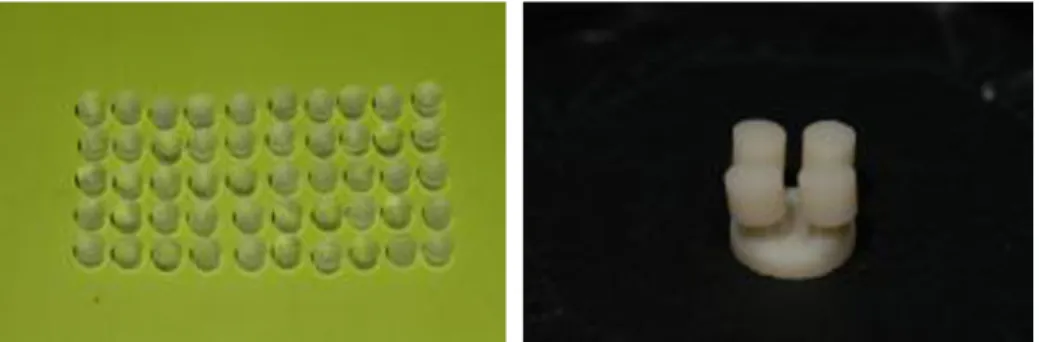
시편제작을 위해 직경 5 mm, 두께 4 mm의 투 명 아크릴릭 레진을 매몰링 기저부에 위치시키 고 통법에 따라 매몰, 소환한 후 Press furnace (IPS Empress EP 600 press furnace, Ivoclar Vivadent, Leichtenstein)를 이용하여 잉곳과 alox plunger를 이용해 열 가압을 시행하였다. 가압이 끝난 후 매 몰재를 제거하고 diamond disc로 주입선을 자른 후 접착면을 400, 800, 1000 grit silicon carbide를 이 용하여 단계적으로 연마하여 도재시편을 완성 하였다(Fig. 1).
3) 치아시편과 도재시편의 접착
직접 접착군(I군)을 포함하여 ALIKETM임시 수 복재를 이용해 brush on technique을 적용한 II군, Luxatemp�를 dough stage에 지대치에 직접 적용한 III군, Fermit�을 광중합시켜 적용한 IV군으로 분 류된 모든 치아 시편을 치면 처리하고 상온의 증 류수에 48시간 보관한 후, 상아질 표면을 35 % 인 산으로 15초 동안 산부식시키고 air-water spray로 10초간 세척한 후, 상아질 결합재로 Excite� (Ivoclar Vivadent, Leichtenstein)을 치면에 고루 바 른 후 광중합 시켰다. 도재시편은 불산 처리 후 접착 전 Monobond-s (Ivoclar Vivadent, Leichtenstein) 을 적용하고, 시편과 치아의 접착을 위해 Variolink II�(Ivoclar Vivadent, Leichtenstein) 레진시멘트를
이용하여 1 kg 정하중기 하에서 치아 상아질 면 에 직각으로 접착을 시행하였으며 4면으로 나누 어 각 면당 20초씩 광중합을 시행하였다.
4) 전단결합강도 측정
상아질에 대한 도재 시편의 전단결합강도를 측정하기 위해 만능시험기(Zwick 1456 41, Zwick, Germany)를 사용하여 하중이 접착면과 평행한 방향으로 전달되도록 지그에 시편을 고정하고 1 mm/min cross head speed의 속도로 하중을 가하 여 도재 시편이 분리되는 시점에서의 최대 전단 하중을 기록하였다(Fig. 2).
5) 파절면의 관찰
파절이 일어난 60개 시편의 표면의 양상을 관 찰하고 미세 구조 변화를 관찰하기 위해 각 군 당 치아 시편을 3개씩 무작위로 선택하여 시편을 7 일간 건조기에 넣어 완전 건조시킨 후 상아질측 파절면의 파절 양상을 확대경(S300 II, Tokyo Kinzoku, Japan) 하에서 관찰하였다.
6) 통계분석
SPSS Ver 12.0을 이용하여 각 군당 평균과 표준 편차를 구하고 P<0.05수준에서 One-way ANOVA 를 이용하였으며 Turkey test로 사후검정을 시행 하였다.
결 과
1) 전단결합강도의 측정
상아질과 도재 시편사이의 전단결합강도는 직 접 접착군이 4.68 MPa로 가장 높게 나타났고 Fermit�을 적용한 군이 3.75 MPa로 가장 낮은 결 합강도를 나타내었으나 통계학적 유의성은 보 이지 않았다(P>0.05, Table I, Fig. 3).
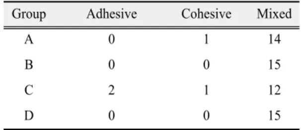
2) 파절양상의 관찰
직접 접착군에서 응집성 파절 양상이 1개, Fermit�적용군에서 접착성 파절양상이 2개 관찰 Fig. 1.Ceramic specimens used in this study.
Fig. 2.Shear bond testing with UTM.
되었으나 모든 실험군에서 대부분 혼합형 파절 의 양상을 보였다(Table II).
고 찰
지대치 삭제 후 임시수복물이나 충전재를 적 용 후, 최종 수복물을 장착하면서 치아 피착면에 대한 처치 및 접착을 시행하는 것이 일반적이다.
그러나 이 과정에서 노출된 상아질 면이 오염될 수 있으며 이는 세라믹 수복물의 상아질 결합을 방해하여 추후 실패 가능성8,9)을 높이게 되므로 Magne 등10)은 이러한 술식의 단점을 보완하고자 지대치 삭제 직후 치면에 상아질 결합제(dentin bonding agent)를 미리 도포하여 피착면을 보호하 는 경우가 전통적으로 나중에 처리한 것보다 결
합강도 면에서 더 우수하였다고 보고하였다. 그 외 임시시멘트 처치 및 인상채득 그리고 핸드피 스 윤활유, 타액, 혈액 등 여러 가지 요인들이 수 복물과 상아질의 결합에 영향을 주는 것으로 보 고되었다.11-14)
임시 수복물은 dough stage의 레진을 지대치 위 에 직접 적합시켜서 수복물을 만드는 직접법과 모형 상에서 외형을 미리 만들고 이를 구강 내에 서 레진 wash-out해서 마무리 짓는 간접법으로 나 뉠 수 있는데 이 과정에서 피할 수 없는 상아질 표면에 직접 접촉하는 레진 모노머 성분이 치은 과 치수같은 연조직에 영향을 주는 것으로 보고 되고,15)임시 수복물을 위한 Polymethyl methacrylate, Polyethyl methacrylate, Epimine, Bis-acryl 등의 재료 에 대한 특성과 Polymethyl methacrylate의 중합과 정에서 발열과 Free monomer의 치수에 대한 유해 성에 대해 여러 연구가 진행되었다.16,17)그러나 임 시수복용 레진의 성분이나 중합과정 또는 그 산 물이 상아질 표면에 변화를 주어 접착성 레진 시 멘트의 접착력에 어떠한 영향을 미치는지에 대 한 연구는 아직 부족한 상황에서 그러한 과정이 접착성 레진 시멘트의 접착력을 약화 시킬 것으 로 예상하고 본 연구를 진행하였으나 통계학적 으로 유의할만한 수준의 결합력 차이는 보이지 않았다.
임시시멘트 사용에 있어서도 Barier18)는 임시 시멘트 사용 시 상아질 표면의 젖음성 감소로 영 구 시멘트의 결합강도에 영향을 미친다고 보고 Table Ⅰ. Results of one-way ANOVA test
Group No. of specimen Mean SD
A 15 4.675a 1.195
B 15 4.344a 0.873
C 15 4.299a 0.839
D 15 3.755a 0.832
Table Ⅱ. Types of bond failure
Group Adhesive Cohesive Mixed
A 0 1 14
B 0 0 15
C 2 1 12
D 0 0 15
7 6 5 4 3 2 1 0
A B C D
Fig. 3. Bond strength of each experimental groups.
하였고, Terata 등19)은 임시 시멘트를 사용 후 기계 적으로 제거한다 해도 주사전자현미경 수준에 서 보면 여전히 잔존 시멘트가 존재하고 이는 인 산으로 식각시켜도 잔여 성분이 치면에 그대로 존재하기 때문에 레진시멘트의 결합력에 영향 을 주는 것으로 보고하였다. Terata 등20)은 레진계 열의 임시수복재가 치아와 레진 시멘트의 결합 강도에 미치는 영향에 대한 연구에서 법랑질에 서는 결합강도에 유의할만한 영향이 없었으나 4-META/MMA-TBB성분의 재료를 제외한 다른 재료는 상아질에서 결합강도를 저하시킨다고 보고하였고, 또한 resin-modified glass-ionomer cement의 결합강도에 임시수복재가 미치는 영향 에 대한 연구21)에서 Fermit�과 자가중합형 레진은 결합강도에 유의할만한 영향이 없었다고 보고 한 반면에, Ganss22)는 그의 연구에서 대조군에 비 해 Fermit�적용 후 dual-curing composite cement 과 수복물의 결합강도가 낮게 나왔다고 보고하기 도 하였다. 본 연구에서는 Fermit를 사용한 군과 그렇지 않은 군간에 유의한 차이가 없어 Terata21) 등의 연구와 비슷한 결과를 보였다.
이상의 제한된 실험 결과를 통해서 볼 때, 피착 면에 재료의 접촉이 일어나는 통상적인 임상과 정이 레진-상아질 결합력에 영향을 주리라 가정 했지만 직접 접착군에 비해 ALIKETM를 접착한 군, Luxatemp�Automix plus를 접착한 군, 그리고 Fermit�을 적용한 군, 모두에서 통계적으로 유의 할만한 차이를 보이지 않아, 임시 수복 및 충전 재료들이 상아질의 도말층에 화학적 변화와 기 계적 영향을 줄 가능성은 적은 것으로 판단되었 다. 그러나 이는 실험실 내에서 Zircate�Prophy Paste를 이용한 상아질 표면의 충분한 세척과 깨 끗한 청결유지가 기여한 바도 클 것으로 사료되 어, 진료 과정 중 여러 여건상 자칫 이러한 과정 이 소홀히 된다면 수복물과 상아질 간의 결합력 이저하될가능성도여전히배제할수없으며, 이에 대한더많은연구가필요할것으로사료되었다.
결 론
본 연구는 접착성 도재 수복물 제작시의 다양 한 임상 과정 중 임시수복재가 접착성 도재 수복 물과 상아질의 전단결합 강도에 미치는 영향에 대해 알아보기 위해 직접 접착군과 ALIKETM적 용군, Luxatemp�적용군, Fermit�적용군으로 나 누어 직접 접착군과 각각의 재료를 적용한 시편 의 도재와의 전단결합강도를 비교 측정함으로 써 다음과 같은 결론을 얻었다.
1. 직접 접착군, ALIKETM적용군, Luxatemp�적 용군, Fermit�적용군 간의 통계학적으로 유 의성있는 상아질과 IPS Empress II 간의 결합 강도에 차이는 없었다.
2. 파절양상은 모든 군에서 대부분 혼합형 양 상을 보였다.
따라서 치과임상에서 임시 충전제 및 수복제 자체가 lithium disilicate glass-ceramic을 접착성 레 진시멘트로 치아 상아질에 접착시킬 경우, 그들 의 영향은 크지 않을 것으로 판단된다.
연구비 지원 및 사의
본 연구는 2012년도 원광대학교 교내연구비 지원에 의해 이루어졌음.
REFERENCES
1. Ho¨land W. Materials science fundamentals of the IPS Empress 2 glass-ceramic. Ivoclar Vivadent Report 1998;12:3-10.
2. Severance G. Introducing a lithium disilicate glass- ceramic: IPS empress 2. Signature 1999;4:1-3.
3. Sorensen JA, Cruz M, Mito WT. Research evaluations of a lithium disilicate restorative system:
IPS Empress 2. Signature 1999;4:4-10.
4. Oh SC, Dong JK, Luthy H, Scharer P. Strength and microstructure of IPS Empress 2 glass-ceramic after different heat treatments. Int J Prosthodont.
2000;13:468-472.
5. Gwinnett AJ. Smear layer : Morphological considerations. Oper Dent 1984;3:3-12.
6. Pashley DH. Clinical correlations of dentin structure and function. J Prosthet Dent 1991;66:777-781.
7. Xavier L, David JB, Glen HJ. Retention of provisional crowns fabricated from two materials with the use of four temporary cements. J Prosthet Dent 1999;81:469-475.
8. Friedman MJ. A 15-years review of porcelain veneer failure-a clinician's observation. Compend Contin Edu Dent 1998;19;625-628.
9. Dumfart H. Porcelain laminate veneers: A retrospective evaluation after 1 to 10years of service:
part II-clinical results. Int J Prosthodont 2000;13;9- 18
10. Magne P, Kim TH, Cascione D, Donovan TE.
Immediate dentin sealing improves bond strength of indirect restorations. J Prosthet Dent 2005;94:511- 519.
11. Xie J, Powers JM. In vitro bond strength of two adhesives to enamel and dentin under normal and contaminated conditions. Dent Mater 1993;9:295- 299.
12. Johnson ME, Burgess JO, Hermesch CB, Buikema DJ. Saliva contamination of dentin bonding agents.
Oper Dent 1994;19:205-210.
13. Paul SJ, Schaerer P. Effect of provisional cements on the bond strength of various adhesive bonding systems on dentin. J Oral Rehabil 1997;24;8-14 14. Kaneshima T, Yatani H. The influence of blood
contamination on bond strength between dentin and an adhesive resin cement. Oper Dent 2000;25:195- 201.
15. Lai YL, Chen YT, Lee SY, Shieh TM, Hung SL.
Cytotoxic effects of dental resin liquids on primary gingival fibroblasts and periodontal ligament cells in vitro. J Oral Rehabil 2004;31:1165-1172.
16. Lui JL, Setcos JC, Phillips RW. Related Articles, Links temporary restorations: a review. Oper Dent 1986;11:103-110.
17. Cho GC, Chee WW. Custom characterization of the provisional restoration. J Prosthet Dent 1993;69:529- 532.
18. Barier RE. Principles of adhesion. Oper Dent Supple 1992;5:1-9.
19. Terata R, Nakashima K, Kubota M, Obara M.
Characterization of enamel and dentin surfaces after removal of temporary cement. Dent Mater J 1994;
13:148-154.
20. Terata R, Yoshinaka S, Nakashima K, Kubota M.
Effect of resinous temporary material on tensile bond strength of resin luting cement to tooth substrate. Dent Mater J 1996;15:45-50.
21. Terata R, Nakashima K, Kubota M. Effect of temporary materials on bond strength of resin- modified glass-ionomer luting cements to teeth. Am J Dent 2000;13:209-211.
22. Ganss C, Jung M. Effect of eugenol-containing temporary cements on bond strength of composite to dentin. Oper Dent 1998;23:55-62.
Effect of Provisional Restorative and Filling Materials on Bond Strength of Adhesive Resin Cement between Lithium Disilicate Glass-Ceramic and Dentin
Sang-Chun Oh, Hun-Bo Sim
Department of Prosthodontics, Dental College, Wonkwang University
The aim of this study was to evaluate the effect of temporary restorative and filling material on bonding strength between lithium disilicate glass-ceramic and dentin. 60 extracted human molars were cross- sectioned at occlusal third and were embedded into self-cure acrylic resin. Then the teeth were randomly divided into four groups of 15 each.
Lithium disilicate glass-ceramic is cemented to dentin as follows: after no any application of the provisional materials (Group A), after application of ALIKETM (GC America Inc.)(Group B), after application of Luxatemp�Automix plus (DMG, Germany)(Group C), after application of Fermit�(Ivoclar Vivadent, Leichtenstein)(Group D). After the specimens were stored in distilled water for 24 hours, the shear bond strength of the specimens were measured using UTM (Zwick 1456 41, Zwick, Germany) at a crosshead speed of 1mm/min. The data were analysed by one-way ANOVA and Tukey HSD tests. There were no statistically significant differences of bond strength among the groups.
Fracture type was showed mixed type of adhesive and cohesive fracture in most of specimens. Within the limitation of this study, bond strength of adhesive resin cement between lithium disilicate glass-ceramic and dentin was not affected by provisional restorative and filling materials. (J Dent Rehab App Sci 2013:29(4):359 - 365)
Key words: Lithium disilicate glass-ceramic, Dentin, Bonding strength, Temporary restorative and filling material
Correspondence to: Sang-Chun Oh
Department of Prosthodontics, College of Dentistry, Wonkwang University 1142 Sanbon-Dong, Gunpo, Gyeonggi-Do, 435-040, Korea
Tel: +82-31-390-2899, Fax: +82-31-390-2777, E-mail: [email protected]
Received: October 7, 2013, Last Revision: November 2, 2013, Accepted: November 15, 2013