• 검색 결과가 없습니다.

Combination of Ischemic Preconditioning and Postconditioning in Isolated and Intact Rat Hearts: Does it Work or Not?

N/A
N/A
Protected

Academic year: 2021

Share "Combination of Ischemic Preconditioning and Postconditioning in Isolated and Intact Rat Hearts: Does it Work or Not?"

Copied!
9
0
0

로드 중.... (전체 텍스트 보기)

전체 글

(1)

Abstract

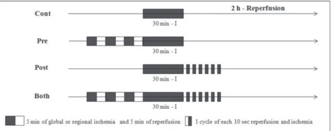
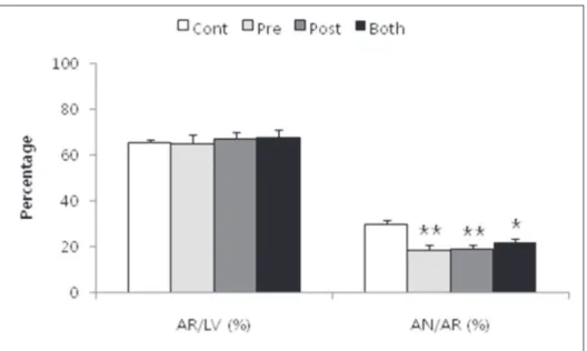
Ischemic preconditioning (Pre) phenomenon was well known as cardioprotective effects and its signaling pathways leading to myocardial protection activated by NO/cGMP pathways. In the meantime, the ischemic postconditioning (Post) in which brief intermittent repetitive interruptions to reperfusion at the onset of timely reperfusion after a prolonged period of ischemia also reduced myocardial injury to an extent comparable to Pre. In isolated working heart experiment, all hearts were subjected to 30 min of regional ischemia and 120 min of reperfusion: Control, no intervention; Pre, 3 cycles of 5-min of global ischemia and 5 min of reperfusion before the ischemic insult, Post; 6 cycles of 10 sec of reperfusion followed by 10 sec of global ischemia immediately after the index ischemia; Both (Pre + Post). Infarct size, coronary flow (CF), heart rate (HR), left ventricular developed pressure (LVDP), and +dP/dt were measured. The infarct size in the Pre (n=7, 18.5% ± 2.1%), Post (n=7, 19.1% ± 1.5%), and Both (n=10, 21.7% ± 1.7%) groups was significantly decreased compared with Control (n=7, 29.9% ± 1.7%). However, there was no additive effect when Pre and Post were combined. There were no significant differences in baseline CF, HR, LVDP, +dP/dt among the groups, and no significant differences in HR, LVDP, +dP/dt in all groups after 2 hours of reperfusion. In conclusion, Pre and Post, Both group improved cardioprotective effects than Control group.

However, combination of ischemic preconditioning and postconditioning showed no additive cardioprotective effects in isolated rat heart models.

Division of Cardiology, Department of Internal Medicine, Keimyung University School of Medicine, Daegu, Institute of Cardiovascular Research, Pusan National University Yangsan Hospital

1

,

Gyeongsangnam-do, Korea

In Gyu Yeo, B.S., Hyoung Seob Park, M.D., Sang Woong Choi, M.D., Han Byul Kim, M.D., Jae Pil Lee, M.D., In Cheol Kim, M.D., Jeong Eun Lee, M.D., Hyuck Jun Yoon, M.D.,

Hyung Seop Kim, M.D., Chang Wook Nam, M.D., Seung Ho Hur, M.D., Yoon Nyun Kim, M.D., Kwon Bae Kim, M.D. Young Ho Jang

1

, M.D.

Combination of Ischemic Preconditioning and Postconditioning in Isolated and Intact Rat Hearts: Does it Work or Not?

Corresponding Author: Hyoung Seob Park, M.D., Department of Internal Medicine, Keimyung University School of Medicine 56 Dalseong-ro, Jung-gu, Daegu 700-712, Korea

Tel : +82-53-250-7473 E-mail : [email protected]

(2)

Introduction

Ischemic preconditioning (Pre) phenomenon was originally introduced by Murry and associates in 1986 [1], and its signaling pathways leading to myocardial protection afforded by Pre is unveiling [2,3]. In the meantime, the ischemic postconditioning (Post) in which brief intermittent repetitive interruptions to reperfusion at the onset of timely reperfusion after a prolonged period of ischemia also reduced myocardial injury to an extent comparable to Pre. It has been suggested that Post is as effective as Pre in limiting infarct size and preserving postischcemic endothelial function [4].

There are similarities and differences between Pre and Post in physiological, cellular and subcellular, and molecular effects [5]. Recently, Downey’s group [6], demonstrated that the combination of Pre and Post resulted in even greater protection than either alone in the in vivo rabbit hearts. The infarct size of combination of Pre and Post declined further to 22% and was significantly less than either Pre or Post alone.

H o w e v e r, V i n t e n-J o h a n s e n’s g r o u p [7], demonstrated that there was no additive cardioprotective effect by Pre and Post in a canine model of regional ischemia and reperfusion. The infarct size was not further reduced when both strategies were sequentially applied, although there was a tendency for the combination to reduce the incidence of reperfusion ventricular fibrillation. In addition, Yellon’s group [8], reported that the combined protective effects on infarct size limitation of Pre and Post did not occur in isolated

perfused rat hearts. Taken together, the effect of the combined intervention of Pre and Post is still controversial.

The cardioprotective phenomena of Pre and Post exert their cardioprotective effects through the recruitment of the phosphatidylinositiol-3-OH kinase (PI3K)-Akt and this protection is mediated t h r o u g h t h e i n h i b i t i o n o f m i t o c h o n d r i a l permeability transition pore (mPTP) opening [9], although the Pre and Post intervene at opposite ends of the ischemic event. However, the strategies may differ in the involvement of p42/44 extracellular signal-regulated kinases (ERK1/2), which is not activated in Pre but may be involved in Post [5,6].

Therefore, the objective of the present study was to address whether the co-application of Pre and Post would have synergic effect and protect heart compared to either intervention alone in isolated perfused rat hearts.

Materials and Methods

1. Chemicals and antibodies

Pentobarbital sodium (Entobar

) will be purchased from Hanlim Pharmacy, Republic of Korea Triphenyltetrazolium chloride (TTC) will be also obtained form Sigma-Aldrich Chemical.

Fluorescent polymer microspheres will be purchased from Duke Scientific Corp., CA, USA.

Other chemicals will be obtained from Sigma Chemical Co., St. Louis, Missouri, USA.

Key Words : Ischemic postconditioning, Ischemic preconditioning, Myocardial ischemia

(3)

2. Isolated perfused rat heart model

Male Sprague-dawley rats weighing 280-330 g obtained from Huco FS Co, Republic of Korea, will be used throughout the experiments. They received 100 mg/kg of pentobarbital sodium and 300 IU of heparin intraperitoneally. Hearts will be isolated and perfused as described previously [10].

Briefly, a midline thoracotomy will be performed and the heart will be quickly excised, arrested in ice-cold heparinized saline, and immediately mounte it on a Langendorff apparatus by the arotic root. The heart will be perfused with modified Krebs-Henseleit solution containing (in mM) 118.5 NaCl, 4.7 KCl, 1.2 MgSO4, 1.8 CaCl

2

, 24.8 NaHCO

3

, 1.2 KH

2

PO

4

, and 10 glucose. The solution will be filtered through a Nargene 2.0 µm microfilter (Nalge Nunc International Corp., USA) and be equilibrated with 95% O2/5% CO

2

.for 30 min before the experiment and pH maintained at about 7.4. The whole system will be heated to 38℃ by means of water jacketing. Perfusion will be performed under a hydrostatic pressure of 100 cmH

2

O by adjusting the height of the reservoir.

To induce regional ischemia, the proximal length of left coronary artery (LCA) will be localized between left atrial appendage and the right ventricular outflow tract followed by the passage of 6-0 polypropylene suture around major trunk of the LCA or its prominent branches. The ends of thread will be passed through a small piece of polyethylene tubing (PE 50) to form a snare. All hearts will be allowed to stabilize for at least 20 min. Ischemia will be induced by pulling the snare and then fix it by clamping the tubing with a small hemostat and confirmed by regional cyanosis and a substantial decrease in left v e n t r i c u l a r d e v e l o p e d p r e s s u r e (LV D P).

Reperfusion will be started by releasing the ends of the suture.

3. Assessment of cardiac function

In isolated hearts, an air-bubble free, Krebs- Henseleit buffer-filled latex balloon connected to a pressure transducer (Baxter, USA) with tubing will be inserted into the left ventricle (LV) through the left atrial appendage. Balloon volume will be adjusted to give a left ventricular end-diastolic pressure (LVEDP) of 5-10 mmHg at the beginning of the experiment. LVDP records with BIOPAC system and will be analyzed using an analysis software (Chart v4.2.4) will be calculated as the difference between left ventricular systolic pressure (LVSP) and LVEDP. Total coronary flow (CF) will be measured by timed collection of the perfusate dripping from the right heart into a graduated cylinder. The HR, LVDP, and CF will be collected at baseline, 30 min of ischemia, and after 2 hrs of reperfusion, respectively.

4. Experimental protocol and exclusion criteria

In isolated working heart experiment, all hearts will be subjected to 30 min of regional ischemia and 120 min of reperfusion and each group consisted of at least 6 hearts. Following stabili- zation period, hearts will be assigned randomly to one of the following four groups: 1) Cont; no intervention either before or after LCA occlusion, 2) Pre; subjected to three 5-min of global ischemia and 5 min of reperfusion before the ischemic insult; 3) Post; comprising 6 cycles of 10 sec of reperfusion followed by 10 sec of global ischemia immediately after the index ischemia, and 4) Both (Pre + Post). These groups are chosen because it is unclear whether the duration of ischemia may affect the additive effect of Pre and Post. The experimental time lines are shown in (Fig. 1).

In isolated hearts, all hearts will be perfused

(4)

within 30-40 sec after excision. The heart perfusion times longer than 60 sec will be excluded from the study. Any hearts with a HR less than 250 beats/min, or CF more than 18 ml/min or less than 8 ml/min at the end of 20 min stabilization will be also excluded. Hearts failing to develop LVSP of more than 80 mmHg when the LVEDP will be kept less than 10 mmHg will be also excluded. Ischemic hearts experiencing ventricular fibrillation (VF) usually revert spontaneously to sinus rhythm. VF lasting more than 45 sec will be treated with thump cardioversion until a perfusing rhythm will be obtained. Rat hearts experiencing irreversible VF more than 2 min despite of cardioversion will be also excluded.

5. Determination of area at risk and infarct size

At the end of each experiment (after 2 hr of reperfusion) in isolated hearts, the LCA perfusion circuit will be reoccluded, and 2-9 µm diluted fluorescent polymer microspheres will be infused

to demarcate the area at risk (AR) as the tissue without fluorescence. The hearts will be weighed, frozen at -20℃ for 1-3 hrs, and cut into transverse slices 2 mm thickness. The slices will be incubated in 1% TTC in sodium phosphate buffer at 37℃ for 20 min. The slices will be immersed in 10%

formalin to enhance the contrast between stained (viable) and unstained (necrotic) tissue and then compress to a uniform 2 mm thickness by placing them (basal side) between two glass plates s e p a r a t e d b y a 2 m m s p a c e. T h e A R o f myocardium will be identified by illuminating the slices with U.V. light. The infracted (unstained) and risk (stained red by TTC) zone regions will be traced on a clear acetate transparent sheet and quantified with Image Tool (UTHSCSA Image Tool, version 3.0). The areas are converted into volumes by multiplying the areas by slice thickness. The area of infarct size will be expressed as a percentage of the AR. All measurements will be performed in a blinded fashion.

Fig. 1. Experimental protocols. Ischemic preconditioning (Pre) hearts subjected to three 5-min periods of global

ischemia and 5-min periods of reperfusion before the 30 or 45 min lethal ischemic insult. Postconditioning (Post)

comprised 6 cycles of 10 sec of reperfusion followed by 10 sec of global ischemia immediately after the index

ischemia.

(5)

6. Statistical analysis

Data will be presented as means ± SEM. Data analysis will be performed with a personal computer statistical software package (SPSS for windows, Release 12.0; SPSS Inc, Chicago, IL, USA). CF and hemodynamic data will be analyzed using two-way analysis of variance (ANOVA) with Bonferroni post-hoc testing. Infarct size and the size of AR will be analyzed between groups using one-way ANOVA with a Student-Newman-Keuls post-hoc test. Differences will be considered to be statistically significant when p values were < 0.05.

Results

1. Infarct Size

Body weight and heart weight were equivalent

among the groups. There were no significant group differences with respect to left ventricular volume, AR volume, and AR/LV ratio among the groups (Table 1). The AR as a percentage of the LV averaged between 65.1% and 67.8% , which indicates that all groups were subjected to equivalent degrees of regional ischemia. The infarct size in the Pre (n=7, 18.5% ± 2.1%), Post (n=7, 19.1% ± 1.5%), and Both (n=10, 21.7% ± 1.7%) groups was decreased significantly compared with Cont (n=7, 29.9% ± 1.7%).

However, there was no significantly additive effect w h e n i s c h e m i c p r e c o n d i t i o n i n g a n d postconditioning were combined (Fig. 2).

2. Coronary flow and cardiodynamic

There were no significant differences in baseline CF, HR, LVDP, +dP/dt

max

among the groups (Table 2). The CF decreased after ischemia

Table 2. Baseline coronary flow and cardiodynamic data

CF: coronary flow, HR: heart rate, LVDP: left ventricular developed pressure.

Table 1. Morphometric data

LV: left ventricular, AR: area at risk.

Group Body Weight (G) Heart Weight (g) LV Volume (㎤) AR Volume (㎤) AR/LV (%) Cont 307.9 ± 14.4 1.52 ± 0.08 0.549 ± 0.019 0.361 ± 0.016 65.6 ± 1.5

Pre 297.9 ± 9.9 1.47 ± 0.04 0.555 ± 0.019 0.358 ± 0.013 65.1 ± 3.9 Post 280.0 ± 7.6 1.43 ± 0.05 0.535 ± 0.023 0.359 ± 0.019 67.3 ± 2.7 Both 311.0 ± 9.4 1.52 ± 0.05 0.524 ± 0.021 0.356 ± 0.023 67.8 ± 3.3

Group CF (mL/min) HR (beats/min) LVDP (mmHg) +dP/dt

max

(mmHg/s)

Cont 18.9 ± 2.4 306.9 ± 6.0 107.6 ± 4.3 3769.8 ± 271.2

Pre 19.8 ± 2.2 315.8 ± 13.8 126.6 ± 9.8 4319.1 ± 252.2

Post 19.7 ± 1.7 293.4 ± 4.3 117.6 ± 4.9 3896.4 ± 211.6

Both 20.1 ± 1.4 321.9 ± 7.8 110.5 ± 5.2 3537.0 ± 243.5

(6)

and declined steadily after reperfusion. But Cont, Pre, Post and Both were no significant differences

(Fig. 3). To determine the functional recovery associated Pre, Post and Both, the percentage Fig. 3. Coronary flow during stabilization, ischemia and reperfusion. BL, baseline; I-30, 30 min after ischemia; R-10, 10 min after reperfusion; R-30, 30 min after reperfusion; R-60, 60 min after reperfusion; R-90, 90 min after reperfusion; R-120, 120 min after reperfusion.

Fig. 2. Bar graph showing area at risk (AR) relative to left ventricular (LV) mass (AR/LV) and area of necrosis (AN)

relative to AR. Infarct size (AN/AR) was significantly reduced Pre, Post, and Both versus Cont. But no significant

differences were found among the three intervention groups. **P<0.005 vs. control, *P<0.05 vs. control.

(7)

changes from the baseline levels in HR, LVDP, +dP/dt

max

after 2 hours of reperfusion were compared. There were no significant differences in HR, LVDP, +dP/dt

max

in all groups. (Fig. 4).

Discussion

Ischemic preconditioning and postconditioning are a very popular topic in the cardiovascular research field. Many previous papers have reported that ischemic preconditioning and postconditioning limits the myocardial infarct size.

In the present study, we demonstrate combination of ischemic preconditioning and postconditioning compare to each ischemic preconditioning and postconditioning in isolated rat heart. The infarct size in the Pre, Post, and Both groups was decreased significantly compared with Cont. But combination of ischemic preconditioning and postconditioning is no significantly additive c a r d i o p r o t e c t i v e e f f e c t t h a n i s c h e m i c preconditioning and postconditioning alone. Also coronary flow and hemodynamic data were no significant differences after 2 hours of reperfusion.

Hausenloy and Yellon [11] reported that increased phosphorylation of Akt and ERK 1/2 i n d u c e c a r d i o p r o t e c t i v e e f f e c t s t h r o u g h reperfusion injury salvage kinase (RISK)-pathway.

The RISK pathway mediates cellular survival through several possible mechanisms, which may include inhibition of mitochondrial permeability transition pore (mPTP) opening. And Hausenloy et al . [12] also reported that ischemic preconditioning activating prosurvival kinases at reperfusion.

D u r i n g t h e i s c h e m i c p r e c o n d i t i o n i n g, mitochondrial ROS were released and PKC was activated. These events reactivate the PI3K-Akt and MEK-1/2–ERK-1/2, which comprise the RISK pathway, in reperfusion.

The protective effects of NO during reperfusion, as well as the contradictory results about this topic have been reviewed by Schulz et al [13]. Penna C et al . [14] reported that ischemic postconditioning reduced infarct size through nitric oxide (NO)/cGMP pathway. They demonstrate for the Fig. 4. The percentage recovery of cardiodynamics

after reperfusion in isolated heart. All groups no significant difference in HR, LVDP, +dP/dt

max

after 2 hours of reperfusion. HR, heart rate (beats/min);

LVDP, left ventricular developed pressure (mmHg);

+dP/dt

max

, maximum positive left ventricular

pressure derivative (mmHg/s).

(8)

first time that NOS and GC play different roles in ischemic postconditioning protection.

Ischemic preconditioning and postconditioning increase phosphorylation of Akt and ERK 1/2, and reduce infarct size [12,15]. We suggest combination of ischemic preconditioning and postconditioning is no significant differences phosphorylation of Akt and ERK 1/2 compare with ischemic preconditioning and postconditioning because of no significant differences infarct size. After 30 min of ischemia, cardiac function was still depressed for variable period of time. This phenomenon reported that cardiac postischemic ventricular dysfunction by Braunwald’s group in 1982 [16].

Combination of ischemic preconditioning and postconditioning was protective, we wondered if this protection could be added to that positive synergy effects. However, no positive synergy effects of combination of ischemic preconditioning and postconditioning. We suggest that because ischemic preconditioning and postconditioning induce cardioprotective effects through the same pathway.

In conclusion, ischemic preconditioning and postconditioning reduces infarct size during ischemia-reperfusion injury through reperfusion injury salvage kinase (RISK)-pathway in isolated rat hearts. However combination of ischemic preconditioning and postconditioning is no significantly additive cardioprotective effect than ischemic preconditioning and postconditioning alone.

References

1. Murry CE, Jennings RB, Reimer KA. Preconditioning with ischemia: a delay of lethal cell injury in ischemic myocardium. Circulation 1986;74:1124-36.

2. Oldenburg O, Qin Q, Kreig T, Yang XM, Philipp S,

Critz SD, et al. Bradykinin induced mitochondrial ROS generation via NO, cGMP, PKG, and mitoKATP channel opening and leads to cardioprotection. Am J Physiol Heart Circ Physiol 2004;286:H468-76.

3. Xu Z, Downey JM, Cohen MV. Timing and duration of administration are crucial for antiinfarct effect of AMP579 infused at reperfusion in rabbit heart. Heart Dis 2003;5:369-71.

4. Zhao ZQ, Corvera JS, Halkos ME, Kerendi F, Wang NP, Guyton RA, et al. Inhibition of myocardial injury by ischemic postconditioning during reperfusion:

comparison with ischemic preconditioning. Am J Physiol Heart Circ Physiol 2003;285:H579-88.

5. Vinten-Johansen J, Zhao Z, Zatta A. Postconditioning;

a new link in nature’s armor against myocardial ischemia-reperfusion injury. Basic Res Cardiol 2005;100:295-310.

6. Yang X, Proctor JB, Cui L, Krieg T, Downey JM, Cohen MV. Multiple, brief coronary occlusions during early reperfusion protect rabbit hearts by targeting cell signaling pathways. J Am Coll Cardiol 2004;44:1103- 10.

7. Halkos ME, Kerendi F, Corvera JS, Wang NP, Kin H, Payne CS, et al. Myocardial protection with postconditioning is not enhanced by ischemic preconditioning. Ann Thorac Surg 2004;78:961-9.

8. Tsang A, Hausenloy D, Mocanu MM, Yellon DM.

Postconditioning: a form of “modified reperfusion”

protects the myocardium by activating the phosphatidylinositiol 3-kinase-Akt pathway. Circ Res 2004;95:230-2.

9. Hausenloy DJ, Tsang A, Yellon DM. The reperfusion injury salvage kinase pathway: a common target for both ischemic preconditioning and postconditioning.

Trends Cardiovasc Med 2005;15:69-75.

10. Park SS, Zhao H, Jang Y, Mueller RA, Xu Z. N6-(3-

lodobenzyl)-adenosine-5’-N-methylcarboxamine

confers cardioprotection at reperfusion at reperfusion

by inhibiting mitochondrial permeability transition

pore opening via glycogen synthase kinase 3β. J

(9)

Pharmacol Exp Ther 2006;318:124-31.

11. Hausenloy DJ, Yellon DM. New directions for protecting the heart against ischaemia-reperfusion injury: targeting the Reperfusion Injury Salvage Kinase (RISK)-pathway. Cardiovasc Res 2004;61(3):448-60.

12. Hausenloy DJ, Tsang A, Mocanu MM, Yellon DM.

Ischemic preconditioning protects by activating prosurvival kinases at reperfusion. Am J Physiol Heart Circ Physiol 2005;288(2):H971-6.

13. Schulz R, Kelm M, Heusch G. Nitric oxide in myocardial ischemia/reperfusion injury. Cardiovasc Res 2004;61(3):402-13.

14. Penna C, Cappello S, Mancardi D, Raimondo S, Rastaldo R, Gattullo D, et al. Post-conditioning reduces infarct size in the isolated rat heart: role of coronary flow and pressure and the nitricoxide/cGMP pathway. Basic Res Cardiol 2006;101(2):168-79.

15. Fujita M, Asanuma H, Hirata A, Wakeno M, Takahama H, Sasaki H, et al. Prolonged transient acidosis during early reperfusion contributes to the cardioprotective effects of postconditioning. Am J Physiol Heart Circ Physio 2007;292(4):H2004-8.

16. Braunwald E, Kloner RA. The stunned myocardium:

prolonged, postischemic ventricular dysfunction.

Circulation 1982;66(6):1146-9.

수치

Fig. 1. Experimental protocols. Ischemic preconditioning (Pre) hearts subjected to three 5-min periods of global  ischemia and 5-min periods of reperfusion before the 30 or 45 min lethal ischemic insult
Table 1.  Morphometric data
Fig. 2. Bar graph showing area at risk (AR) relative to left ventricular (LV) mass (AR/LV) and area of necrosis (AN)  relative to AR

참조

관련 문서

2) Serum Cortisol, ACTH 3) Serum TSH and Free T4 4) Serum Prolactin and GH 5) Serum 25OHD and PTH.. What is the most common laboratory. abnormality expected in this

Basic aspects of AUTOSAR architecture and methodology Safety mechanisms supported by AUTOSAR.. Technical safety concepts supported by AUTOSAR Relationship to ISO

GDP impact of COVID-19 spread, public health response, and economic policies. Virus spread and public

After first field tests, we expect electric passenger drones or eVTOL aircraft (short for electric vertical take-off and landing) to start providing commercial mobility

Today, it will be cloudy in the afternoon, and it’s going to rain at night.. On Saturday, it will be colder, and the rain will

1 John Owen, Justification by Faith Alone, in The Works of John Owen, ed. John Bolt, trans. Scott Clark, &#34;Do This and Live: Christ's Active Obedience as the

Anoxic

Utility of T1-and T2-weighted high-resolution vessel wall imaging for the diagnosis and follow up of isolated posterior inferior cerebellar artery dissection with