© Copyright
Keimyung University School of Medicine 2017 계명의대학술지 제36권 1호
65
Keimyung Med J Vol. 36, No. 1, June, 2017
Apocrine hidrocystoma is a benign tumor arising from apocrine sweat gland. It presents as a solitary translucent nodule, usually on the face, head, and neck. However it is extremely rare for the apocrine hidrocystoma to arise on the extremities, and no case of apocrine hidrocystoma on lower extremity has been reported in Korean dermatologic literature. Herein, we report a case of 63-year-old male apocrine hidrocystoma that developed in ankle area with a review of the literature.
Keywords: Ankle, Apocrine hidrocystoma
서 론
아포크린땀샘낭종(apocrine hidrocystoma)은 1964년 Mehregan [1]이 처음 보고한 아포크린땀샘의 분비부에서 발생하는 양성종양으로 병변은 대개 단발성의 낭포성 결절로, 피부색이나 적갈색 또는 짙은 푸른색을 띈다. 신체의 어느 부위에서나 발생할 수 있으나 주로 얼굴, 머리, 목 및 체간에 많이 발생한다. 사지에는 매우 드물게 발생하며[2], 특히 하지에 발생한 예는 국내 문헌에 보고된 바가 없었다.
저자들은 비호발부위인 발목에 발생한 아포크린땀샘낭종을 경험하고 국내 문헌에 보고된 적이 없는 드물고 흥미로운 예라고 생각하여 문헌고찰과 함께 보고하는 바이다.
Received: March 21, 2017 Revised: April 27, 2017 Accepted: May 31, 2017
Corresponding Author: Joon Soo Park, M.D., Department of Dermatology, The Catholic University School of Medicine, Daegu, Korea 3056-6, Daemyung-4-Dong, Nam-gu, Daegu 42472, Korea
Tel: +82-53-650-4161 E-mail: [email protected]
• The authors report no conflict of interest in this work.
Department of Dermatology, The Catholic University of Daegu School of Medicine, Daegu, Korea
Osung Kwon, M.D., Yong Woo Choi, M.D., Kyung Duck Park, M.D., Hyun Chung, M.D., Joonsoo Park, M.D.
A Case of Apocrine Hidrocystoma on the Ankle
대구가톨릭대학교 의과대학 피부과학교실
권오승·최용우·박경덕·정현·박준수
발목에 발생한 아포크린땀샘낭종 1례
66
계명의대학술지 제36권 1호 2017증 례
63세 남자가 좌측 발목에 발생한 검푸른색의 낭포성 결절을 주소로 내원하였다. 병변은 약 20년 전 발생하였으며, 크기가 점점 증가하는 양상을 보였다.
과거력 및 가족력상 특이병력은 없었다. 신체진찰상 피부소견 외 특이소견은 없었다. 피부 소견상 좌측 발 목 에 검 푸 른 색 을 띄 는 낭 포 성 결 절 이 관찰되었다(Fig. 1). 내원 당일 시행한 전혈구검사, 혈청 생화학검사 및 전해질검사상 특이 사항 없었다.
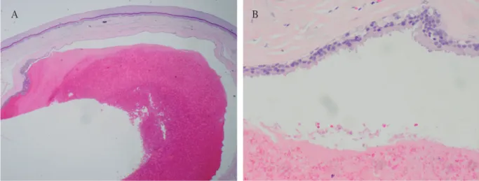
좌측 발목의 병변에서 시행한 조직검사상 진피에 부 정 형 의 호 산 성 물 질 로 이 루 어 진 낭 포 가 관찰되었으며(F i g. 2A), 낭포의 벽은 특징적인 단두분비를 보이는 원주세포(Fig. 2B)로 이루어져 있 었 다 . 분 비 세 포 는 P A S 염 색 에 양 성 반 응 을
보였으며(Fig. 3A), diastase에 저항하고(Fig. 3B), CEA염색에 양성반응을 보였다(Fig. 3C).
내원 당일 병변에서 절제생검을 시행하였으며, 이후 재발 없이 추적관찰 중이다.
고 찰
아포크린땀샘낭종은 대게 반구형의 투명한 낭포성 결절로 나타난다. 색은 대부분 투명하며, 푸른색 또는 적갈색을 띠기도 한다. 아포크린땀샘낭종이 푸른색을 띠는 것은 분비과립에 멜라닌이나 혈철소가 함유되어 있거나, lipofucin 침착에 의한 Tyndall 현상에 의한 것이라고 보고되고 있다[3]. 본 증례의 경우, 조직생검 소견에서 멜라닌이나 혈철소 등이 관찰되지 않은 점으로 미루어 Tyndall 현상에 의한 색조 변화로 생각된다.
아포크린땀샘낭종은 신체의 어느 부위에서나 발생할 수 있으나 주로 얼굴, 머리, 목 및 체간에 많이 발생한다. Anzai 등[2]은 일본에서 발생한 아포크린 땀샘낭종 167례를 분석하면서, 그 중 123례(73.7%)가 얼굴이나 머리에 발생하였고, 하지에는 11례(6.5%)만 발생하였다고 보고하였다. 아포크린땀샘낭종의 발생기전에 대해서는 아직 정확히 밝혀진 바가 없다.
아포크린땀샘은 발생학적으로 모낭에서 기원한 상피아(epithelial bud)에서 유래한다. 태아 단계에서 아포크린땀샘의 분포는 모낭이 존재하는 부위 전체에 걸쳐 존재할 수 있지만, 재태 6개월 경 대부분 흡수되고 출생 시에는 겨드랑이, 외이도, 눈꺼풀, 유방 등에만 한정되어 존재하게 된다. Mehregan [1]은 이 과정에서 미처 흡수 되지 못한 아포크린땀샘의 잔 여 물 이 본 질 환 의 발 생 과 연 관 이 있 다 고 주장하였다. 아포크린땀샘낭종의 발생기전에 대한 또 다른 가설로는 다분화성 세포(pluripotent cell)의 분화설이 존재하는데, Pfeifer 등[4]은 모낭 주위에 존재하는 다능성 세포가 아포크린땀샘으로 분화하여 발생한다고 주장하였다. 본 질환이 대부분 중년 이 후 에 발 생 하 는 점 과 출 생 시 존 재 하 는 아포크린땀샘부위가 아닌, 모낭이 많이 분포하는 얼굴과 머리에 호발하는 점으로 미루어 후자의 Fig. 1. The picture shows a dark blue colored cystic
nodule on the ankle of the left foot.
67
발목에 발생한 아포크린땀샘낭종 1례가능성이 더 높다고 생각한다[5,6]. 본 증례에서와 같이 발목 부위에 발생한 보고가 드문 이유 또한 같은 이유에서라고 추측된다.
아포크린땀샘낭종의 특징적인 조직학적 소견은 진피에 존재하는 한 개이상의 낭포성 공간에 존재하며 낭포내로 특징적인 단두분비를 보이는 원주세포가 관 찰 된 다 는 것 이 다 . 분 비 세 포 는 PA S 염 색 에 양성반응을 보이며, diastase처리한 후에도 저항성을 보인다. 본 증례에서도 이와 같은 조직 소견을 관찰할 수 있었으며, 면역조직화학염색상 CEA에 양성반응을 보였다. Sugiyama 등[7]은 아포크린땀샘낭종과 아포크린낭샘종(a p o c r i n e a d e n o c y s t o m a)이 아포크린땀샘에서 발생하는 양성종양을 일컫는 용어로 혼용되고 있으나, 두 질환 사이에는 명확한
차이가 존재하며, 구분되어서 사용되어야 한다고 주장하였다. 아포크린낭샘종에는 조직학적으로 진성 유두돌기가 존재하며, 비정형성 세포들과 유사분열이 관찰되므로 아포크린땀샘낭종과 감별할 수 있다. 본 증 례 에 서 는 위 와 같 은 소 견 이 관 찰 되 지 않 아 , 아포크린땀샘낭종으로 진단하였다.
감별질환으로는 표피낭종, 기저세포암, 모반 등이 있다[8,9]. 특히 에크린땀샘낭종과의 감별이 중요한데, 조직학적으로 아포크린땀샘낭종에서 단두 분비를 보이는 분비세포를 관찰함으로써 두 질환을 감별할 수 있다. 아포크린땀샘낭종은 피부절제를 통해 치료가 가능하며, 크기가 작은 경우나 다발성인 경우 CO
2laser를 통한 제거술도 시행된다[10].
아포크린땀샘낭종은 전신에서 발생할 수 있으며, Fig. 2. A cystic spac is filled with amorphous eosinophilic materials in the dermis (A;H&E, ×10) and the cyst is wall is lined with columnar epithelial cells showing decapitation secretion (B;H&E, ×100).
A B
Fig. 3. The granules are PAS-positive (A;PAS, ×100), resistant to diastase (B;DPAS, ×100) and CEA-positive (C;CEA, ×100).
A B C
68
계명의대학술지 제36권 1호 2017다양한 색조를 띄는 경우가 많아 다른 질환과 감별이 어려울 수 있다. 하지만 특징적인 조직소견을 보여 쉽게 진단이 가능하고 별다른 합병증 없이 단순 절제로 치료가 가능한 질환이므로, 임상의사의 질환에 대한 인식이 중요하다. 하지만 아직 발생기전이 명확하지 않으며, 비호발부위의 증례 발표가 적어 더 많은 증례 발표를 통해 원인과 분포에 대한 연구가 이루어져야 할 것이다.
참 고 문 헌