• 검색 결과가 없습니다.

Severe Acute Fibrinous and Organizing Pneumonia with Acute Respiratory Distress Syndrome

N/A
N/A
Protected

Academic year: 2021

Share "Severe Acute Fibrinous and Organizing Pneumonia with Acute Respiratory Distress Syndrome"

Copied!
5
0
0

로드 중.... (전체 텍스트 보기)

전체 글

(1)

Tuberc Respir Dis 2011;71:368-372

CopyrightⒸ2011. The Korean Academy of Tuberculosis and Respiratory Diseases. All rights reserved.

Severe Acute Fibrinous and Organizing Pneumonia with Acute Respiratory Distress Syndrome

Hyo Jae Kang, M.D.

1

, Sun Mi Choi, M.D.

1

, Yun Jeong Jeong, M.D.

1

, Jong Sun Park, M.D.

2

, Sei Won Lee, M.D.

2

, Ho Il Yoon, M.D., Ph.D.

2

, Jae Ho Lee, M.D., Ph.D.

2

, Choon-Tack Lee, M.D., Ph.D.

2

, Young-Jae Cho, M.D., M.P.H.

2

1

Division of Pulmonary and Critical Care Medicine, Department of Internal Medicine and Lung Institute, Seoul National University College of Medicine, Seoul,

2

Division of Pulmonary and Critical Care Medicine, Department of Internal Medicine and Lung Institute, Seoul National University Bundang Hospital, Seoul National University College of Medicine, Seongnam, Korea Acute fibrinous and organizing pneumonia is a newly recognized pattern of acute lung injury. A 49-year-old female presented with a cough and worsening dyspnea on exertion. She had no history of smoking and no specific past medical history except exposure of home humidifier containing sterilizer. A chest computed tomography scan showed patchy consolidation with fibrosis in the right lower lobe and ill-defined centrilobular ground glass opacity in both lungs. The pathological findings were patchy areas of lung parenchyma with fibrin deposits in the alveolar ducts and alveoli, and fibrin balls with hemosiderin deposition in the alveolar spaces. The histological pattern of our case is differentiated from diffuse alveolar damage by the absence of hyaline membranes, and from eosinophilic pneumonia by the lack of eosinophils. In our case, the patient was treated with corticosteroid pulse therapy. However, the clinical course became aggravated and she died within two weeks.

Key Words: Acute Respiratory Distress Syndrome; Organizing Pneumonia, Cryptogenic; Inhalation Exposure

Address for correspondence: Young-Jae Cho, M.D., M.P.H.

Division of Pulmonary and Critical Care Medicine, Department of Internal Medicine and Lung Institute, Seoul National University Bundang Hospital, Seoul National University College of Medicine, Gumi-dong, Bundang-gu, Seongnam 463-707, Korea

Phone: 82-31-787-7058, Fax: 82-31-787-4051 E-mail: lungdrcho@gmail.com

Received: Jun. 27, 2011 Accepted: Aug. 19, 2011

Introduction

Acute lung injury (ALI) may occur secondary to an extensive number of direct or indirect pulmonary insults and consequently results in hypoxic respiratory failure

1

. Most cases of clinical ALI will show a specific histo- logical feature, diffuse alveolar damage (DAD). In 2002, Beasley et al.

2

reported acute fibrinous and organizing pneumonia (AFOP) as an unusual type of ALI. The his- tological pattern of AFOP is differentiated from DAD by the absence of hyaline membranes, and from eosino- philic pneumonia (EP) by the lack of eosinophils. They

noted several conditions associated with AFOP, includ- ing collagen-vascular diseases, malignancies, drugs, and infections. However, few cases of AFOP have been re- ported until now in Korea. Herein, we report an extra- ordinary case of AFOP in a healthy patient.

Case Report

Patient: A 49-year-old female.

Chief complaint: Worsening of dyspnea on exertion for one month.

Present illness: A 49-year-old female visited our hos- pital emergency room (ER) due to worsening of dys- pnea. Four weeks ago, she slept under a home ultra- sonic humidifier turned on, containing sterilizer for sev- eral days. After several days of that event, she devel- oped dry cough and dyspnea on exertion. She visited another hospital and checked the chest radiography.

The considerable differential diagnoses were bronchioli-

tis obliterans with organizing pneumonia (BOOP) or

(2)

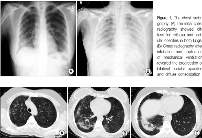
Figure 1. The chest radio- graphy. (A) The initial chest radiography showed dif- fuse fine reticular and nod- ular opacities in both lungs.

(B) Chest radiography after intubation and application of mechanical ventilation revealed the progression of bilateral nodular opacities and diffuse consolidation.

Figure 2. Chest computed tomography scan. (A) Upper lung field, (B) middle lung field and (C) lower lung field. The CT scan showed patchy consolidation with fibrosis in the right lower lobe and ill-defined centrilobular ground glass opacity in the both lungs. CT: computed tomography.

nonspecific interstitial pneumonia (NSIP). She was giv- en oral prednisone (60 mg/day) and empirical anti- biotics for about three weeks, but suffered a progressive worsening of dyspnea.

Past medical history: The patient had no specific past medical history.

Social history: The patient had no history of smoking and alcohol consumption.

Physical examination: At ER, the patient was alert with a body temperature of 98.8

o

F, heart rate of 96 beats/min, respiratory rate of 22/min, blood pressure of 116/74 mm Hg and oxygen saturation at 80.2% on room air. There was no fever or peripheral edema. Cardiac and abdominal examinations were normal. Lung auscul- tation revealed fine inspiratory crackles in both basal lung fields.

Initial laboratory findings: Complete blood cell count

showed a white blood cell count of 15.13×10

3

/μL (normal, 4.0∼10.0×10

3

/μL), a hemoglobin concen- tration of 15.0 g/dL (normal, 14∼18 g/dL), and a plate- let count 282×10

3

/μL (normal, 130∼450×10

3

/μL).

The differential count showed neutrophils at 67%, lym- phocytes at 21%, and monocytes at 8.3%. The C-re- active protein level was 1.1 mg/dL (normal, ≤0.5 mg/dL). Her serum sodium, potassium, creatinine, mag- nesium, calcium, and liver function test were all within normal limits. Blood and sputum cultures and gram staining were all negative. Urinary S. pneumoniae Ag was negative.

Radiologic findings: The initial chest radiography

showed diffuse fine reticular and nodular opacities in

both lungs (Figure 1A). The chest radiograph after in-

tubation and application of mechanical ventilation

changed the progression of initial findings with diffuse

(3)

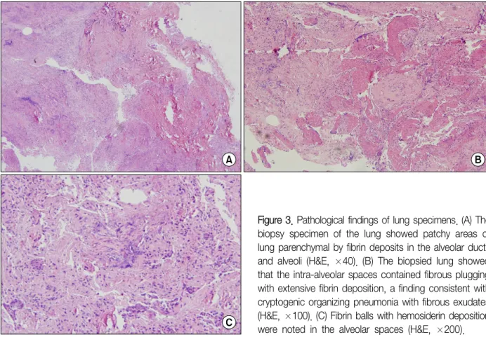
Figure 3. Pathological findings of lung specimens. (A) The biopsy specimen of the lung showed patchy areas of lung parenchymal by fibrin deposits in the alveolar ducts and alveoli (H&E, ×40). (B) The biopsied lung showed that the intra-alveolar spaces contained fibrous plugging with extensive fibrin deposition, a finding consistent with cryptogenic organizing pneumonia with fibrous exudates (H&E, ×100). (C) Fibrin balls with hemosiderin deposition were noted in the alveolar spaces (H&E, ×200).

consolidations (Figure 1B). Chest computed tomogra- phy scan at ER demonstrated patchy consolidations with fibrosis in the right lower lobe and ill-defined centril- obular ground glass opacity in the both lungs (Figure 2).

Bronchoscopic findings: Bronchoscopy on first day of hospitalization did not reveal any endobronchial lesions or abnormal secretions. Bronchioalveolar lavage (BAL) was done in the right lower lobe postero-basal segment.

Microbiologic cultures for common respiratory virus, bacteria and fungus containing Pneumocystis jirovecii were all negative. The BAL fluid consisted of 34% mac- rophages, 30% neutrophils, and 16% lymphocytes.

There was no evidence of malignant cells.

Pathological findings: The pathological finding show- ed patchy areas of lung parenchymal by fibrin deposits in the alveolar ducts and alveoli. The lung parenchyma shows intra-alveolar spaces containing fibrous plugging with extensive fibrin deposition, a finding consistent with cryptogenic organizing pneumonia with fibrous exudates. Fibrin balls with hemosiderin deposition are

noted in the alveolar spaces (Figure 3). The character- istic intra-alveolar fibrin ball and lack of hyaline mem- brane are the predominant histological features of AFOP.

Hospital course: On first hospital day, the patient de- veloped a respiratory rate of 40 per min after the bron- choscopy and a high fever up to 103.1

o

F and the de- mand for oxygen increased. Thus, she was started on broad spectrum antibiotics including 3rd cephalosporin and quinolone. Additionally, she transferred to the med- ical intensive care unit for close monitoring. She sub- sequently required intubation and started on mechanical ventilator care.

On second hospital day, the patient underwent

wedge biopsies of the right upper lobe and right lower

lobe through the video-assisted thoracoscopic surgery

(VATS). After establishing the diagnosis of AFOP, the

patient was started on intravenous corticosteroid pulse

therapy (methylprednisolone, 1 g/day) for three days

and continued with maintenance therapy (60 mg/day).

(4)

However, the PaO

2

/FiO

2

ratio gradually decreased with an increase in ventilator requirements (FiO

2

0.95). The arterial blood gas analysis showed permissive hyper- capnia (PaO

2

75.7, PCO

2

51.4, pH 7.252). The patient could not be weaned off the ventilator and chest radio- graph was aggravated, and nitric oxide gas was applied after all.

On 4th hospital day, the family of the patient con- sented to wait for a lung transplantation and an ex- tracorporeal membrane oxygenator (ECMO) was in- serted to her due to persistent hypoxemia and hyper- capnia under full ventilator settings. However, during the vascular procedure, the patients lost massive amounts of arterial blood and went into hypovolemic shock. The patient was successfully resuscitated from hypovolemic shock. But the patient's hospital course deteriorated rapidly.

On 9th hospital day, the family of the patient refused to wait for a lung transplantation and wanted removal of ECMO. On 13th hospital day, she died due to respira- tory failure and multiple organ failure.

Discussion

AFOP was first described by Beasley et al. in 2002

2

. In their paper, tissue histopathology of AFOP revealed intra-alveolar fibrin in the form of fibrin balls and organ- izing pneumonia with patchy distributions

1,2

. The differ- ential diagnosis of AFOP is primarily with DAD and EP.

The AFOP pattern differs from that of DAD in that the fibrin is organized into balls within the alveolar spaces and classic hyaline membranes are not seen. And the histological pattern of AFOP differs from the pattern of EP by the lack of prominent eosinophils

2

.

Since it is difficult to diagnose AFOP by small biopsy, a confident diagnosis of AFOP requires surgical lung bi- opsy

2

. In our case, VATS biopsy was done and the pathological findings showed patchy areas of the lung parenchyma by fibrin deposits in the alveolar ducts and alveoli. Fibrin ball is a characteristic finding of AFOP and presented as dense and red coalescent masses on hematoxylin-eosin (H&E) staining. Finally, there was no

evidence of hyaline membranes in alveoli in our case, the final pathologic diagnosis was consistent with AFOP.

In previous studies about AFOP, the onset of dyspnea and non-productive cough of less than two months du- ration were observed. Moreover, the most common ra- diological findings of AFOP showed bilateral basilar in- filtrates

2

. In this case, the patient had similar symptoms and radiological findings. In addition, AFOP has been associated with infections, connective tissue disorders, occupational hazards, drug exposures, and so on

2-5

. Recently, additional cases of AFOP have been reported in patients with HIV and Pneumocystis jirovecii pneu- monia, hematopoetic stem cell transplantation, and sys- temic lupus erythematosus

6-8

.

However, some patients had no identifiable predis- posing condition or association with AFOP

2

. Our patient did not appear to be associated with any known sig- nificant exposures or past medical diseases. However, one possibility of exposure was the home ultrasonic hu- midifier, which contained a sterilizer. For several days, the patient slept with the humidifier turned on, and the symptoms of dyspnea on exertion and cough appeared a few days later. In some previous studies, humidifiers have been associated with inhalation lung injury, for ex- ample, hypersensitivity pneumonitis

9,10

. And some cases were reported about humidifier-associated diseases like extrinsic allergic alveolitis and toxic aveolitis

11,12

. How- ever, it is not clear that humidifier have been associated with AFOP in this case.

The prognosis of AFOP appears to have two distinct patterns of disease progression: one is a fulminant ill- ness with rapid progression to death and the other is a subacute and, less fulminate course from which the patient recovers eventually

2

. Approximately, 30% of pa- tients with AFOP require mechanical ventilation and all patients who needed mechanical ventilation died.

Overall, AFOP showed poor prognosis with up to 50%

mortality

2

.

Some previous case reports showed the successful

treatment of AFOP with corticosteroid and immuno-

suppressant agents

13-15

, such as the combination of cy-

clophosphamide and prednisone

13

or mycophenolate

(5)

and methylprednisolone

14

. In our case, the patient was treated with intravenous corticosteroid pulse therapy (methylprednisolone, 1 g/day) for three days. However, the clinical course was aggravated and lung trans- plantation was planed. Despite the proper treatment for AFOP, the patient had a poor response and outcome to steroid therapy.

References

1. Beasley MB. The pathologist's approach to acute lung injury. Arch Pathol Lab Med 2010;134:719-27.

2. Beasley MB, Franks TJ, Galvin JR, Gochuico B, Travis WD. Acute fibrinous and organizing pneumonia: a his- tological pattern of lung injury and possible variant of diffuse alveolar damage. Arch Pathol Lab Med 2002;

126:1064-70.

3. Damas C, Morais A, Moura CS, Marques A. Acute fi- brinous and organizing pneumonia. Rev Port Pneumol 2006;12:615-20.

4. Balduin R, Giacometti C, Saccarola L, Marulli G, Rea F, Bartoli M, et al. Acute fibrinous and organizing pneumonia in a patient with collagen vascular disease

"stigma". Sarcoidosis Vasc Diffuse Lung Dis 2007;24:

78-80.

5. Prahalad S, Bohnsack JF, Maloney CG, Leslie KO. Fatal acute fibrinous and organizing pneumonia in a child with juvenile dermatomyositis. J Pediatr 2005;146:289- 92.

6. Yokogawa N, Alcid DV. Acute fibrinous and organizing pneumonia as a rare presentation of abacavir hyper- sensitivity reaction. AIDS 2007;21:2116-7.

7. Heo JY, Song JY, Noh JY, Yong HS, Cheong HJ, Kim WJ. Acute fibrinous and organizing pneumonia in a patient with HIV infection and Pneumocystis jiroveci pneumonia. Respirology 2010;15:1259-61.

8. Lee SM, Park JJ, Sung SH, Kim Y, Lee KE, Mun YC, et al. Acute fibrinous and organizing pneumonia fol- lowing hematopoietic stem cell transplantation. Korean J Intern Med 2009;24:156-9.

9. Hariri LP, Unizony S, Stone J, Mino-Kenudson M, Sharma A, Matsubara O, et al. Acute fibrinous and or- ganizing pneumonia in systemic lupus erythematosus:

a case report and review of the literature. Pathol Int 2010;60:755-9.

10. Daftary AS, Deterding RR. Inhalational lung injury asso- ciated with humidifier "white dust". Pediatrics 2011;

127:e509-12.

11. Patterson R, Mazur N, Roberts M, Scarpelli D, Semerdjian R, Harris KE. Hypersensitivity pneumonitis due to humidifier disease: seek and ye shall find. Chest 1998;114:931-3.

12. Baur X, Behr J, Dewair M, Ehret W, Fruhmann G, Vogelmeier C, et al. Humidifier lung and humidifier fever. Lung 1988;166:113-24.

13. Müller-Wening D, Koschel D, Stark W, Sennekamp HJ.

Humidifier-associated disease in the general popula- tion. Dtsch Med Wochenschr 2006;131:491-6.

14. Bhatti S, Hakeem A, Torrealba J, McMahon JP, Meyer KC. Severe acute fibrinous and organizing pneumonia (AFOP) causing ventilatory failure: successful treatment with mycophenolate mofetil and corticosteroids. Respir Med 2009;103:1764-7.

15. Cho JY, Lee HK, Lee SS, Lee HK, Lee YM, Lee HP,

et al. A case of acute fibrinous and organizing pneu-

monia. Tuberc Respir Dis 2006;61:479-83.

수치

Figure  1.  The  chest  radio- radio-graphy. (A) The initial chest radiography  showed   dif-fuse fine reticular and  nod-ular opacities in both lungs.
Figure 3. Pathological findings of lung specimens. (A) The  biopsy  specimen  of  the  lung  showed  patchy  areas  of  lung parenchymal by fibrin deposits in the alveolar ducts  and  alveoli  (H&E,  ×40)

참조

관련 문서

(Background) Gallbladder wall thickening(GWT) and gallbladder contraction are often observed in patients with acute hepatitis.. The incidence of acute hepatitis A

The measured values for benzene in DMF were compared with the The measured values for benzene in DMF were compared with the reference data, and

 Although van der Waals equation is still less accurate at high

There was a significant increase in new bone formation in the group in which toothash and plaster of Paris and either PRP or fibrin sealants were used, compared with the groups

- 각종 지능정보기술은 그 자체로 의미가 있는 것이 아니라, 교육에 대한 방향성과 기술에 대한 이해를 바탕으로 학습자 요구와 수업 맥락 등 학습 환경에 맞게

Haemorragic fever with renal syndrome (HFRS) caused by Hantanvirus has been one of the principal acute febrile disease in Korea. Hantaviruses are carried by numerous

The benefits and harms of intravenous thrombolysis with recombinant tissue plasminogen activator within 6 h of acute ischaemic stroke (the third

The purpose of this study was to analyze the impaction pattern of the impacted mandibular third molar and the relationship with the inferior alveolar nerve