https://doi.org/10.22889/KJP.2021.52.2.84
84
Aspergillus fumigatus 발효 추출물의 멜라닌 합성 억제 효과
송태양1*·김창원1·강미옥1·갈상완2·황을문1
1
㈜서린바이오사이언스,2
진주산업대학교 미생물공학과The Melanin Synthesis Inhibitory Effect of Aspergillus fumigatus Fermented Extract
Tae-Yang Song1*, Chang Won Kim1, Mi Ok Kang1, Sang Wan Gal2, and Eul Moon Hwang1
1
Seoulin Bioscience Co., Ltd, KOREA BIO PARK, SeongNam 13488, Korea2
Department of Microbiological Engineering, Jinju National University JinJu 52725, KoreaAbstract − This study was carried out to investigate the melanin synthesis inhibitory effect of Aspergillus fumigatus fermented extract. In this study, we revealed the effects of A. fumigatus fermented extract on melanin contents, mushroom tyrosinase activ- ity, and expression levels of mRNA and proteins of melanogenesis-related gene in B16F10 melanoma cells. A. fumigatus fer- mented extract inhibited both melanin contents and tyrosinase activity. In addition, the expression level of mRNA or proteins of melanogenesis was down-regulated in the A. fumigatus fermented extract treated B16F10 cells with dose-dependent manner.
Moreover, when the clinical test was conducted, it was confirmed that the use of the fermented extract of A. fumigatus for 8 weeks improved skin brightness 1.586 times brighter and skin melanin 1.331 times better compared to the control product.
Taken together, our results suggest that A. fumigatus fermented extract has melanogenesis inhibitory effect and whitening activ- ity, thus it showed the possibility for using as a functional whitening cosmetic resource.
Keywords − Aspergillus fumigatus, Melanin, Tyrosinase, Cosmetic resource
피부색은 melanin 색소, 베타카로틴, 혈액 등에 의해 결정 되는데 그 중에서 가장 영향이 큰 요인이 melanin 색소이다.
1)
Melanin 색소는 피부 표피의 맨 아래에 위치한 기저층에 존 재하는 melanocyte에서 만들어지며 melanin 생성은 자외선 이나 염증 등 자극에 의해 촉진된다.2,3)
또한 과도한 melanin 축적은 과색소 현상을 유발하게 되어 기미나 주근깨의 생 성 등과 같은 미용적인 측면에서 문제로 인식되고 있다.Melanin 생합성은 microphthalmia-associated transcription factor(MITF), tyrosinase, tyrosinase-related protein-1(TRP- 1), tyrosinase-related protein-1(TRP-2)등과 같은 효소들이 조절자로 작용하여 복합적인 과정을 통해 형성된다.
4,5)
따라 서 미백효과를 검증하기 위해서 위 효소들의 길항작용을 통 한 melanin 색소 침착 방지 및 melanin 생성 억제 여부를 검증하는 것이 중요하다.6-8)
미백효과를 위해서 개발된 arbutin, kojic acid, azelaic acid와 같은 tyrosinase 길항 물질 들이 주로 사용되었으나9,10)
세포독성, 피부자극, 제형 안전성 등의 부작용 사례가 많아지고 안전성에 대한 문제가 대
두되어
11-13)
한약 및 약용식물등을 이용한 다양한 천연 물질의 미백소재 연구가 확대되고 있다.
14,15)
Aspergillus fumigatus는 흙과 비료 속에서 발육하는 내열 성의 진균이다.
16)
Ascomycetes에 속하는 진균으로 유성생식에 의해 주머니 모양의 자낭속에 자낭포자를 만들며 자연계에 널리 분포되어 있다.17)
공기를 통해 사람의 폐, 비동, 부비 동, 각막, 외이도를 침범하는 아스페르길루스증을 일으키는 1차병원체로 인식되고 있으나, 배양을 통해 여러 종의 항생 물질인 fumigacin 및 gliotoxin을 형성하는데 또한 사용되고 있다.18)
1980년에 Aspergillus fumigatus의 멜라닌 색소 분해 능력을 확인하기 위하여 8주 동안 배양을 하였고 멜라닌 분 해 능력을 증명하였다는 연구가 처음 진행이 되었으나 과 학적으로 정밀한 Aspergillus fumigatus의 미백 효능 연구는 현재까지 미비한 실정이다.19)
본 연구는 천연에서 분리한 Aspergillus fumigatus 미생물 과 천연물 소재의 곡물배지를 이용 및 배양하여 기능성 화 장품의 천연 물질의 미백소재로서 화학소재에 비해 안전성 과 낮은 부작용을 극대화 시켰다. 또한 Aspergillus fumigatus
*교신저자(E-mail):[email protected] (Tel): +82-31-628-3090
발효 추출물을 이용한 멜라닌 억제 효과를 in vitro 부터 임 상까지 효과를 조사하여, 기능성 미백 화장품 소재로서의 가능성을 확인한 것이다.
재료 및 방법
시료의 추출 − 본 연구에서 사용된 Aspergillus fumigatus 는 단일 균주로 분리하였고, 5.8S ribosomal DNA internal transcribed spacer 1(ITS)의 DNA 시퀀싱에 의존하여 동정을 실시하였으며, 동정 결과 균주는 A. fumigatus의 ITS와 90%
일치되는 결과로 나타나 A. fumigatus로 확인되었다. 균주는 진주산업대학교로부터 전달 받아 사용하였다. A. fumigatus 를 배양하기 위해 Table I에서와 같이, 3가지 조건의 곡물 배지를 제조 및 고온 멸균 후 상온에서 균주를 접종하고 0.09 g force, 30
o
C에서 pH 4.5 ± 1.0 범위로 9일간 진탕 배 양하였다. 배양이 끝난 배지는 거즈를 이용해 1차적으로 배 지와 균사체로 분리하였고, 분리된 배지는 filter를 이용해 2 차 여과를 하여 배양액 추출물을 제조하였다. 배양액 추출 물은 감압농축하여 농축물 상태로 4o
C에 실험 전까지 보관 하였다. 분리된 균사체는 homogenizer(BioSpec Products Inc., OK, USA)를 이용해 잘게 갈아준 뒤, 80% acetone을 첨가하여 overnight으로 균사체 내 성분들을 추가로 추출하 였다. 그리고 1660 g force, 15분간 원심분리 후, 상층액만 분리하여 감압 농축 하여 균사체 농축물을 얻어내었다. 이 후 4o
C에 실험 전까지 보관하였다. 각 농축물은 실험에 이 용 시 DMSO(Dimethyl sulfoxide)에 녹여 농도를 조정한 다 음 사용하였다.세포 배양 및 시약 − B16F10와 B16F0 mouse melanoma cell(마우스 흑색종 세포)는 American Type Culture Collection (Manassas, USA)로부터 구입하여 CO
2
세포배양기(37o
C, 5%CO
2
)에서 배양하였다. 10% fetal bovine serum(FBS)와 1%penicillin/streptomycin(HyClone, USA)이 첨가된 dulbecco’s modified eagles medium(DMEM, HyClone, USA) 배지를 사용하여 24시간 주기로 계대배양 하였다. 시약 3-(4,5- dimethylthiazol-2-yl)-2,5-diphenyltetrazolium bromide(MTT),
mushroom tyrosinase, arbutin, kojic acid는 Sigma chemical Co.(St. Louis, MO, USA)에서 구입하여 사용하였다.
세포 생존율 및 세포독성 측정 − 세포 생존율 및 세포 독 성은 3-(4,5-dimethylthiazol-2-yl)-2,5-diphenyltetrazolium bromide(MTT) Assay방법을 통하여 측정하였다. B16F0 세 포를 96 well plate에 5 × 10
3
cells/well로 분주하여 37o
C, 5% CO2
incubator에서 24시간 동안 배양한 후, 시료를 농도 별로 조제하여 200 nM alpha-melanocyte stimulating hormone (α-MSH)와 함께 처리하고 72시간동안 배양하였다. 대조군은 시료 및 200 nM α-MSH 처리 없이 배양하였다. 이 후, 0.5 mg/ml의 최종 농도로 MTT solution을 첨가하여 37o
C에서 4시간 동안 반응시킨 후, MTT solution이 포함된 media를 제거하고 각 well 당 DMSO 100 μl를 처리하여 살아있는 세포와 반응하여 생성된 formazan 침전물을 용해시켰다.ELISA reader(CLARIOstar Plus, BMG LABTECH, Germany) 기를 이용하여 550 nm에서 흡광도를 측정하였다.
멜라닌 생합성 저해율 측정 − B16F0 세포를 6 well culture plate에 5 × 10
4
cells/well로 분주하여 24시간 배양 후 200 nM α-MSH와 추출물을 농도별로 처리하여 72시간동 안 더 배양하였다. 이후 각 well의 배지를 모두 제거하고 phosphate-buffered saline(PBS)로 세척한 다음, trypsin- (EDTA)로 처리하여 세포를 microtube에 회수한 후 10%DMSO가 첨가된 1 N NaOH 300 μl를 처리하여 60
o
C에서 1시간 동안 반응시켜 melanin을 완전히 용해 시킨 다음 405 nm에서 흡광도를 측정하였다. 실험은 3번 수행하였으며, 이 에 따른 평균값과 표준오차는 Microsoft Excel program을 이용하여 분석하였다.Tyrosinase 저해 활성 측정 −67 mM sodium phosphate buffer (pH 6.8) 80μl에 10mM L-DOPA(L-3,4-dihydroxyphenylalanine, Sigma, USA)를 녹인 기질액 40 μl와 발효 추출물 40 μl 혼 합액을 만들고, 이 혼합액에 200 U/ml mushroom tyrosinase (Sigma, USA) 40μl을 첨가하여 37
o
C에서 10분간 반응시켜 반응액 중에 생성된 DOPA chrome을 492 nm에서 흡광도를 측정하였다. Tyrosinase 저해 활성은 시료 용액의 첨가군과 무첨가군의 흡광도 감소율로 나타내었다.저해율(%) = (1 – A / B) × 100, A = 발효 추출물 첨가군의 흡강도, B = 무첨가군의 흡강도
quantitative RT-PCR(qRT-PCR)을 통한 mRNA 발현 측정 − B16F10 세포를 100 mm culture plate에 6 × 10
5
cells로 분주하여 24시간 동안 배양한 후 시료를 농도 0.1~10%로 처리하여 24시간 더 배양하였다. 이후 각 배지를 제거한 후 trizol lysis 과정, chloroform 및 isopropanol 반응 과정, EtOH-Diethylpyrocarbonate 처리 및 건조과정을 거쳐 Total RNA를 추출하였다. Oligo dT primer와 추출한 RNA(2 ug)로 반응 후 reaction buffer, MgCl2
, PCR nucleotide mix, reverse Table I. Nutrient compositions of grain mediumGrain medium compositions
Grain medium 1.
Grain medium 2.
Grain medium 3.
PDB 0.1% - 0.1%
Collagen 2% 2% 2%
Potato starch 0.5% 0.5% 0.6%
Sugar 0.5% 0.5% 0.5%
MSG 0.1% 0.1% 0.1%
Hemp seed - 1% -
Glutinous rice flour - 1% -
transcriptase를 첨가하여 cDNA를 합성하였다. PCR tube에 Go Flexi DNA polymerase, primer, cDNA를 첨가하여 qRT-PCR(PIKOReal 96, Thermo Scientific, Ma, USA)로 분석 정량 하였고 밴드로 재 확인하였다. 조건은 94
o
C에서 30초, 60o
C에서 45초, 72o
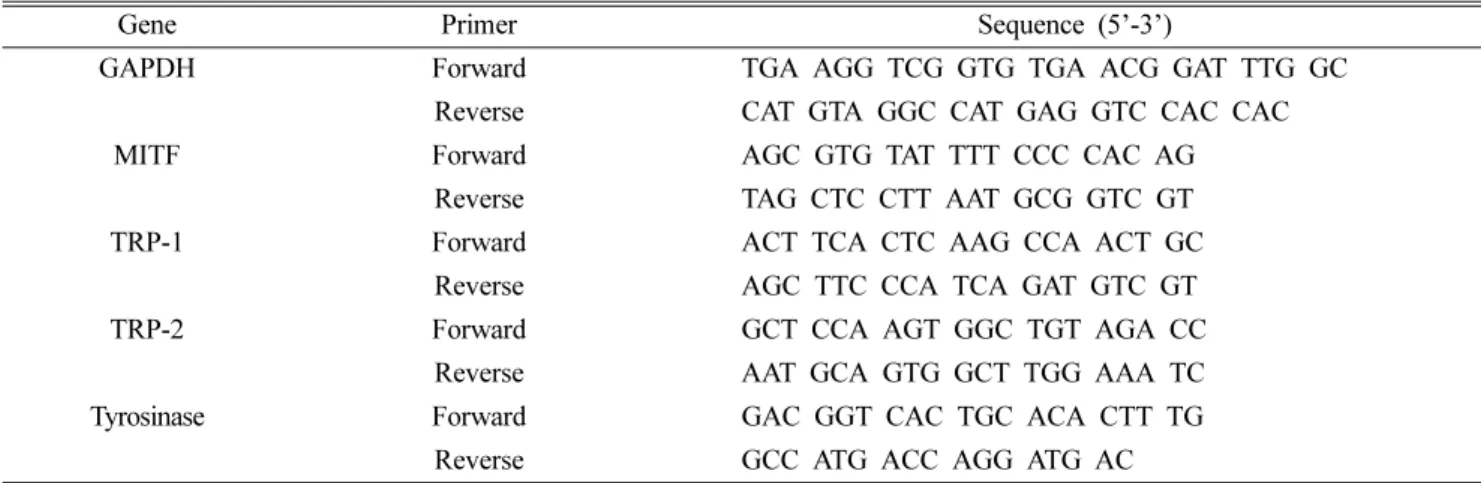
C에서 45초, 40회 반복하여 반응 시켰다. qPCR에 사용된 primer 서열은 Table II와 같다.Western blot을 통한 단백질 발현 측정 − B16F10 세포를 100 mm culture plate에 6 × 10
5
cells로 분주하여 24시간 동 안 배양한 후 시료를 농도 별로 처리하여 24시간 더 배양 하였다. Radio-immunoprecipitation(RIPA) buffer로 cell을 용해한 후 BCA protein assay kit를 사용하여 정량 하였다.20 ul의 단백질을 10% SDS-PAGE상에서 전기영동하여 분 리한 후 분리된 단백질을 PVDF membrane에 옮겨 5% skim milk로 blocking을 실시하였고 1차 antibody를 4
o
C에서 overnight으로 반응시켰다. 이때 사용된 1차 antibody는 다 음과 같다. Anti-MITF(Santa Cruz Biotechnology, 1:1000), anti-tyrosinase(Santa Cruz Biotechnology, 1:1000), anti-TRP- 1(Santa Cruz Biotechnology, 1:1000), anti-TRP-2(Santa Cruz Biotechnology, 1:1000). 이후 Tris-buffered saline-Tween 20 으로 세척하고 2차 antibody를 실온에서 2시간 붙인 다음 3회 세척하여 Davinch-ChemiTM
Imager CAS-400SM(DAVINCH Chemi, Korea) 기기를 이용하여 단백질 발현 양을 확인하 였다.인체적용시험을 통한 미백 기능성 평가 − 시험에 참가한 피실험자는 얼굴 좌우에 과색소침착증상이 있고 시험대상
자 제외기준을 만족하는 20~60세의 성인 여성 26명이다. 피 실험자들의 특징은 Table III과 같다. 8주 동안 피부 밝기 측 정 및 피부 멜라닌 측정을 통해 미백 개선효과에 대한 기기 적 평가를 Mexameter MX18(Courage+Khazaka electronic GmbH, Germany)를 이용해 수행하였다. 연구기간은 2020 년 6월 1일부터 2020년 7월 28일까지 8주간 진행하였다. 각 피실험자 별 실험 처치 적용 기간은 8주를 적용하였고 측 정값의 산출은 4주(실험 중간시점), 8주(실험 종료시점)로 나뉘어 진행되었다. 본 시험은 A. fumigatus가 포함된 cream 의 미백 기능성 평가 인체적용시험(이중맹검, 무작위배정, 음성대조, half-test)이란 시험제목으로 피엔케이피부임상연 구센터㈜에서 표준시험방법(SOP)에 따라 진행하였다. 본 시 험에서 사용된 소재는 곡물 배지를 이용하여 만들어진 A.
fumigatus 배양액을 KB코스매틱에서 cream으로 제작하여 만들어졌다.
결과 및 고찰
곡물배지에서 배양 한 A. fumigatus로부터 발효 추출물 정제 과정 − 균주의 분리는 오징어 먹물을 식물 정원의 토 양에 뿌린 후, 일정기간이 경과하여 멜라닌 분해 능력이 있 는 토양에서 분리한 미생물을 스크리닝하여 A. fumigatus를 천연에서 분리하였고, 환경 호르몬이나 유전자 변형에 따른 화장품 성분에 대한 문제점이 심각하게 대두되면서 천연물 소재의 곡물배지를 이용하여 실험을 진행하였다. 3가지 조 Table II. The sequence of primers used for real-time quantitative PCR
Gene Primer Sequence (5’-3’)
GAPDH Forward TGA AGG TCG GTG TGA ACG GAT TTG GC
Reverse CAT GTA GGC CAT GAG GTC CAC CAC
MITF Forward AGC GTG TAT TTT CCC CAC AG
Reverse TAG CTC CTT AAT GCG GTC GT
TRP-1 Forward ACT TCA CTC AAG CCA ACT GC
Reverse AGC TTC CCA TCA GAT GTC GT
TRP-2 Forward GCT CCA AGT GGC TGT AGA CC
Reverse AAT GCA GTG GCT TGG AAA TC Tyrosinase Forward GAC GGT CAC TGC ACA CTT TG
Reverse GCC ATG ACC AGG ATG AC
Table III. Characteristics of participants
Factors Division and frequency (%)
Age 20s 30s 40s 50s 60s
0 (0%) 2 (7.7%) 8 (30.7%) 16 (61.6%) 0 (0%)
Skin type Very dry Dry Moderate Greasy Very Greasy
10 (38.4%) 9 (34.6%) 6 (23.2%) 1 (3.8%) 0 (0%)
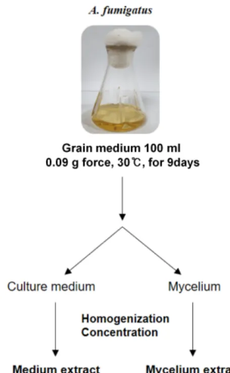
건의 곡물배지에서 9일간 A. fumigatus를 배양하고 배양액 과 균사체를 분리하여 배양액은 감압농축하여 곡물배지 배 양액 추출물을 정제하였고, 균사체는 homogenizer를 이용해 잘게 갈아준 뒤, 80% acetone을 첨가하여 하루 동안 균사 체 내 성분들을 추가로 추출하고 감압농축하여 곡물배지 균 사체 추출물을 정제하였다(Fig. 1).
A. fumigatus 발효 추출물의 세포독성 확인 − 3가지 곡 물배지 조건에서 배양한 A. fumigatus 발효 추출물의 세포 독성을 확인하기 위해 배양액 및 균사체 추출물들을 50, 100 μg/ml 농도로 B16F0(mouse melanocyte)에 72시간 동안 처 리 한 후 MTT assay를 실시하였다. 멜라닌 생성 세포인 B16F0(mouse melanocyte)에 대한 A. fumigatus 배양액 및 균사체 발효 추출물의 세포독성을 확인한 결과 3가지 조건 의 곡물배지에서 배양한 모든 A. fumigatus 발효 추출물 처 리 군에서 세포 독성이 전혀 나타나지 않는 것을 관찰하였 다(Fig. 2A). 세포 독성이 없는 것으로 볼 때 피부에 대한 자극적인 부작용이 없는 것으로 판단이 되어 화장품 소재 로서의 가능성을 확인 할 수 있었다. 추가 실험은 세포 독 성을 보이지 않는 100 μg/ml 농도에서 멜라닌 생성 억제 효 Fig. 1. Schematic shows the experimental workflow for extraction
of A. fumigatus.
Fig. 2. Effect on cell viability and melanin contents for using extracts of A. fumigatus. (A) Effect of medium and mycelium extracts on the cytotoxicity. Cytotoxic effect of extracts was evaluated using MTT assay. (B) Inhibition of melanogenesis of B16F0 mouse melanocyte treated with the medium and mycelium extracts of grain medium after 72h. Arbutin (Arb) was used as positive control.
Error bars represent S.D., n = 3 (*P-value < 0.05).
과를 확인하였다.
A. fumigatus 발효 추출물의 세포내 멜라닌 생성 억제 효과 − 멜라닌 생성세포인 B16F0(mouse melanocyte)에 A.
fumigatus 배양액 및 균사체 발효 추출물의 과색소 침착 억 제능을 확인하기 위하여 멜라닌 생성 억제 실험을 진행하
였다. B16F0를 24시간 배양 후, α-MSH 200 nM 과 곡물배 지에서 배양한 A. fumigatus 시료(6가지, 배양액 및 균사체 발효 추출물)를 함께 처리하고 72시간 후 각 시료의 멜라닌 생성 억제력을 비교하였다. α-MSH를 단독으로 처리한 세 포에서 melanin 생성이 증가하였으며, 모든 곡물배지 균사 체 추출물에서 melanin 생성이 억제되었다. 특히, 1번 조건 의 곡물배지 배양액 추출물 특이적으로 기존 미백물질로 알 려진 양성대조군 arbutin
20-22)
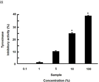
과 비교하였을 때 유의한 수준 으로 높은 억제 활성을 확인하였다(Fig. 2B). 따라서 1번 조 건의 곡물배지 배양액 추출물이 화장품 원료로서 최적임을 검증하였다. 이후 실험은 배양액 발효 추출물을 이용하여 진행하였다.A. fumigatus 배양액 발효 추출물의 tyrosinase 활성 억 제 효과 − A. fumigatus 발효 추출물의 in vitro tyrosinase 활 성 억제를 확인한 결과, 시료 농도 의존적으로 활성 저해 효 과를 확인하였다. 효소 tyrosinase은 인체내 멜라닌 생합성 초기 결정 단계에 중요한 요소이며, 이 효소 활성 저해는 멜 라닌 생성 저해를 의미한다.
23,24)
Fig. 3에서 보는 바와 같이 추출물을 10% 농도 첨가 시 25%의 tyrosinase 효소의 활성 억제를 보였으며, 최종 100% 농도 처리시 40% 억제됨을 관찰하였다. 위와 같은 결과로 A. fumigatus 발효 추출물이 효소 활성을 감소시켜 멜라닌 생성 억제에 직접 관여함을 Fig. 3. Effect of using extracts of A. fumigatus on mushroomtyrosinase activity. There were measured by absorbance at 492 nm after incubation with various concentration of A. fumigatus.
Error bars represent S.D., n = 3. Significance of differences was evaluated (*P-value < 0.05).
Fig. 4. The relation between treated B16F10 mouse melanocyte and the mRNA expression level of melanogenesis-related gene in A. fumigatus-extract. B16F10 mouse melanoma cell treated with various concentration of extract for 72 hr. The mRNA levels of MITF, tyrosinase, TRP-1, and TRP-2 were analyzed by qRT-PCR. Kojic acid was used as positive control. The mRNA levels were normalized to the level of GAPDH and shown as relative values. Error bars represent S.D., n = 3 (*P-value < 0.05).
입증하였다. 이는 피부 흑화나 색소침착 등을 방어할 수 있 는 기능성 미백화장품 소재로 이용 가능하리라 생각된다.
A. fumigatus 발효 추출물에 의한 멜라닌 생성 관련 전 사인자 및 단백질 발현 억제 − 멜라닌 생성 기전에 있어, α-MSH의 전사인자인 MITF와 신호전달 기전의 중요한 매 개자인 tyrosinase, TRP-1 그리고 TRP-2는 멜라닌 생성 과 정에서 매우 중요한 인자들로 멜라닌 생성 억제 기전에 사 용되는 시료의 효능 평가의 표적으로 많이 활용이 되고 있
다.
25-27)
MITF, tyrosinase, TRP-1 그리고 TRP-2 억제는 멜라닌 생성 억제와 직결되어 있고 A. fumigatus 발효 추출물 이 멜라닌 생성 억제에 직접 관여함을 입증하여, 인자들의 mRNA 및 단백질 수준에서 발현이 억제되는지 qPCR 및 Western blot 방법으로 검증하였다. 두 실험 모두, B16F10 세포에 α-MSH를 처리하여 멜라닌을 과 발현 시키고 A.
fumigatus 발효 추출물 0.1, 1, 10% 농도를 처리하여 MITF, tyrosinase, TRP-1 그리고 TRP-2 발현양을 확인한 결과, 추 출물의 농도 의존적으로 발현이 감소함을 확인하였다(Fig.
4, 5). 즉, A. fumigatus 발효 추출물이 인자들의 transcription 레벨과 translation 레벨에서 발현을 억제함을 규명하였다.
특히, TRP-1과 TRP-2의 발현이 추출물 10% 농도 만으로도 기존 미백물질로 알려진 kojic acid(양성대조군)
10)
와 비교 시 현저히 감소함을 확인 할 수 있었다(Fig. 4C, D). qPCR의 경우 GAPDH로 보정하고 겔전기영동을 통해 재확인 하였 으며, Western blot의 경우 β-actin로 보정하고 정량화 및 수 치화를 통해 재확인 하였다. 이러한 결과는 A. fumigatus 발효 추출물이 melanin 합성에 관여하는 유전자의 발현 조절을 통해 melanin 생성을 억제하는 것으로 보인다. 그러나 melanin 생 합성에 관여하는 근본적인 A. fumigatus 발효 추출물 성분 Fig. 5. Effect of A. fumigatus-extract on expression level of melanogenesis-related protein in B16F10 mouse melanoma cell. Whole- cell lysates of B16F10 cells were used for western blot analysis with antibodies against MITF, tyrosinase, TRP-1, and TRP-2. β- actin served as loading control. Kojic acid was used as positive control. Error bars represent S.D., n = 3. Significance of differences was evaluated (*P-value < 0.05).에 대한 연구는 현재 미비한 실정으로 미백 기능을 나타낼 수 있는 화학적 성분 분석에 대한 추가적인 연구가 필요하다.
A. fumigatus 발효 추출물이 함유된 화장품 원료의 미 백효과 − 곡물배지 조건에서 배양한 A. fumigatus 발효 추출 물의 천연물질 미백소재 활용성을 검증하고 향후 미백화장 품으로 개발하기 위한 인체적용시험을 진행하였다. 인체 피 부 일차 자극 시험은 기 진행하여 무자극으로 판정되어(data not shown), 현 시험은 미백(피부 밝기, 피부 멜라닌) 개선 에 대한 효과를 평가하고자 시행하였다. 시험은 (주)피엔케 이피부임상연구센터에 위탁하여, 시험대상자 선정 및 제외 기준을 만족하는 20~60세의 성인 여성 26명을 대상으로 A.
fumigatus 발효 추출물이 주성분인 cream으로 이중맹검, 무 작위배정, 및 음성대조 실험을 진행하였다. 피부 밝기는 spectrophotometer를 이용하여 측정을 3회 실시한 후 사용 전, 사용 4주 후, 사용 8주 후의 평균값으로 피부 밝기 평 가를 진행하였다. 피부 멜라닌은 Mexameter MX18을 이용 하여 측정하였고, 동일한 색소침착 부위를 측정하여 사용 전, 사용 4주 후, 사용 8주 후의 피부 멜라닌 값을 평가하 였다. Fig. 6에서 보는 바와 같이, A. fumigatus 발효 추출물 이 주성분인 cream을 8주 사용으로 대조제품 대비 피부 밝 기 1.586배 개선, 피부 멜라닌 1.331배 개선에 도움을 주는 것으로 확인되어 A. fumigatus 발효 추출물의 기능성 미백 화장품 소재로서의 가능성을 인체적용시험을 통해 입증하 였다(Fig. 6). 실험자들의 특성은 Table III에 표기 하였다.
결 론
본 연구는 A. fumigatus 발효 추출물을 이용한 멜라닌 억 제 효과를 in vitro 부터 임상까지 효과를 조사하여, 기능성
미백 화장품 소재로서의 가능성을 확인한 것이다. 천연에서 분리한 A. fumigatus 미생물과 천연물 소재의 곡물배지를 이 용한 배양은, 기능성 화장품의 천연 물질의 미백소재로서 화학소재에 비해 안전성과 낮은 부작용을 극대화 시켰다.
Aspergillus fumigatus 발효 추출물의 세포독성 결과만 보더 라도, 3가지 조건의 곡물배지에서 배양한 모든 발효 추출물 에서 세포독성이 전혀 발견되지 않았고, 기 진행한 인체 피 부 일차 자극 시험에서 또한 무자극으로 판정이 되었다. α- MSH가 처리된 B16F0 melanocyte를 이용한 멜라닌 생성 억 제 효과의 경우, 곡물배지 배양액 추출물 특이적으로 미백 제로 알려진 양성대조군 arbutin 과 비교하였을 때 유의한 수준으로 높은 멜라닌 생성 억제 활성을 확인하여 과색소 침착 억제능을 검증하였으며, 인체내 멜라닌 초기 생성에 중요한 요소인 tyrosinase 효소의 활성 또한 억제함을 규명 하였다. 멜라닌 생성 과정에서 매우 중요한 단백질인 MITF, tyrosinase, TRP-1 그리고 TRP-2 발현양을 확인한 결과, 추 출물의 농도 의존적으로 발현이 감소함을 확인하여 A.
fumigatus 발효 추출물이 인자들의 transcription 레벨과 translation 레벨에서 발현을 억제함을 규명하였다. 그러나 melanin 합성에 관여하는 유전자의 발현을 조절하는 A.
fumigatus 발효 추출물 성분에 대한 연구는 미비하여 화학 적 성분 분석에 대한 추가적인 연구는 필요하다. 최종, 천 연 미백소재 활용성을 검증하고 향후 미백화장품으로 개발 하기 위해 인체적용시험을 진행 한 경우 또한, A. fumigatus 발효 추출물 8주 사용으로 대조제품 대비 피부 밝기 1.586 배 개선, 피부 멜라닌 1.331배 개선에 도움을 주는 것으로 확인되었다. 따라서 A. fumigatus 발효 추출물은 세포독성은 없고 미백효과는 높은 기능성 미백 화장품 소재로서의 가 능성을 제시하였다.
Fig. 6. Verified clinical effectiveness of cosmetic products containing A. fumigatus-extract. Twenty-six female adults (mean age 45 ± 10 year) were enrolled, all participants used cosmetic product including A. fumigatus-extract. (A) The change of skin brightness (L- value) was measured by spectrophotometer after 4 and 8 weeks of use. (B) The change of skin melanin (M.I, Melanin Index) was measured by Mexameter after 4 and 8 weeks of use. Error bars represent S.D., n = 3. Significance of differences was evaluated (*P- value < 0.05 by Mann-Whitney U test, **P-value < 0.05 by independent T test).
사 사
본 연구는 2020년도 중소기업 네트워크형 기술개발사업의 연구비 지원을 받아 수행되었음(과제번호 S2689413).
인용문헌
1. Maranduca, M. A., Branisteanu, D., Serban, D. N., Braniste- anu, D. C., Stoleriu, G., Manolache, N. and Serban, I. L.
(2019) Synthesis and physiological implications of melanic pigments. Oncol. Lett. 17: 4183-4187.
2. Bonaventure, J., Domingues, M. J. and Larue L. (2013) Cel- lular and molecular mechanisms controlling the migration of melanocytes and melanoma cells. Pigment Cell Melanoma Res. 26: 316-325.
3. D’Mello, S. A., Finlay, G. J., Baguley, B. C. and Askarian- Amiri, M. E. (2016) Signaling pathways in melanogenesis.
Int. J. Mol. Sci. 17: 1144.
4. Serre, C., Busuttil, V., and Botto, J. M. (2018) Intrinsic and extrinsic regulation of human skin melanogenesis and pig- mentation. Int. J. Cosmet. Sci. 40: 328-347.
5. Kang, M. K., Lee, Y. E., Woo, W. H. and Mun, Y. J. (2014) Commelina communis Ledeb inhibits melanin synthesis in alpha-MSH-stimulated B16F10 Cells. J. Physiol. & Pathol.
Korean Med. 28: 506-511.
6. Tsao, Y. T., Huang, Y. F., Kuo, C. Y., Lin, Y. C., Chiang, W.
C., Wang, W. K., Hsu, C. W. and Lee, C. H. (2016) Hinokitiol Inhibits melanogenesis via AKT/mTOR signaling in B16F10 mouse melanoma cells. Int. J. Mol. Sci.17: 248.
7. Wu, P. Y., You, Y. J., Liu, Y. J., Hou, C. W., Wu, C. S., Wen, K. C., Lin, C. Y. and Chiang, H. M. (2018) Sesamol Inhibited melanogenesis by regulating melanin-related signal trans- duction in B16F10 cells. Int. J. Mol. Sci. 19:1108.
8. Park, S. H., Kim, D. S., Kim, W. G., Ryoo, I. J., Lee, D. H., Huh, C. H., Youn, S. W., Yoo, I. D. and Park, L. C. (2004) Terrein: a new melanogenesis inhibitor and its mechanism.
Cell Mol. Life Sci. 61: 2878-2885.
9. Tai, A., Ohno, A. and Ito, H. (2016) Isolation and char- acterization of the 2,2′-azinobis (3-ethylbenzothiazoline-6- sulfonic acid) (ABTS) radical cation-scavenging reaction products of arbutin. J. Agric. Food Chem. 64: 7285-7290.
10. Saeedi, M., Eslamifar, M. and Khezri, K. (2019) Kojic acid applications in cosmetic and pharmaceutical preparations.
Biomed. Pharmacother. 110: 582-593.
11. Burnett, C. L., Bergfeld, W. F., Belsito, D. V., Hill, R. A., Klaassen, C. D., Liebler, D. C., Marks, J. G. Jr., Shank, R. C., Slaga, T. J., Snyder, P. W. and Andersen, F. A. (2010) Final report of the safety assessment of kojic acid as used in cos- metics. Int. J. Toxicol. 29: 244-273.
12. Battaini, G., Monzani, E., Casella, L., Santagostini, L. and Pagliarin, R. (2000) Inhibition of the catecholase activity of
biomimetic dinuclear copper complexes by kojic acid. J. Biol.
Inorg. Chem. 5: 262-268.
13. Chang, C. T., Chang, W. L., Hsu, J. C. and Shih, Y. (2013) Chemical composition and tyrosinase inhibitory activity of Cinnamomum cassia essential oil. Bota. Stud. 54: 10-17.
14. Kim, K. Y. and Lee, N. K. (2014) Herbal extracts research trend that have effects on melanin production and control.
Kor. J. Aesthet. Cosmetol. 12: 453-461.
15. Park, J. M. and Kim, K. J. (2010) The anti-wrinkle effects and whitening effects of Galla Rhois. J. Kor. Ori. Med. Opht.
Otol. 23: 135-148.
16. Latgé, J. P. and Chamilos, G. (2020) Aspergillus fumigatus and Aspergillosis in 2019. Clin. Microbiol. Rev. 33: e00140-18.
17. Gastebois, A., Clavaud, C., Aimanianda, V. and Latgé, J. P.
(2009) Aspergillus fumigatus: cell wall polysaccharides, their biosynthesis and organization. Future Microbiol. 4: 583-595.
18. Park, H. S. and Yu, J. H. (2016) Developmental regulators in Aspergillus fumigatus. J. Microbiology. 54: 223-231.
19. Luther, J. P. and Lipke, H. (1980) Degradation of melanin by Aspergillus fumigatus. Appl. Environ. Microbiol. 40: 145-155.
20. Sugimoto, K., Nishimura, T., Nomura, K., Sugimoto, K. and Kurik, T. (2014) Inhibitory effects of α-arbutin on melanin synthesis in cultured human melanoma cells and a three- dimensional human skin model. Biol. Pharm. Bull. 27: 510-514.
21. Lim, Y. J., Lee, E. H., Kang, T. H., Ha, S. K., Oh, M. S., Kim, S. M., Yoon, T. J., Kang, C., Park, J. H. and Kim, S. Y. (2009) Inhibitory effects of arbutin on melanin biosynthesis of α- melanocyte stimulating hormone-induced hyperpigmentation in cultured brownish guinea pig skin tissues. Arch. Pharm.
Res. 32: 367-373.
22. Inoue, Y., Hasegawa, S., Yamada, T., Date, Y., Mizutani, H., Nakata, H., Matsunaga, K. and Akamatsu, H. (2013) Anal- ysis of the effects of hydroquinone and arbutin on the dif- ferentiation of melanocytes. Biol. Pharm. Bull. 36: 1722-1730.
23. Hearing, V. J. and Ekel, T. M. (1973) Mammalian tyrosinase.
A comparison of tyrosine hydroxylation and melanin for- mation. Biochem. J. 157: 549-557.
24. Agar, N. and Young, A. R. (2005) Melanogenesis: a pho- toprotective response to DNA damage? Mutation Research.
571: 121-132.
25. Abdel-Malek, Z., Swope, V. B., Suzuki, I., Akcali, C., Harriger, M. D., Boyce, S. T., Urabe, K. and Hearing, V. J. (1995) Mito- genic and melanogenic stimulation of normal human melanocytes by melanotropic peptides. Proc. Natl. Acad. Sci. USA. 92: 1789-1793.
26. Fuller, B. B. and Meyskens, F. L. Jr. (1981) Endocrine responsiveness in human melanocytes and melanoma cells in culture. J. Natl. Cancer Inst. 66: 799-802.
27. Takechi, Y., Hara, I., Naftzger, C., Xu, Y. and Houghton, A.
N. (1996) A melanosomal membrane protein is a cell surface target for melanoma therapy. Clin. Cancer Res. 2: 1837-1842.
(2021. 5. 28 접수; 2021. 6. 15 심사; 2021. 6. 22 게재확정)