pISSN 1225-6552, eISSN 2287-7630 http://dx.doi.org/10.7853/kjvs.2012.35.4.283
< Original Article >
Veterinary Service
Available online at http://kjves.org
*Corresponding author: Seong-Guk Kim, Tel. +82-53-326-0013, Fax. +82-53-326-1066, E-mail. ksk8719007@korea.kr
경북지역 환돈 유래 Streptococcus suis 의 PFGE 패턴 실태 조사
김성국*ㆍ김영환ㆍ이홍영ㆍ윤문조
경상북도가축위생시험소
PFGE patterns of Streptococcus suis isolates from diseased pigs in Gyeongbuk province, Korea
Seong-Guk Kim*, Young-Hoan Kim, Hong-Young Lee, Mun-Jo Yun
Gyeongbuk Veterinary Service Laboratory, Daegu 702-911, Korea
(Received 27 Septemper 2012; revised 15 December 2012; accepted 20 December 2012)
Abstract
Streptococcus(S.) suis is a pathogen, causing meningitis, septicemia and sudden death in weaning piglets as well as fattening pigs. Using multiplex PCR method based S. suis capsular genes, 61 S. suis isolates was classified as serotypes 2, 7, 9 and untypable. Genotyping of S. suis isolates was analysed by PFGE pattern with treated Sma I restricted enzyme. Of the 61 S. suis, 25 (40.9%) were serotype 2, 6 (9.8%) were serotype 7, 5 (8.2%) were serotype 9, and 25 (40.9%) were untypable, respectively. Twenty four PFGE patterns were detected in this study and also PFGE patterns were classified according to serotype;
serotype 2 was classified as 6 genotypes, serotype 7 was 5 genotypes, serotype 9 was 3 genotypes, and untypable was 11 genotypes, respectively.
Key words : Streptococcus suis, Multiplex PCR, Serotype, PFGE, Genotype
서 론
Streptococcus(S.) suis는 Gram 양성의 연쇄상구균으 로 면양혈액 첨가배지에서 주로 α-용혈을 일으키고 상부호흡기관(특히 편도 및 비강)에 주로 상재하며 돼지를 사육하는 대부분 나라에서 질병을 일으키는 원인균이다(Marois 등, 2007). 돼지에서 S. suis는 주로 16주령 이하에서 질병을 일으키며(Reams 등, 1996) 뚜렷한 증상없이 폐사하거나 뇌막염에 의한 운동실 조, paddling, 안구진탕 등의 신경증상을 동반한 전구 증상을 나타내고(Clifton-Hadley 등, 1986) 관절염, 패 혈증, 폐렴 및 드물게 심내막염, 비염, 유산, 질염 등 을 유발한다(Sanford와 Tilker, 1982; Sihvonen 등, 1986).
외형적으로 건강한 돼지의 비강, 편도, 상부호흡기에 존재하다가 질병 진행에 따라 생식기와 소화관에서 도 검출이 되며 보균돈의 유입에 의한 발병이 주를 이룬다(Mwaniki 등, 1994; Higgins와 Gottschallk, 1999).
S. suis는 35개의 협막혈청형으로 구분되고 형별에 따
른 병원성의 차이가 인정되나 주로 전 세계적으로 2 형이 많이 분리되고 있으며(Staats 등, 1997), Vecht 등 (1996)이 네덜란드에서 조사한 예에서도 병변별로 2 형이 많이 분리되었으며 Kataoka 등(1993)이 일본의 환돈에서 뇌막염과 폐 병변에서 각각 38%와 33%의 2형 분리율을 보고한 바 있다. 국내에서는 So 등 (1995)이 도축돈의 폐병변에서 분리한 S. suis의 혈청 을 조사한 결과 2형이 32.1%이었다고 보고한 바 있 으며, Koo 등(2002)은 multiplex PCR을 통한 형별에서 2형이 38%라고 보고한 바 있으며 Jung 등(1998)은PCR 기법을 이용한 S. suis의 신속 동정 방법을 제시 하였다.
세균 분류방법은 생물형, 혈청형, 파지형, 약제내성 형 등이 많이 이용되고 있으나 분자생물학의 발전과 더불어 유전형 및 분자형 등의 방법이 최근 들어 많 이 연구되고 있다. 어떤 형태의 균 분류방법을 이용 하든 높은 감별력과 반복된 실험에서 동일한 결과를 얻을 수 있는 재현성이 반드시 수반되어야 올바른 방 법이라 할 수 있다(Chu 등, 1986). 질병 발생 시 원인 체의 분리와 함께 유입경로를 조사하여 질병의 예방 및 조기 종식을 위한 역학관련 조사를 위해서는 보다 세분화되고 분별력 있는 분류방법이 필요하다. Pulsed- field gel electrophoresis (PFGE)는 세균을 용해해 유출 된 DNA에 대하여 제한효소처리한 후 전기영동시켜 전기파장을 다각도에서 전달하여 절단된 DNA 분절 의 형태학적 양상을 비교하는 것으로 전기파의 시간 및 DNA 절단제한효소의 종류 등 여러 조건에 따라 다양한 DNA 분절 형태를 관찰할 수 있고, 우수한 감 별력과 재현성이 뛰어나 다양한 역학관련 자료를 제 공하고 높은 유전적 상관관계를 제시함으로써 여러 유전형별 중에서도 gold standard로 인식되고 있다 (McPeek 등, 1986; Olson, 1989). Luey 등(2007)은 역 학적으로 관련성이 없는 환자에서 분리한 22 S. suis 를 대상으로 항생제 감수성 시험, multilocus sequenc- ing typing (MLST), PFGE를 실시하고 분별력을 조사 한 결과 PFGE의 분별력이 가장 높다고 보고하였으 며, Berthelot-Hérault 등(2002)이 사람과 돼지 유래 S.
suis의 PFGE를 실시한 결과 분리주의 유전적 근연관
계를 평가하기에 유용하여 역학적 자료로 사용성이 높다고 평가하였다.이번 연구에서는 경북지역 양돈장에서 의뢰된 환 돈에서 분리된 S. suis를 대상으로 PCR에 의한 협막 혈청형별을 실시하였으며 PFGE를 이용하여 유전형 별을 시도하여 그 결과를 토대로 역학적 추적조사의 실험실 분석의 기초자료로 이용하기 위한 유용성을 확인하기 위해 실험을 실시하고 그 결과를 보고하고 자 한다.
재료 및 방법
실험균주 및 혈청형
2004년부터 2009년 사이에 경북지역 양돈장에서 병성감정 의뢰된 환돈의 뇌 및 폐장 등의 실질장기에 서 분리된 61개 S. suis 분리균주를 대상으로 실험에
사용하였으며 Wisselink 등(1999, 2002)의 방법에 따 라 PCR기법을 이용하여 혈청형을 분류하였다.
PFGE에 의한 유전형별
PFGE는 plug 제작, 균주용해, 제한효소 처리, 전기 영동의 실시, 염색, 상관관계 분석의 단계로 나누어 수행하였으며, 미국 질병통제센터(CDC)에서 운영 중 인 PFGE network인 'PulseNet'에서 사용되는 표준 실 험법(2006)과 Luey 등(2007)의 방법을 응용하여 다음 과 같이 실시하였다. 먼저 분리주의 분절 DNA 크기 를 측정하기 위해 표준 DNA marker로써 Salmonella Braenderup ATCC BAA-664주를 사용하였으며(Hunter 등, 2005), lysostaphin (1.6 mg/ml, Sigma, USA)과 lyso- zyme (0.94 mg/ml, Sigma, USA) 첨가 3 ml의 cell sus- pension TE buffer (100 mM Tris, 100 mM EDTA, pH 7.5)에 실험균주를 접종하고 탁도를 조정한 후 10분 간 실온에 정치하였고 proteinase K (20 mg/ml, Sigma, USA) 10 ml를 첨가하여 동량의 plug mold (Biorad, USA)에 분주하여 4oC에서 굳혀 agarose plug를 제작 하였다.
Genomic DNA 분절을 위한 제한효소는 Sma I (Takara, Japan)을 사용하였으며 plug 절편을 gel cast- ing stand에 올려놓은 후 준비한 1% Seakem gold agarose를 분주하고 CHEF Mapper XA PFGE system (Biorad, USA)에서 6V/cm, 120°, switch time 1.2∼30초 의 반응조건으로 20시간 동안 전기영동을 하였고, 25 ml ethidium bromide (10 mg/ml, Bioneer, Korea) 첨가 한 500 ml 멸균증류수에 30분간 염색하고 UV trans- illuminator상에서 DNA 분절을 확인한 후 화상장치 (Gel Doc XR, Biorad, USA)를 이용하여 촬영하였다.
분석프로그램인 Fingerprinting II Informatix software (Biorad, USA)를 이용하여 유전자수준에서의 상동성 을 분석하는 절차를 따랐다. DNA 분절의 위치는 1%
허용범위를 적용하였고 개별 DNA 분절의 분자량은 표준 marker의 DNA 분절 lane에 기초하여 정렬하였 다. 균주 간의 clustering은 unweighted pair group meth- od of average linkage (UPGMA)에 의해 dendrogram을 작성하였다.
결 과
Multiplex PCR에 의한 61개 S. suis 분리균주의 협
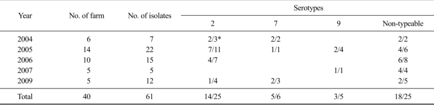
Table 1. Serotype of S. suis isolates in year
Year No. of farm No. of isolates Serotypes
2 7 9 Non-typeable
2004 6 7 2/3* 2/2 2/2
2005 14 22 7/11 1/1 2/4 4/6
2006 10 15 4/7 6/8
2007 5 5 1/1 4/4
2009 5 12 1/4 2/3 2/5
Total 40 61 14/25 5/6 3/5 18/25
*Number of farm/Number of isolates.
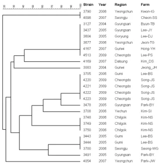
Fig. 1. PFGE after Sma I digestion of 61 S. suis isolated from dis- eased pig. A dendrogram was generated by Dice analysis (optimization 0.5%, band tolerance 1%) and cluster analy- sis with unweighted pair group method with arithmetic average, using Fingerprinting II Informatix software (Biorad, USA).
Fig. 2. Dendrogram of S. suis serotype 2 isolated from diseased pig by PFGE pattern (n=25).
막혈청형 결과는 Table 1과 같다. 실험균주 61주 중 2형이 25주(40.9%)로 가장 많았으며, 7형이 6주 (9.8%), 9형이 5주(8.2%)이었고 PCR에 의해 형별되 지 않은 분리주가 25주(40.9%)로 나타났다. 동일농 장에서는 모두 동일한 혈청형의
S. suis가 분리되었
다.S. suis의 유전형별을 위해 제한효소 Sma I으로 처리하고 PFGE를 실시하여 분리주의 DNA 분절 양 상을 분석한 결과 모두 24가지의 유전형으로 분류 되었다(Fig. 1). 혈청형 2형의 경우 총 6가지의 유전 형으로 분류되었으며 Seo-SJ 농장의 경우 2004년에 분리된 2주와 2005년에 분리된 4주가 모두 동일한 혈청형이었고 유전형도 동일한 것으로 확인되었으
나, 반면 Song-JS 농장의 동일한 혈청형 2형의 분리 주가 분리 연도에 따라 유전형이 다른 것으로 나타 났다(Fig. 2). 혈청형 7형을 대상으로 유전자 분석 패턴을 조사한 결과 6가지 유전형으로 분류되었고 Kim-BD 동일농장에서 2007년과 2009년에 각각 분 리된 동일혈청형에 대해서 다른 유전형으로 분류되 는 것으로 나타났다(Fig. 3). 혈청형 9형인 5주의 S.
suis에 대해 유전형을 조사한 결과 3가지 유전형으
로 분류되었으며 2005년도에 동일농장(Lee-JW, Ha-JD) 에서 분리된 9형의 S. suis는 각각 동일한 유전형으 로 확인되었다(Fig. 4). 이번 실험에서 multiplex PCR 법에 의해 혈청형이 확인되지 않은 25주의 S. suis 에 대해 PFGE에 의한 유전적 상관관계를 조사한 결과 11가지의 유전형으로 분류되었으며 해당하는 18개 농장 중에서 3주 이상 분리된 3호에 대해서 유전형을 조사한 결과 2호(Song-JS, Kim-Ns)는 동일 한 유전형으로 확인되었으나 2005년과 2006년에 각Fig. 3. Dendrogram of S. suis serotype 7 isolated from diseased pig by PFGE pattern (n=6).
Fig. 5. Dendrogram of non-typeable S. suis isolated from diseased pig by PFGE pattern (n=25).
Fig. 4. Dendrogram of S. suis serotype 9 isolated from diseased pig by PFGE pattern (n=5).
각 분리된 Lee-BS 농장은 유전형이 서로 다른 것으 로 나타났다(Fig. 5).
고 찰
분자유전학의 발달과 함께 병원체에 대한 DNA 수 준의 형별을 위한 여러 가지 방법이 널리 이용되고 있 으며, 그 중에서 PFGE법은 세균의 전체 염색체 DNA 를 대상으로 제한효소를 처리하여 일반적으로 40 Kb에 서 1,000 Kb의 DNA 분절을 염색된 gel상에 나타내어 분자형을 할 수 있는 "Gold standard"로 인식되어 널리 이용되고 있다. Luey 등(2007)은 S. suis serotype 2의 subtyping을 위해 PFGE를 실시하고 항생제 감수성 결 과, 병원성 관련인자 및 MLST 성적과 비교한 결과 PFGE에서 9가지 유전형으로 분류되었으며 시험법의 표준화로 실험실간 분리주에 자료를 공유함으로써 유전적 상관관계 분석이 가능할 것으로 기대하였다.
이번 실험에서는 총 61주의 S. suis에서 24가지 유전 형으로 분류되었고 PCR에 의한 혈청형별을 실시한 후 혈청형별 유전학적 상관관계를 분석한 결과 2형, 7형, 9형, 기타 형별에서 각각 6가지, 6가지, 3가지, 11가지 유전형으로 분류되어 지역별, 시기별, 농장 간 분리된
S. suis의 관련성을 비교 분석하는데 도움
이 될 것으로 생각한다. Berthelot-Hérault 등(2002)은 프랑스 내 돼지와 외국사람 유래 123개 S. suis 분리 균주를 대상으로 PFGE를 실시하여 분석한 결과 크게 3가지 유전형으로 분류되었으며 실험의 재현성이 우 수하고 결과해석이 용이하며 역학관련 자료로 사용될 수 있다고 보고한 바 있다. 또한, Vela 등(2003)은 스페인의 양돈장에서 분리된 S. suis를 대상으로 혈청 형별과 두 가지 제한효소를 사용하여 각각 PFGE에 의한 유전형별을 실시한 결과 serotype 9가 67.4%로 가장 많았으며 serotpye 2는 14.8%라고 보고하였고 PFGE에 의한 유전형은 총 302주가 60가지의 유전형 으로 분류되며 동일 농장 내에서도 여러 형태의 유전 형이 존재한다고 보고하였다. 이번 실험에서는 경북 지역 40개 양돈장에서 분리된 61주를 대상으로 PCR 에 의한 혈청형별을 시도한 결과 2형이 41%를 차지 하여 So 등(1995)이 도축돈의 폐렴 병변부에서 분리 한 S. suis 2형이 32.1%라는 성적보다는 다소 높은 것 으로 조사되었으나 Koo 등(2002)이 도축돈을 대상으 로 조사한 결과인 38%와는 유사한 성적으로 나타나 국내에서 발생하는 돼지의 S. suis에 의한 연쇄상구균 증은 대부분이 serotype 2에 의한 것으로 알 수 있고 PCR에 의한 혈청형별과 PFGE를 병행하여 실시함으 로써 동일농장내 발생한 감염증에 대해 유입경로의 분석과 차단을 위한 기초자료로 활용이 가능할 것으 로 생각한다. 이번 실험에서 혈청형별로 PFGE에 대 한 비교분석에서 동일농장에서 분리된 동일 혈청형 의 경우 분리연도에 따라 유전형이 다른 예가 일부 확인되었다. Seo-SJ 농장은 분리연도에 상관없이 동 일한 유전형으로 조사되었고 Song-JS, Kim_BD 및 Lee-BS 농장에서는 분리연도가 다른 경우에 동일한
혈청형에서도 유전형이 다른 것으로 확인되었다. 이 러한 예가 동일 농장 내 균주 간 발병시기의 경과에 따른 농장 내 균주의 유전변환에 의해 질병이 재발한 것인지 아니면 감염돈의 신규입식 등으로 외부 유입 에 따른 새로운 발병인지를 밝혀낼 수 있는 새로운 유전자 검사법의 도입이 필요하리라고 생각한다.
결 론
경북지역 환돈에서 분리된 61주의 S. suis를 대상으 로 PCR에 의한 혈청형별과 PFGE에 의한 유전형별을 실시한 결과는 다음과 같았다. Multiplex PCR을 이용 한 혈청형 분류 결과 S. suis 1형은 확인되지 않았고, 2형이 25주(41%), 7형이 6주(9.8%), 9형이 5주(8.2%) 이었고 형별되지 않은 경우가 25주(41%)로 나타났으 며 분리주는 분리농장별 동일혈청형만 존재하는 것 으로 확인되었다. PFGE에 의한 유전적 상관관계를 분석한 결과 전체적으로 24가지의 유전형으로 분류 되었으며 혈청형별로는 2형이 6 genotype, 7형이 5 genotype, 9형이 3 genotype, 비형별 균주가 11 geno- type으로 유전형별되었다.
참 고 문 헌
Berthelot-Hérault F, Marois C, Gottschalk M, Kobisch M. 2002.
Genetic diversity of Streptococcus suis strains isolated from pigs and humans as revealed by pulsed-field gel electrophoresis. J Clin Microbiol 40: 615-619.
Chu G, Vollrath D, Davis RW. 1986. Separation of large DNA molecules by contour-clamped homogeneous electric fields. Science 234: 1582-1585.
CDC, Pulsenet. 2006. Standardized laboratory protocol for molec- ular subtyping of Yersinia pestis by PFGE.
http://www.cdc.gov/pulsenet/protocols.
Clifton-Hadley FA, Enright MR, Alexander TJ. 1986. Survival of Streptococcus suis type 2 in pig carcases. Vet Rec 118:
275.
Higgins R, Gottschalk M. 1999. Streptococcal diseases. pp.
563-578. In: Straw BE, D'Allaire S, Mengeling WL, Taylor DJ(ed.). Diseases of swine. 8th ed. Iowa State University Press, Ames, Iowa.
Hunter SB, Vauterin P, Lambert-Fair MA, van Duyne MS, Kubota K, Graves L, Wrigley D, Barrett T, Ribot E.
2005. Establishment of a universal size standard strain for use with the PulseNet standardized pulsed-field gel electrophoresis protocol: converting the national da-
ta-bases to the new size standard. J Clin Microbiol 43:
1045-1050.
Jung BY, Jung SC, Kim JY, Park YH, Kim BH. 1998.
Polymerase chain reaction for a rapid and specific identi- fication of Streptococcus suis. Korean J Vet Res 38:
771-776.
Kataoka Y, Sugimoto C, Nakazawa M, Morozumi T, Kashiwazaki M. 1993. The epidemiological studies of Streptococcus suis infections in Japan from 1987 to 1991. J Vet Med Sci 55: 623-626.
Koo KM, Lim JH, Koh HB. 2002. The detection of Streptococcus suis 1(+14), 2(+1/2), 7 and 9 from pneumonic lungs slaughtered pigs by a multiplex PCR. Korean J Vet Res 42: 495-504.
Luey CK, Chu YW, Cheung TK, Law CC, Chu MY, Cheung DT, Kam KM. 2007. Rapid pulsed-field gel electrophoresis protocol for subtyping of Streptococcus suis serotype 2.
J Microbiol Methods 68: 648-650.
Marois C, Le Devendec L, Gottschalk M, Kobisch M. 2007.
Detection and molecular typing of Streptococcus suis in tonsils from live pigs in France. Can J Vet Res 71:
14-22.
McPeek FD Jr, Coyle-Morris JF, Gemmill RM. 1986. Separation of large DNA molecules by modified pulsed field gra- dient gel electrophoresis. Anal Biochem 156: 274-285.
Mwaniki CG, Robertson ID, Trott DJ, Atyeo RF, Lee BJ, Hampson DJ. 1994. Clonal analysis and virulence of Australian isolates of Streptococcus suis type 2.
Epidemiol Infect 113: 321-334.
Olson MV. 1989. Separation of large DNA molecules by pulsed-field gel electrophoresis. A review of the basic phenomenology. J Chromatogr 470: 377-383.
Reams RY, Harrington DD, Glickman LT, Thacker HL, Bowersock TL. 1996. Multiple serotypes and strains of Streptococcus suis in naturally infected swine herds. J Vet Diagn Invest 8: 119-121.
Sanford SE, Tilker ME. 1982. Streptococcus suis type II-asso- ciated diseases in swine: observations of a one-year study. J Am Vet Med Assoc 181: 673-676.
Sihvonen L, Kurl DN, Salmela P. 1986. Infection with Streptococcus suis serotypes 1 and 2 in the same dis- eased pig. Acta Vet Scand 27: 626-628.
Staats JJ, Feder I, Okwumabua O, Chengappa MM. 1997.
Streptococcus suis: past and present. Vet Res Commun 21: 381-407.
So SH, Kim BH, Cho GJ. 1995. Biochemical characterisitics and capsular serotypes of Streptococcus suis isolated from pneumonic lungs of slaughter pigs. Korean J Vet Res 35: 297-306.
Vecht U, Wisselink HJ, Stockhofe-Zurwieden N, Smith HE.
1996. Characterization of virulence of the Streptococcus suis serotype 2 reference strain Henrichsen S 735 in newborn gnotobiotic pigs. Vet Microbiol 51: 125-136.
Vela AI, Goyache J, Tarradas C, Luque I, Mateos A, Moreno MA, Borge C, Perea JA, Domínguez L, Fernández-
Garayzábal JF. 2003. Analysis of genetic diversity of Streptococcus suis clinical isolates from pigs in Spain by pulsed-field gel electrophoresis. J Clin Microbiol 41:
2498-2502.
Wisselink HJ, Joosten JJ, Smith HE. 2002. Multiplex PCR assays for simultaneous detection of six major serotypes and two virulence-associated phenotypes of Streptococcus
suis in tonsillar specimens from pigs. J Clin Microbiol 40: 2922-2929.
Wisselink, HJ, Reek FH, Vecht U, Stockhofe-Zurwieden N, Smits MA, Smith HH. 1999. Detection of virulent strains of Streptococcus suis type 2 and highly virulent strains of Streptococcus suis type 1 in tonsillar specimens of pigs by PCR. Vet Microbiol 67: 143-157.