가자( Terminalia chebula Retz.) 열매 메탄올 추출물의 Histone Acetyltransferase 활성 저해에 따른 항전립선암 효과
이 유 현 수원대학교 식품영양학과
The Inhibitory Effect on Androgen Receptor-Dependent Prostate Cancer Cell Growth by Anti-Histone Acetyltransferase Activity from
Terminalia chebula Retz. Fruit Methanol Extract
Yoo-Hyun Lee
Dept. of Food Science and Nutrition, University of Suwon, Gyeonggi 445-743, Korea
ABSTRACT The inhibitory effect of histone acetyltransferase from methanol extract of Terminalia chebula Retz.
fruit (TCME) was investigated in prostate cancer cell. TCME significantly inhibited histone acetyltransferase (HAT) activity by over 50% at 100 μg/mL concentration. TCME treatment repressed androgen receptor (AR) mediated tran- scription, mRNA level of AR target genes, prostate specific antigen (PSA) and NKX-3.1, as well as AR acetylation.
Finally, the prostate cancer cell viability was dramatically reduced by TCME treatment at 0~100 μg/mL concentration.
These results indicated that TCME, as a potent HAT inhibitor, could suppress prostate cancer cell growth by AR mediated transcription regulation.
Key words: Terminalia chebula, prostate cancer, histone acetyltransferase, androgen receptor, acetylation
Received 16 September 2013; Accepted 26 September 2013 E-mail: [email protected], Phone: 82-31-229-8194
서 론
전립선암은 최근 우리나라에서 가파른 발병률의 증가를 보이는 남성암으로 미국에서는 남성사망의 두 번째 원인으 로 꼽히고 있다(1). 전립선암 발병에는 노화와 육식 위주 식 습관에 따른 동물성지방 과다섭취 등의 식생활이 영향을 미 치는 것으로 알려져 있다(2,3).
전립선암의 치료는 호르몬 요법이 전형적으로 사용되는 데(4), 안드로젠은 남성호르몬으로 생식계의 발달, 유지, 성 장에 필수적이며, 이 호르몬의 수용체인 안드로젠 수용체 (androgen receptor, AR)는 전립선암의 발달과 깊은 연관 이 있다(5). 최근 안드로젠 수용체(androgen receptor, AR) 의 아세틸화, 인산화 등의 전사 후 변형(post transcrip- tional modification)이 전립선암세포의 성장과 관련이 있다 고 밝혀지면서(6) 암과 연관된 히스톤 탈아세틸화 효소 (histone deacetylase, HDAC) 및 히스톤 아세틸 전이효소 (histone acetyltransferase, HAT) 등의 히스톤 변형 효소 에 관한 연구가 진행되고 있다. p300, p/CAF, TIP60 등의 HAT 단백질은 AR을 직접적으로 아세틸화하고, 이는 co- activator binding을 강화하여(7), 결국 AR을 핵으로 translocation 함으로써 전립선암세포의 성장을 촉진시킨
다고(4) 보고되었다. 그러므로 HAT 활성의 억제를 이용한 AR 아세틸화 조절은 새로운 전립선암 치료제 개발에서 중 요한 기작이 될 수 있으며, 이러한 활성을 지닌 천연물의 발굴이 기존의 호르몬 치료와 병행할 수 있는 보완적인 치료 제 개발의 기반이 될 수 있다.
가자는 사군자과(Cambretaceae)에 속하는 Terminalia chebula Retz.의 성숙한 과실을 건조한 것으로 약용으로 사 용되고 있다(8). 가자의 성분으로는 chebulic acid, chebu- lagic acid, gallic acid, tannin 등이 있다고 알려져 있으며 (8), 항균(9), 항암(10), 항염 및 항산화 활성(11), 항당뇨 활성(12)이 있다고 알려져 있다. 본 연구에서는 가자 메탄올 추출물이 HAT 활성을 억제하고 AR의 아세틸화를 감소시 킴으로써 전립선암세포의 성장을 저해하는 것을 밝히고, 전 립선암 예방 치료의 소재 개발의 기초자료를 제공하고자 한 다.
재료 및 방법
실험재료
본 연구에서 사용한 가자(Terminalia chebula Retz.)의 열매 추출물은 2007년 한국추출물은행(Daejeon, Korea) 으로부터 구매하였으며 4°C에 보관하며 시료로 사용하였 다. 세포배양에 사용된 배지, 혈청, 항생제 등은 Gibco-BRL (Grand Island, NY, USA)에서 구입하였다. HAT activity
colorimetric assay kit는 Biovision(Mountain View, CA, USA)에서 구입하였으며, Easy-spin total RNA extraction kit는 Intron(Seoul, Korea)에서 구입하여 사용하였다.
Lipofectamine 2000 transfection reagent는 Invitrogen (Carlsbad, CA, USA)에서 구입하여 사용하였으며, 항체는 Upstate Biotechnology(Charlottesville, VA, USA)와 Santa Cruz Biotechnology(Santa Cruz, CA, USA)에서 구 입하여 사용하였다. 본 실험에 사용된 pGL3-PSA con- struct는 연세대학교 의과대학 김경섭 교수님이 제공하였으 며, androgen responsive element(ARE)를 포함하고 있는 androgen dependent reporter construct이며, pGL5 SV40은 control vector로 사용되었다.
세포배양
LNCaP, DU145는 RPMI1640 배지에서, MCF-7 세포주 는 DMEM에서 10% fetal bovine serum(FBS), 1% anti- biotics/antimycotics를 첨가하여 37°C, 5% CO2 조건의 습 윤한 incubator에서 배양되었다.
HAT 활성의 측정
HeLa 세포로부터 얻은 nuclear extract(NE)로(13) HAT 활성을 측정하였다. HAT 활성의 측정 중 HAT activity kit 를 이용한 활성의 측정은 Biovision의 protocol을 따랐다.
특이적인 HAT 단백질인 p300과 CBP 단백질 활성 측정을 위한 전처리 과정은 다음과 같다. NE는 anti-p300(Upstate Biotechnology)와 anti-CBP(Santa Cruz Biotechnology) polyclonal antibody를 사용하여 immunoprecipitation을 하였다. 150 μg의 NE를 상기의 antibody 및 protein A/G PLUS agarose bead와 함께 4°C에서 18시간 반응시킨 후, HAT buffer(50 mM pH8.0 tris, 10% glycerol, 0.1 mM EDTA)로 2회 세척하고 HAT 활성을 측정하였다. Autor- adiography의 경우는 다음과 같이 시행하였다. NE를 HAT buffer와 함께 30°C에서 60분간 1 μL의 4.7 Ci/mmol [3H]acetyl coenzymeA와 함께 반응시키고 SDS/PAGE (15% gel)에서 분리시킨 후 autoradiography로 분석하였 다.
Reporter assay
LNCap 세포주를 6 well plate에 500,000 cells/well의 밀도로 분주하고, 16시간이 지난 후 reporter assay를 위하 여 transfection을 시행하였다. Transfection을 위하여 6 well plate 당 pGL3-PSA 0.5 μg과 pGL5 SV40 1 ng을 사용하였으며, 시행 후 24시간 동안 배양하였다. Androgen 첨가에 따른 세포 내 전사반응을 보기 위하여 10% char- coal-stripped FBS를 첨가한 RPMI1640 배지로 교환한 뒤 48시간 동안 배양을 지속한 후, 합성 androgen인 50 nM R1881(Sigma-Aldrich Co., St. Louis, MO, USA)을 가자 메탄올 추출액 TCME 0, 50, 100 μg/mL와 함께 첨가하였
다. 다시 16시간 후, 세포를 수집하여 파쇄하고 dual luci- ferase assay(Promega Co., Madison, WI, USA)를 시행하 였다.
Total RNA의 추출과 real time PCR
Real time PCR 시행을 위한 total RNA 추출을 위하여, LNCaP 세포를 500,000 cells/well의 밀도로 6 well plate 에 분주하고 10% FBS를 첨가한 RPMI1640 배지에서 배양 하였다. 세포가 90% confluence에 도달하였을 때, 세포를 PBS로 2회 세척하고 10% charcoal-stripped FBS를 첨가 한 RPMI-1640 배지로 교환하고 다시 배양하였다. 48시간 후, 합성 androgen인 R1881을 50 nM을 첨가하고, 다시 6시간 후 가자 메탄올 추출액 TCME 0, 25, 50 100 μg/mL 의 농도로 함께 첨가하였다. 16시간 후, 세포를 수집하고 total RNA를 추출하고 cDNA를 합성하였다.
Real time PCR 분석은 SYBR Green PCR Master mix reagent를 이용하여(ABI Prism 7700 sequence detection system, Applied Biosystem, Foster city, CA, USA) 다음 과 같은 조건으로 시행하였다. 모든 결과는 GAPDH에 따라 상대적으로 표현하였다. PCR은 94°C에서 2분, 94°C에서 30초, 55°C에서 30초, 72°C에서 30초, 그리고 72°C에서 다시 10분간 27 cycle로 시행되었다. 본 분석에 사용된 프 라이머는 다음과 같다. PSA는 5'-AGTCTGAGGAGGTGT TCTGGTG-3'과 5'-GAGGTCGTGGCTGGAGTCATCA -3', NKX 3.1은 5'–CTGTCAGCCCCTGAACGG-3' 및 5' –AACCATAT CTTCACTTGGGTCTCC-3'이 사용되었으 며, GAPDH는 5'-CGCGGGGCTCTCCAGAACATCATCC -3'과 5'-CTCCGACGCC TGCTTCACCACCTTCTT–3' 이 사용되었다. 모든 반응은 삼 반복으로 시행되었으며, relative expression 및 SD value는 comparative method 를 사용하여 계산하였다.
Immunoprecipitation과 western blot
100 mm plate에 배양된 LNCaP 세포가 90% con- fluence에 도달하였을 때, 10% charcoal stripped FBS를 첨가한 phenol free RPMI1640 배지로 교환하였다. 48시간 후, 50 nM R1881과 TCME를 농도별로 처리하고 16시간이 지난 후 회수하고 RIPA buffer에 lysis하였다. Lysate는 1 μL의 monoclonal anti-Ac lysine(Santa cruz Biotechnol- ogy)에 protein A/G PLUS agarose bead에 4°C에서 overnight 처리하였다. 결과물을 10% SDS gel에 전기영동 을 시행하고, AR antibody로 반응하고 enhanced chem- iluminescence kit(ECL kit, Pierce Antibodies, Rock- ford, IL, USA)를 이용하여 확인하였다.
MTT assay
두 종류의 전립선암세포인 LNCaP과 DU145를 96 well plate에 10,000 cells/well로 분주하고 유방암세포주인
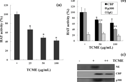
Fig. 1. TCME has a HAT inhibitory activity on various concentration of TCME. (a) Histone acety- lase assays were performed with nuclear extracts, (b) with immunoprecipiteated products (anti-CBP, anti-p300 and IgG) and (c) with radiolabeled acety- lated histones. Results represent the mean values with SD of three independent experiments. *P<0.05 or **P<0.01 is assessed as significant differences between the untreated group and TCME treated group.
MCF-7은 5,000 cells/well로 분주하여 배양하였다. 배양 24시간 후, PBS로 2회 세척을 하고 FBS가 포함되지 않은 배지로 교체한 후 TCME를 0~0.2 mg/mL의 농도로 처리하 여 배양하였다. 48시간이 지난 후, 3-(4,5-dimethylthiazol- 2-yl)-2,5-diphenyltetrazolium bromide(MTT, Sigma- Aldrich Co.)를 배지에 첨가하여 2시간 동안 배양하고 형성 된 blue formazan을 DMSO에 녹여 570 nm와 630 nm에서 microplate reader를 사용하여 세포생존률을 측정하였다.
통계분석
본 연구에서는 최소 3반복에 대한 평균 및 표준편차로 표시하였으며, 각 실험군을 대조군에 대한 백분율로 나타내 었다. 각 실험군간의 통계적 유의성은 Student's t-test를 사용하여 검정하였다. 통계검정은 SigmaStat(version 3.5, Systat Software Inc., San Jose, CA, USA)를 이용하였다.
결과 및 고찰
TCME의 HAT 활성 저해
가자의 메탄올 추출물(TCME)의 HAT 활성 저해 효과를 측정한 결과는 Fig. 1A와 같았다. 추출물을 처리하지 않은 정상군과 비교하였을 때, 25 μg/mL부터 100 μg/mL까지 농도의존적으로 저해하였으며, 모든 농도는 정상군에 대하 여 유의적으로 저해활성을 보이는 것으로 나타났다(P<0.05).
특히 50 μg/mL부터는 50% 이상 저해효과를 보였다. 특이 적인 HAT 단백질에 관한 저해효과는 Fig. 1B와 C의 결과에 서 볼 수 있듯이, 높은 효과를 보인 50과 100 μg/mL를 대상 으로 TCME를 처리하였을 때 CBP와 p300에서 유의적인 저해효과를 보였다. 특히 p300의 경우 50% 이상의 저해를 보였으며, 반면에 IgG의 값은 변화가 없었던 것과 대조적으 로 나타났다. 이같은 결과는 TCME가 HAT 저해활성을 갖 고 있으며, HAT 단백질 중 p300에서 특이적인 활성을 보인 다고 생각된다.
천연물을 기반으로 한 HAT 활성 저해제는 꾸준히 연구되 고 있는데, gallic acid(14), EGCG(15), garcinol(16) 등은 암과 연관된 천연물 기반 HAT 저해제로 밝혀져 있다. 특히 전립선암에서 HAT 활성은 히스톤의 아세틸화뿐 아니라 AR의 아세틸화를 촉진하여 전사조절에 영향을 미치고 암의 발달을 유도한다(6,15).
TCME의 AR 매개 전사 활성의 억제
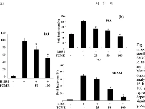
TCME 추출물이 androgen 의존적인 전립선암세포인 LNCaP에서 AR 매개 전사활성조절에 미치는 영향을 증명 하기 위하여, ARE(androgen responsive element) 부분을 가지고 있는 androgen dependent reporter construct를 세포에 transfection 시킨 후 luciferase 활성을 측정하였다 (Fig. 2A). LNCaP 세포는 48시간 동안 charcoal stripped 배지를 처리한 후 합성 androgen인 R1881을 18시간 동안 처리하였으며, TCME 처리군은 R1881 처리 시, 50 및 100 μg/mL의 농도로 TCME를 처리하였다. 그 결과 R1881만 처리한 군과 비교할 때, TCME의 각 처리 농도에서 유의적 으로 낮은 활성을 보였으며, 특히 100 μg/ mL의 농도에서는 40% 이상의 저해율을 보였다.
다음으로 endogenous AR target gene인 PSA 및 NKX 3.1의 mRNA 발현에 미치는 영향을 real time PCR로 확인 하였다(Fig. 2B). 역시 상기와 같은 조건에서 실험을 시행한 결과, R1881 처리군에서는 PSA 및 NKX3.1 양쪽 모두 높은 mRNA 발현을 보였으며, TCME가 농도의존적으로 발현을 저해했고 100 μg/mL의 처리군에서는 아무것도 처리하지 않은 정상군과 유사한 수준을 보임을 확인할 수 있었다. 이 같은 결과들에서, TCME는 AR 매개 전사를 억제한다고 생 각되며, AR target gene의 mRNA 발현이 저해된다고 생각 된다.
TCME 처리군의 AR 아세틸화 저해
TCME 처리가 AR 아세틸화에 미치는 영향을 확인하기
Fig. 2. TCME repressed AR-mediated tran- scription activation. (a) LNCaP cells were tran- siently transfected with pGL3-PSA and pGL5 SV40. Cells were treated with or without R1881 and TCME (0, 50, 100 μg/mL). Results are presented as relative luciferase activity.
Mean values were calculated from three in- dependent experiments. (b) Real time PCR analysis of PSA and NKX3.1 were performed 16 h after treatments with TCME (0, 25, 50, 100 μg/mL), with or without R1881. Results represent the mean values with SD of three in- dependent experiments. *P<0.05 is assessed as significant differences between the untreated group and TCME treated group.
Fig. 3. Androgen receptor acetylation was significantly reduced by TCME. LNCaP cells were treated with or without R1881, alone or in combination. Cell lysates were immunoprecipitated with anti-ac-lysin and blotted with anti-AR.
Fig. 4. Effect of TCME on various cancer cell growth. LNCaP, DU145, and MCF-7 were treated with serum-free medium con- taining TCME (0~100 μg/mL) for 48 h. Results represent the mean value with SD of three independent experiments.
위하여, 48시간 동안 charcoal stripped 배지에 배양된 LNCaP 세포에 R1881과 농도별 TCME를 처리하고 18시간 동안 다시 배양하고 회수된 세포를 lysis 시켜 사용하였다.
Lysate는 특이적인 lysine residue에 아세틸화 된 정도를 측정하기 위하여 anti-acetyl lysine으로 immunopre- cipitation을 시행하고, anti-AR로 western blot을 시행하 였다. Fig. 3의 결과와 같이 R1881을 처리하였을 때 아세틸 화가 증가하는 반면, TCME를 처리했을 때 농도별로 아세틸 화가 감소하고 있음을 확인할 수 있다.
HAT 단백질은 histone 이외에 AR과 같은 비히스톤 단백 질도 아세틸화 시키는 것으로 알려져 있으며(17), HAT 단 백질은 AR의 핵으로의 translocation을 통하여 전립선암세 포의 성장을 촉진한다고(4) 보고되고 있다. Lee 등(15)의 연구에서 녹차의 EGCG가 AR의 아세틸화를 저해하여 핵으 로의 translocation을 억제했다고 제안했으며, 본 연구에서 도 이러한 AR의 아세틸화의 저해는 전립선암의 성장 억제 로 연결될 것이라고 생각된다.
TCME의 전립선암세포 성장 억제효과
TCME가 안드로젠 의존적인 전립선암세포인 LNCaP과 다른 세포주에서 보이는 암세포 성장 억제효과를 비교하기 위하여 실시하였다. 실험에 사용된 세포주는 유방암세포주 인 MCF-7, 안드로젠 비의존적 전립선암세포주인 DU145 및 이 실험에서 사용된 LNCaP 등으로 TCME 0~200 μg/
mL의 농도로 24시간 동안 처리하여 MTT assay를 실시하 였다. LNCaP의 경우 농도별로 억제효과를 볼 수 있었으며, 100 μg/mL의 농도에서 40% 정도 cell viability가 억제되는 결과를 얻을 수 있었으며, MCF-7과 DU145의 경우 동일 농도 구간에서는 TCME 처리에 따른 차이를 보이지 않았다.
상기의 결과에서 언급했듯이, AR의 아세틸화 억제는 Fig.
4에서 안드로젠 의존성 전립선세포의 성장을 억제했다고 생
각된다.
요 약
본 연구에서는 Terminalia chebula Retz.의 성숙열매인 가 자 메탄올 추출물(TCME)을 이용하여 HAT 활성을 억제함
으로써 AR의 아세틸화 감소를 유도하여 전립선암세포의 성 장을 억제하였다. TCME의 처리는 HAT 활성을 100 μg/mL 의 농도에서 50% 이상 저해하였으며, p300과 CBP 등의 특이적 HAT 단백질에서도 유의적인 저해활성을 보였다.
TCME를 0~100 μg/mL로 안드로젠 수용체 의존적 전립선 암세포인 LNCaP에 처리한 결과, reporter assay에서 AR 매개 전사를 억제하고 AR target gene의 mRNA 발현을 억 제하였다. 동일 농도에서 AR의 아세틸화가 감소한 결과를 보였으며, 결국 전립선암세포주의 성장억제를 유도하였다.
이같은 결과에서 가자 메탄올 추출물은 HAT 활성을 억제하 며, AR의 아세틸화를 감소시킴으로써 효과적인 전립선암 치료소재로 개발될 수 있는 가능성이 있음을 제안한다.
REFERENCES
1. Manoharan M, Ramachandran K, Soloway MS, Singal R.
2007. Epigenetic targets in the diagnosis and treatment of prostate cancer. Int Braz J Urol 33: 11-18.
2. Kang HI, Kim JY, Cho HD, Park KW, Kang JS, Seo KI.
2010. Resveratrol induces apoptosis in primary human pros- tate cancer cells. J Korean Soc Food Sci Nutr 39: 1119- 1125.
3. Moon JM, Seok GH, Cho SI. 2012. Antiproliferative effect of Schisandrae fructus extract on PC-3 human prostate can- cer cells. Kor J Herbology 27: 17-23.
4. Yoon HG, Wong J. 2006. The corepressors silencing media- tor of retinoid and thyroid hormone receptor and nuclear receptor corepressor are involved in agonist- and antago- nist-regulated transcription by androgen receptor. Mol En- docrinol 20: 1048-1060.
5. Kang Z, Jänne OA, Palvimo JJ. 2004. Coregulator recruit- ment and histone modifications in transcriptional regulation by the androgen receptor. Mol Endocrinol 18: 2633-2648.
6. Gong J, Zhu J, Goodman OB Jr, Pestell RG, Schlegel PN, Nanus DM, Shen R. 2006. Activation of p300 histone ace- tyltransferase activity and acetylation of the androgen re- ceptor by bombesin in prostate cancer cells. Oncogene 25:
2011-2021.
7. Fu M, Rao M, Wang C, Sakamaki T, Wang J, Di Vizio
D, Zhang X, Albanese C, Balk S, Chang C, Fan S, Rosen E, Palvimo JJ, Jänne OA, Muratoglu S, Avantaggiati ML, Pestell RG. 2003. Acetylation of androgen receptor enhan- ces coactivator binding and promotes prostate cancer cell growth. Mol Cell Biol 23: 8563-8575.
8. Park JO. 2008. Effects of methanol extract from Terminalia chebulae on renal and pulmonary toxicities induced by para- quat in rats. J Life Sci 8: 129-135.
9. Lee KS, Kim SH, Sim KC, Park CS, Shin YS. 1997. Antimi- crobial activity of Terminalia chebula Retz. extract of against intestinal pathogens. Korean J Food & Nutr 10: 559- 563.
10. Achari C, Reddy GV, Reddy TC, Reddanna P. 2011. Chebu- lagic acid synergizes the cytotoxicity of doxorubicin in hu- man hepatocellular carcinoma through COX-2 dependant modulation of MDR-1. Med Chem 7: 432-442.
11. Bag A, Kumar Bhattacharyya S, Kumar Pal N, Ranjan Chattopadhyay R. 2013. Anti-inflammatory, anti-lipid per- oxidative, antioxidant and membrane stabilizing activities of hydroalcoholic extract of Terminalia chebula fruits.
Pharm Biol Posted online on 5 Sep 2013.
12. Silawat N, Gupta VB. 2013. Chebulic acid attenuates ische- mia reperfusion induced biochemical alteration in diabetic rats. Pharm Biol 51: 23-29.
13. Eliseeva ED, Valkov V, Jung M, Jung MO. 2007. Characte- rization of novel inhibitors of histone acetyltransferases.
Mol Cancer Ther 6: 2391-2398.
14. Choi KC, Lee YH, Jung MG, Kwon SH, Kim MJ, Jun WJ, Lee J, Lee JM, Yoon HG. 2009. Gallic acid suppresses lip- opolysaccharide-induced nuclear factor-κB signaling by pre- venting RelA acetylation in A549 lung cancer cells. Mol Cancer Res 7: 2011-2021.
15. Lee YH, Kwak J, Choi HK, Choi KC, Kim S, Lee J, Jun W, Park HJ, Yoon HG. 2012. EGCG suppresses prostate cancer cell growth modulating acetylation of androgen re- ceptor by anti-histone acetyltransferase activity. Int J Mol Med 30: 69-74.
16. Oike T, Ogiwara H, Torikai K, Nakano T, Yokota J, Kohno T. 2012. Garcinol, a histone acetyltransferase inhibitor, ra- diosensitizes cancer cells by inhibiting non-homologous end joining. Int J Radiat Oncol Biol Phys 84: 815-821.
17. Fu M, Wang C, Zhang X, Pestell R. 2004. Acetylation of nuclear receptors in cellular growth and apoptosis. Biochem Pharmacol 68: 1199-1208.