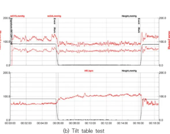
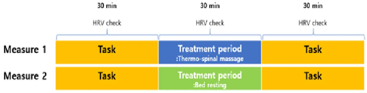
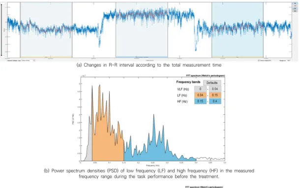
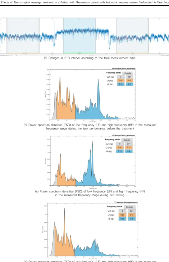
• 검색 결과가 없습니다.

Effects of Thermo-spinal massage treatment in a Patient with Rheumatism patient with Autonomic nervous system Dysfunction: A Case Report

N/A
N/A
Protected

Academic year: 2021

Share "Effects of Thermo-spinal massage treatment in a Patient with Rheumatism patient with Autonomic nervous system Dysfunction: A Case Report"

Copied!
10
0
0

로드 중.... (전체 텍스트 보기)

전체 글

(1)

Effects of Thermo-spinal massage treatment in a Patient with Rheumatism patient with Autonomic nervous system Dysfunction:

A Case Report

Jun Hyun Choi

1

, Jong-Hoo Lee

2

, Yong-Soon Yoon

2*

1

Resident of Department of Rehabilitation Medicine, Presbyterian (Jesus) Medical Center

2

Physiatrist of Department of Rehabilitation Medicine, Presbyterian (Jesus) Medical Center

자율신경 기능 이상이 동반된 류마티즘 환자에 대한 온열-척추 마사지 치료 효과 : 증례 연구

최준현

1

, 이정후

2

, 윤용순

2*

1

전주 예수병원 재활의학과 전공의

2

전주 예수병원 재활의학과 전문의

Abstract Rheumatoid arthritis (“RA”), whose characteristics are chronic inflammation and chronic pain, accompanies autonomic nervous system(ANS) dysfunction. In particular, ANS dysfunction in patients with chronic musculoskeletal pain was found to have increased the local pain intensity while lowering the pain threshold, thereby negatively influencing pain. It is reported that thermo-spinal massage affects relief of chronic pain and recovery of ANS in patients with chronic musculoskeletal pain. Therefore, we report a case of rheumatoid patients with chronic pain and ANS dysfunction, who experienced recovery of ANS dysfunction and pain reduction by applying thermo-spinal massage treatment.

Key Words : Convergence, Thermal, Massage, Thermo-spinal massage device, Rheumatoid arthritis, Autonomic nervous system, Chronic pain

요 약 만성통증을 동반한 류마티즘 환자에서 자율신경계 이상(autonomic nervous system dysfunction)이 동반되 기도 한다. 특히 만성통증 환자에서 자율신경계 이상은 국소 통증 강도 증가 및 통증 역치 감소를 발생하여 만성 근골 격계 통증에 악영향을 미친다고 확인되었다. 이런 만성 근골격계 통증 환자에 온열-척추 마사지 치료를 실시한 실험에 서 통증경감과 자율신경계 회복이 되었다고 보고되고 있다. 그래서 우리는 만성 통증과 자율신경계 이상이 동반된 류마 티즘 환자에 온열-척추 마사지 치료를 적용하였고 자율신경기능의 회복과 통증의 감소를 경험한 사례가 있어 보고하고 자 한다.

주제어 : 융합, 온열, 마사지, 온열-척추 마사지기, 류마티즘, 자율신경계, 만성통증

*Corresponding Author : Yong-Soon Yoon([email protected]) Received July 2, 2020

Accepted August 20, 2020 Revised August 5, 2020

Published August 28, 2020

(2)

1. Introduction

Rheumatoid arthritis (RA) is an autoimmune disease characterized by local invasion of the synovial joint, and systemic inflammatory response and chronic pain. In addition, many studies reported autonomic dysfunction characterized by decreased parasympathetic activity, increased sympathetic activity, and reduced baroreflex sensitivity in RA patients[1].

Such autonomic dysfunction has also been reported in patients with chronic musculoskeletal pain. In these patients, an abnormal interaction between the nociceptive and autonomic nervous systems (ANS) was observed. The increase in sympathetic activity was found to have increased the local pain intensity while lowering the pain threshold, thereby negatively influencing chronic musculoskeletal pain[2].

The popular methods used to treat such cases of chronic musculoskeletal pain are thermal and massage treatments. Thermal treatment was reported to be effective for vasodilation, facilitated blood circulation, active metabolism, and functional improvement in tissues via parasympathetic activation[3]. Massage treatment, on the other hand, was reported to have an effect on improving physiological balance and controlling pain via sympathetic and parasympathetic activations[4,5].

In a recent study, simultaneous thermospinal massage treatment was reported to lower stress and reduce pain via parasympathetic activation, with more outstanding effects than massage treatment alone[6].

This study thus applied thermospinal massage treatment with a reported effect on reduced pain via ANS regulation in patients with rheumatism who had autonomic dysfunction and reports the consequent positive effect on pain control in a single case.

2. Case Report

A 43-year-old female patient who had been diagnosed as having RA 14 years before was transferred from the division of rheumatology to our department for pain control. The patient complained of arthralgia caused by RA in the metacarpophalangeal and proximal interphalangeal joints from the first to the fourth fingers of both hands, shoulder pain on both sides, neck pain, tension-type headache, and intermittent dizziness. The shoulder pain on both sides, neck pain, and headache showed mean visual analog scale (VAS) scores of 5–6 in daily activities, with a pattern of aggravation to VAS scores of 7–9 several times during the day.

To identify the other causes of the pain, additional tests (brain magnetic resonance imaging [MRI], C-spine MRI, shoulder MRI, and electromyography) were performed, but without unusual findings. To identify the cause of intermittent dizziness, a vestibular function test was performed at the division of otorhinolaryngology, but no abnormality was found.

However, the results of the sympathetic skin response and tilt table tests showed that the condition accompanied an autonomic dysfunction (as shown Fig. 1).

(a) Sympathetic skin response

(3)

Fig. 2. Changes in mean visual analog scale (VAS) score and number of opioids used during the 20-week treatment period.

Variable Normal range Units 1 week 10 weeks 20 weeksㄴ

WBC 4∼10.5 10

3

/uL 2.5 4.9 3.1

CRP 0∼0.3 mg/dL 0.01 0.02 0.01

RAF 0∼15 IU/mL 43.23 NT 45.80

Anti-CCP 0∼4.9 U/mL 113.3 NT 125.6

ASO 0∼160 IU/mL 278.1 NT 336.0ㄴ

CRP: C-Reactive Protein, RA: Rheumatoid Factor, Anti-CCP: Anti-Cyclic Citrullinated Peptide antibody, ASO: Anti-streptolysin 'O Table 1. Blood Sampling Test

(b) Tilt table test

Fig. 1. (a) The result shows delayed onset latency. (b) Within 3 min of standing, a decrease in systolic blood pressure by ≥20 mmHg or diastolic blood pressure by ≥10 mmHg was observed (baseline blood pressure [BP], 105/58 mmHg and heart rate [HR], 71 beats per minute; head-up tilt BP, 82/48 mmHg and HR, 89 beats per minute; and head-down tilt BP, 135/77 mmHg and HR, 78 beats per minute)

The initial blood test results showed that the white blood cell count and C-reactive protein level in cerebrospinal fluid were normal, while

the measurements of the levels of the RA factors, anti-CCP, and antistreptolysin O were higher than the normal levels (as shwon Table 1).

For rheumatism control, the patient had been given injections of a disease-modifying anti-rheumatic drug (DMARD; tocilizumab 400 mg/20 ml, Actemra 400 mg). For pain control, the patient received celecoxib 200 mg (Celebrex 200 mg) and ultracetER (acetaminophen 650 mg with tramadol hydrochloride 75 mg) twice a day.

Especially, during a pain shock when the pain level suddenly increased to VAS 7~10, the patient had been receiving an opioid (oxycodone hydrochloride 10 mg with naloxone hydrochloride dihydrate 5.45 mg, Targin 10/5 mg) as necessary.

Despite the continuous pain control

medication and repetitive administration of the

opioid medication, the shoulder pain on both

sides, neck pain, headache, anxiety, and

insomnia persisted. To ease the pain and

increase the stability of the patient’s condition, a

(4)

Task: Typing using a computer for 30 minutes in a sitting posture.

Fig. 3. Flowchart of the measurements

device for simultaneous thermospinal massage treatment was recommended. The patient used the thermospinal massage device (CGM MB-1401;

Ceragem, South Korea) for 40 min twice a day during the 20-week treatment period.

The change in pain level was measured weekly using the VAS, and the change in medication was recorded. The first notable change was observed after 10 weeks. The overall pain was reduced.

Average weekly pain level decreased from VAS 5

~ 9 to VAS 3 or below. Also, the amount of opioid administered during a pain shock decreased from 3 ~ 5 times a week to twice a week. After 12 weeks, the pain level was reduced to a VAS score of 1–3, without the previously observed pain increase during the day; thus, the opioid medication was withdrawn (as shown Fig.

2).

In addition, dizziness was reduced, and the sleep pattern was improved. After the 20 weeks of using the device for the simultaneous thermospinal massage treatment, heart rate variability (HRV) was analyzed to examine the continuous change of the ANS in relation to the use of the device, as the analysis has been reported to be a noninvasive method with high levels of reliability and reproducibility[7].

To measure the HRV, a polar HR10 Bluetooth heart rate strap (Polar Electro, Kempele, Finland), which has verified accuracy, and the free smartphone application Elite HRV(Ashville, North Carolina, USA) were used[8]. To analyze the

measured HRV, the Kubios version 3.4.0 heart rate variability software (Biosignal Analysis and Medical Imaging Group, Department of Physics, University of Kuopio, Kuopio, Finland) was used.

In the HRV analysis, time- and frequency-domain analyses were used. For the former, the SDNN (standard deviation of the node-to-node intervals), RMSSD (square root of the mean squared difference of successive R-R intervals), and stress index were measured for the evaluation of parasympathetic activity. For the latter, the total power (TP) that reflects the overall ANS activity, the low frequency (LF) values that indicate the sympathetic activity, the high frequency (HF) values that indicate the parasympathetic activity, and the LF/HF ratio that shows the overall balance between sympathetic and parasympathetic activities were estimated[9].

HRV was measured in a quiet room between

4:00 pm and 6:00 pm. The patient participated in

the experiment after taking an adequate rest on

the previous day. For Measurement 1, the patient

was given a 30-min task of typing using a

computer in a sitting posture before and after

the treatment. During the 30-min treatment time,

simultaneous thermospinal massage treatment

was performed. HRV was measured before,

during, and after the treatment. In Measurement

2, only the 30-min treatment time varied from

that in Measurement 1, during which time the

patient was asked to maintain a comfortable bed

(5)

resting posture for 30 min without simultaneous thermospinal massage treatment (as shown Fig. 3).

From the data obtained from each phase, namely before, during, and after the treatment, the first and last 5 min were removed, and the selected 20 min in the middle was analyzed.

For Measurement 1, the SDNN and RMSSD increased during the simultaneous thermospinal massage treatment, whereas the LF decreased, HF increased, and LF/HF ratio decreased. For the two measurements (all phases), the most significant change was observed in the HRV measured during the simultaneous thermospinal massage treatment. This coincided with the result of the study of Lee et al., who reported the effects of simultaneous thermospinal massage treatment in patients with chronic pain. The result in this study is thus presumed to indicate an improvement in the parasympathetic activity during the simultaneous thermospinal massage treatment.

The measurements after the treatment showed an increase in parasympathetic activity as compared with those before the treatment, but when compared with those during the treatment, the parasympathetic activity was found to have slightly decreased (as shown Fig. 4, Table 2).

Variable Before Thermo-Massage After

Mean RR(ms) 812 998 826

Mean HR(bpm) 74 60 73

SDNN(ms) 25.1 35.6 29.4

RMSSD(ms) 23.7 41.2 27.6

Stress Index 15.8 10.3 13.5

SDNN: Standard Deviation of the Node to Node intervals, RMSSD:

square Root of the Mean Squared difference of Successive R-R Distance.

Table 2. Measure 1: Thermo-spinal massage treatment, (a) Time-domain results

Variable Before Thermo-Massage After

Total Power(ms

2

) 524 1247 712

Power-LF(ms

2

) 286 381 357

Power-HF(ms

2

) 195 782 306

LF/HF ratio 1.468 0.487 1.170

LF: Low Frequency , HF: High Frequency (b) Frequency-domain results

For Measurement 2, the SDNN and RMSSD increased during bed rest, while the LF decreased, HF increased, and LF/HF ratio decreased, but the levels of the changes were lower than those in Measurement 1 with simultaneous thermospinal massage treatment. In addition, the measurement after the treatment showed an increase in sympathetic activity as compared with that before the treatment (as shwon Fig. 5, Table 3).

Mean RR(ms) 856 1002 822

Mean HR(bpm) 70 60 73

SDNN(ms) 33.1 32.8 30.6

RMSSD(ms) 33.0 36.0 26.7

Stress Index 12.0 9.8 13.8

SDNN: Standard Deviation of the Node to Node intervals, RMSSD:

square Root of the Mean Squared difference of Successive R-R Distance.

Table 3. Measure 2: Bed rest treatment.

(a) Time-domain results

Variable Before Thermo-Massage After

Total Power(ms

2

) 954 934 872

Power-LF(ms

2

) 441 336 456

Power-HF(ms

2

) 440 552 328

LF/HF ratio 1.000 0.609 1.388

LF: Low Frequency , HF: High Frequency.

(b) Frequency-domain result

(6)

(a) Changes in R-R interval according to the total measurement time

(b) Power spectrum densities (PSD) of low frequency (LF) and high frequency (HF) in the measured frequency range during the task performance before the treatment.

(C) Power spectrum densities (PSD) of low frequency (LF) and high frequency (HF) in the measured frequency range during the simultaneous thermospinal massage treatment.

(d) Power spectrum densities (PSD) of low frequency (LF) and high frequency (HF) in the measured frequency range during the task performance after the treatment.

Fig. 4. Measure 1: Thermo-spinal massage treatment. (a),(b),(c), and (d)

(7)

(a) Changes in R-R interval according to the total measurement time.

(b) Power spectrum densities (PSD) of low frequency (LF) and high frequency (HF) in the measured frequency range during the task performance before the treatment

.

(c) Power spectrum densities (PSD) of low frequency (LF) and high frequency (HF) in the measured frequency range during bed resting.

(d) Power spectrum densities (PSD) of low frequency (LF) and high frequency (HF) in the measured frequency range during the task performance after the treatment

Fig. 5. Measure 2: Bed rest treatment.

(8)

The result of HRV analysis showed the highest parasympathetic activity during the simultaneous thermospinal massage treatment, the level of which was maintained after the treatment (as shown Fig. 4, Table 2).

3. Discussion

Ferguson et al. reported that the inflammatory cytokines produced as a result of rheumatism influence the circumventricular organs via blood to cause sympathetic acceleration[10]. Adlan et al. reported that the increase in C-reactive protein level related to aggravated rheumatism was associated with the decrease in parasympathetic activity, whereas Tracey et al.

reported that the inflammatory response was regulated by the parasympathetic activity[11,12].

However, the case reported in this study showed steady levels of inflammatory or rheumatoid factors, without any change in blood test results while the patient was receiving continuous administration of a DMARD (as shown Table 1). Thus, in the present case, the increase in parasympathetic activity was presumed to have directly regulated the inflammatory cytokines or rheumatism factors, thereby controlling pain.

Various studies have reported a correlation between chronic pain in the central nervous system and ANS[13,14]. Lovick et al. reported on the effect of the periaqueductal gray substance (PAG), one of the key players in the control of endogenous pain. Initially, for a rapid response to avoid the nociceptive stimuli, the activation of dorsal PAG leads to sympathetic activation. To suppress the sympathetic activation and control the pain caused by the nociceptive stimuli, the ventral PAG is activated. Ventral PAG activation leads to parasympathetic activation, and the serotonergic network of the descending pain inhibitory system that passes by the nucleus

raphe magnus and noradrenergic network in relation to the locus ceruleus is activated.

Through this, the release of somatostatin and substance P from the spinal cord is inhibited.

Pain control in this process was reported to show an opiate analgesic effect[15].

In addition, Megan et al., in a study on the association between chronic muscle pain and the ANS, reported that the exercise pressor reflex (EPR) of the muscle leads to an increase in sympathetic activity through the AD and C nerve fibers.16 Based on this, the simultaneous thermospinal massage treatment in the present case was thought to stimulate the muscle surrounding the spine; thereby, the EPR of the spinal muscle is generated, with resulting stimulation of the descending pathway of the dorsal PAG (dPAG), which led to an immediate sympathetic acceleration. To suppress the sympathetic acceleration, parasympathetic activation is induced and the ventral PAG (vPAG) is activated to produce an opiate analgesic effect via the serotonergic and noradrenergic networks.

This is presumed to be the reason for the reduced pain in the reported case after the 20-week repeated simultaneous thermospinal massage treatment.

Furthermore, in Measurement 1, the

parasympathetic activity was shown to have been

increased even after the simultaneous

thermospinal massage treatment as compared

with the time before the treatment, and the

increased level was maintained. However, in

Measurement 2, the sympathetic activity was

shown to have been accelerated after bed rest. In

accordance with the finding of Christopher et al.,

this is regarded as indicative of the restoration of

the physiological balance of the ANS, as the

repeated stimuli from the simultaneous

thermospinal massage on the muscle surrounding

the spine during the treatment cause an

alternation between sympathetic and

parasympathetic activations[4].

(9)

Despite several limitations, including the fact that only a single case is reported, HRV was not measured prior to the initiation of the 20-week simultaneous thermo-spinal massage treatment, and HRV was measured two times, the findings of the present case imply the need for further research on the effects of simultaneous thermospinal massage treatment on other types of chronic pain in addition to that in rheumatism with autonomic dysfunction.

REFERENCES

[1] A. M. Adlan, G. Y. Lip, J. F. Paton, G. D. Kitas & J. P.

Fisher. (2014). Autonomic function and rheumatoid arthritis—A systematic review. Seminars in Arthritis and Rheumatism, 44(3), 283-304.

DOI : 10.1016/j.semarthrit.2014.06.003

[2] D. Hallman & E. Lyskov. (2012). Autonomic regulation in musculoskeletal pain. IntechOpen . 35-62.

DOI :10.5772/51086

[3] S. Inoue & M. Kabaya (1989). Biological activities caused by far-infrared radiation. International Journal of Biometeorology, 33(3), 145-150.

DOI : 10.1007/BF01084598

[4] C. A. Moyer, J. Rounds & J. W. Hannum (2004). A meta-analysis of massage therapy research.

Psychological bulletin, 130(1), 3-18.

DOI : 10.1037/0033-2909.130.1.3

[5] Y. D. Kim, Y. S. Chang, D. H. Choi, H. J. Lee & K. S.

Tae. (2010). Evaluation of immediate effects of an electrical massager on stress relaxation using the heart rate variability (HRV). Journal of the Korean Society for Precision Engineering, 27(6), 75-81.

[6] D. W. Lee, J. H. Park, S. N. Eom, D. W. Kim, S. H. Cho, C. Y. Ko & H. S. Kim. (2012). Effects of Combined Stimulus on Stress Relief. Journal of Biomedical Engineering Research, 33(4), 194-201.

[7] Electrophysiology, T. F. O. T. E. S. O. C. T. N. A. S. O.

P. (1996). Heart rate variability: standards of measurement, physiological interpretation, and clinical use. Circulation, 93(5), 1043-1065.

DOI : 10.1161/01.CIR.93.5.1043

[8] K. E. Speer, S. Semple, N. Naumovski & A. J. McKune.

(2020). Measuring Heart Rate Variability Using Commercially Available Devices in Healthy Children:

A Validity and Reliability Study. European Journal of Investigation in Health, Psychology and Education, 10(1), 390-404.

DOI : 10.3390/ejihpe10010029

[9] M. V. Kamath & E. L. Fallen, (1993). Power spectral analysis of heart rate variability: a noninvasive signature of cardiac autonomic function. Critical reviews in biomedical engineering, 21(3), 245-311.

[10] L. A. De Luca Jr, J. V. Menani & A. K. Johnson. (2014).

Circumventricular Organs: Integrators of Circulating Signals Controlling Hydration, Energy Balance, and Immune Function. Neurobiology of Body Fluid Homeostasis: Transduction and Integration . Boca Raton : CRC Press/Taylor & Francis.

[11] A. M. Adlan, J. J. V. van Zanten, G. Y. Lip, J. F. Paton, G. D. Kitas & J. P. Fisher. (2017). Cardiovascular autonomic regulation, inflammation and pain in rheumatoid arthritis. Autonomic Neuroscience, 208, 137-145.

DOI : 10.1016/j.autneu.2017.09.003

[12] K. J. Tracey. (2002). The inflammatory reflex. Nature, 420(6917), 853-859.

DOI : 10.1038/nature01321

[13] T. Schlereth & F. Birklein. (2008). The sympathetic nervous system and pain. Neuromolecular medicine, 10(3), 141-147.

DOI :10.1007/s12017-007-8018-6

[14] W. Jänig. (2003). Relationship between pain and autonomic phenomena in headache and other pain conditions. Cephalalgia, 23(1 suppl), 43-48.

DOI : 10.1046/j.1468-2982.2003.00573.x

[15] T. A. Lovick (1991). Interactions between descending pathways from the dorsal and ventrolateral periaqueductal gray matter in the rat. In The midbrain periaqueductal gray matter . pp. 101-120.

Boston : Springer.

[16] M. N. Murphy, M. Mizuno, J. H. Mitchell & S. A. Smith.

(2011). Cardiovascular regulation by skeletal muscle reflexes in health and disease. American Journal of Physiology-Heart and Circulatory Physiology, 301(4), H1191-H1204.

DOI : 10.1152/ajpheart.00208.2011

최 준 현(Jun Hyun Choi) [정회원]

․ 2016년 2월 : 계명대학교 의학과(의학 사)

․ 2016년 3월 ~ 2017년 3월 : 예수병 원 인턴

․ 2017년 3월 ~ 현재 : 예수병원 재활의 학과 전공의

․ 관심분야 : 소아재활, 근골격계 재활

․ E-Mail : [email protected]

(10)

이 정 후(Jong-Hoo Lee) [정회원]

․ 2010년 2월 : 전남대학교 의학과(의학 학사)

․ 2011년 9월 ~ 2015년 8월 : 전남대 학교 재활의학과 전공의 수료

․ 2016년 3월 ~ 현재 : 예수병원 재활 의학과 과장

․ 관심분야 : 통증, 뇌신경, 재활

․ E-Mail : [email protected]

윤 용 순(Yong-Soon Yoon) [정회원]

․ 1994년 2월 : 충남대학교 의학과(의학 사)

․ 1997년 2월 : 충남대학교 의학과(의학 박사)

․ 2010년 3월 ~ 현재 : 예수병원 의료 기기임상시험 센터장

․ 2002년 3월 ~ 현재 : 예수병원 재활 의학과장

․ 관심분야 : 뇌신경재활, 소아재활, 근골격계 재활

․ E-Mail : [email protected]

수치

Fig.  2.  Changes  in  mean  visual  analog  scale  (VAS)  score  and  number  of  opioids  used  during  the  20-week  treatment  period.
Fig.  3.  Flowchart  of  the  measurements
Table 2. Measure 1: Thermo-spinal massage treatment, (a)  Time-domain  results
Fig.  4.  Measure  1:  Thermo-spinal  massage  treatment.  (a),(b),(c),  and  (d)
+2

참조

관련 문서

In this study, a titanium surface was modified with acrylic acid (AA) using a plasma treatment and immobilized with bioactive arginine- glycine-aspartic acid (RGD) peptide,

This study reports sensitivity enhancement method of a temperature sensor based on fiber Bragg grating (FBG) in combination with an auxiliary materials with a

Sports Massage Using Thumb Pressure on the Effects on Subjective Neck

Compromised CD4+ CD25(high) regulatory T-cell function in patients with relap sing-remitting multiple sclerosis is correlated with a reduced frequency of

The feed is commonly a solution in a solvent like ethanol or t-butanol, and the nonsolvent is water..

Conclusion: We found that aroma foot reflex massage enhance on sleep, reduce depression levels in elderly with dementia patients. therefore this intervention can be utilized

For my study, this being diagnosed with prostate cancer receiving treatment in patients with complementary and alternative therapies for their experience

This study aims to suggest a basic data for the activation of aromatherapy pimple treatment program with the pimple skin care and treatment by testing the actual