Contrast Enhancement for X-ray Images Based on Combined Enhancement of Scaling and Wavelet Coefficients
Chun-Joo Park*, Do-Il Kim*, Do-Yoon Jang*, Han Been Yoon†, Bo-Young Choe*, Ho Kyung Kim†, Hyoung-Koo Lee*
*Department of Biomedical Engineering, College of Medicine, The Catholic University of Korea, Seoul,
†School of Mechanical Engineering, Pusan National University, Busan, Korea
An applied technique of contrast enhancement for X-ray image is proposed which is based on combined enhancement of scaling and wavelet coefficients in discrete wavelet transform space. Conventional contrast enhancement methods such as contrast limited adaptive histogram equalization (CLAHE), multi-scale image contrast amplification (MUSICA) and gamma correction were applied on scaling coefficients to enhance the contrast of an original. In order to enhance the detail as well as reduce the blurring caused by up scaling of contrast modified scale coefficients from lower resolution, the sigmoid manipulation function was used to manipulate wavelet coefficients. The contrast detail mammography (CDMAM) phantom was imaged and processed to measure the image line profile of results and contrast to noise ratio (CNR) comparatively. The proposed technique produced better results than direct application of various contrast enhancement methods on image itself. The proposed method can enhance contrast, and also suppress the amplification of noise components in a single process. It could be useful for various applications in medical, industrial and graphical images where contrast and detail are of importance.
Key Words: X-ray image enhancement, Wavelet transform, Contrast enhancement
This study was supported by a grant of the Seoul R&BD Program (10550).
This work was supported by the Korea Science and Engineering Foundation (KOSEF) grant funded by the Korea Government (MOST).
Submitted June 23, 2008, Accepted September 7, 2008
Corresponding Author: Hyoung-Koo Lee, Department of Biomedical Engineering, College of Medicine, The Catholic University of Korea, 505, Banpo-dong, Seocho-gu, Seoul 137-040, Korea.
Tel: 02)590-2415, Fax: 02)590-2425 E-mail: [email protected]
Corresponding Author: Ho Kyung Kim, School of Mechanical Engineering, Pusan National University, San 30, Jangjeon-dong, Geumjeoug-gu, Busan 609-735, Korea.
Tel: 051)510-3511, Fax: 051)518-4613 E-mail: [email protected]
INTRODUCTION
Optimized visual interpretation with contrast enhancement is of critical importance in diagnostic radiology. Conventionally, adaptive contrast limited histogram equalization (AHE); con- trast limited adaptive histogram equalization (CLAHE) and multi-scale image contrast amplification (MUSICA) are most widely known techniques to perform contrast enhancement of
diagnostic images. Recently, various techniques using enhance- ment function in wavelet transform space have been actively proposed.1,2) Most of previous methods based on wavelet trans- form are focused on the enhancement of wavelet coefficients.
Since the wavelet coefficients contain high frequency compo- nents of an original image in its respective directions (horizon- tal, vertical, and diagonal), the manipulation on wavelet co- efficient will provide enhancement of detail contrast (more re- lated to sharpness). It is the scaling function which determines the contrast enhancement of an original. In the conventional methods to enhance contrast via wavelet transform, manipu- lation was done only upon wavelet coefficients which are re- lated to more like sharpness and resolution. In this study, we propose an applied technique of contrast enhancement for X-ray images which is based on the simultaneous enhancement of scaling and wavelet coefficients in discrete wavelet trans- form space. The concept of the proposed method is that if two components of approximation (scaling) and detail (wavelet) co- efficients are controlled in combination, the contrast as well as detail can be enhanced. Since scaling coefficients are low
∑ ∑∑
∞=
+
=
k j j k
k j j k
j
j k x d k x
c x f
0 0
0( ) ( ) ( ) ( )
)
( ϕ , ψ ,
, )
( c c
c
X X if M P
X X M X a x
y ⎟ <
⎠
⎜ ⎞
⎝
= ⎛ Fig. 1. The resultant decomposition of wavelet transform of
two-dimension.
passed signal components which contain less amount of noise from the higher resolution, the conventional contrast enhance- ment method applied on scaling coefficients would provide less amount of noise amplification than the direct application on the image. We applied various contrast enhancement algo- rithms on scaling coefficients: CLAHE, MUSICA and gamma correction, which were compared with the direct application on images. Comparative evaluations in terms of contrast and con- trast-to-noise ratio (CNR) on contrast detail mammography phantom showed that the new technique would be useful in contrast enhancement of various diagnostic images. The pro- posed method is also flexible to various gray level images and applicable to other contrast enhancement algorithms.
MATERIAL AND METHODS
1. Discrete wavelet transform
The wavelet series expansion of function f(x) of one di- mension into relative wavelet Ψ(x) and scaling function φ(x) is defined as,
(1) where j0 is an arbitrary starting scale, and cj0(k) and dj(k) are referred to as the approximation or scaling coefficients and de- tail or wavelet coefficients respectively. Fig. 1 refers to single level decomposition of wavelet transform. For each higher scale j≥j0 in the second sum, a finer resolution is added to the approximation.3)
2. Enhancement on scaling coefficients
As stated above, the scaling coefficient can be considered as approximation function from higher resolution. It is a low pass filtered image which determines the total contrast of the
original. The mathematical expression of the proposed method can be denoted as
(2) where L is the number of levels in the transform, function g and k are sigmoid function and conventional enhancement techniques to enhance contrast, respectively.
Followings are methods used to enhance the contrast of scaling coefficients
1) Contrast limited adaptive histogram equalization (CLAHE): AHE is an excellent contrast enhancement method for both natural and medical and other initially non-visual images. It is an applied method of histogram equalization (HE) of which, an image is divided into m by m contextual regions.
In AHE, an image is divided into sub-blocks of same size which do not overlap, and transformation functions are ob- tained by calculating the cumulative sum of histogram of each contextual region.
CLAHE is an additive method of AHE, where histogram is restricted up to clip limit. AHE produces excellent results in the signal component of an image, but noise in the image is also enhanced. This effect is severe when HE is performed in relatively homogeneous contextual region. The clip limit re- stricts the concentrated region in the histogram and hence pre- vents the excessive amplification of contrast in intensity con- centrated regions.4)
2) Multi-scale image contrast amplification (MUSICA):
The MUSICA algorithm is a different method to enhance con- trast by equalizing the detail components of an original image.
Unlike histogram equalization, which reshapes the image histo- gram by direct gray value manipulation, the MUSICA algo- rithm applies to signal frequency variations. The basic algo- rithm is as follows
1. Decomposition of an original image into a Laplacian pyr- amid representing local detail at all scales.
2. Modification of pyramid values according to power function.
3. Apply the inverse of the decomposition operator to attain a contrast enhanced image.
The power function used for modification is as follows.
(3)
{ }
∑
== L − i
i D i V i H
i g g k
g w t Enhancemen
1
1 (ψ ), (ψ ), (ψ ), (ψ )
Xc
X if M P
X X M X a x
y ⎟⎟⎠ ≥
⎜⎜ ⎞
⎝
= ⎛ ) (
{ }
∑
== L − i
i D i V i H
i g g
g w t Enhancemen
1
1 (ψ ), (ψ ), (ψ ),φ
{
( )} { [ (
( )) ] ( )[ ( ) ] }
⎪⎭
⎪⎬
⎫
⎪⎩
⎪⎨
⎧
+
−
−
−
= <
else b x y y c sigm b x y y c sigm Max a
T x y if x x y
y
g r
i r
i r i
r s r i r
r i
i ( , ) ( , ) ,
, ,
, ,
ψ
ψ ψ ψ
( )
y eysigm −
= + 1
1 (4)
where,
−M<x<M, Xc<<M
and x represents coefficients of Laplacian pyramid of re- spective resolution level, M is the maximum coefficient value, a is the amplification factor and p is the degree of non- linearity. In order to limit the amplification of noise, threshold is used as the transition point between linear to power function.5)
3) Gamma correction: The gamma corrections have the basic form
G
( )
x,y =cf(x,y)γ (5)where c and γ are positive constants. The algorithm is per- haps the simplest way of showing the contrast enhancement, since it is based upon simple manipulation of look up table (LUT) of an original image. As it can be suggested from the above equation, γ>1 would show wider range of bright part and γ<1 would show wider range of dark part in the output gray level than its original. Thus, the gamma correction can be useful for the general-purpose of contrast enhancement.3) 3. Enhancement on wavelet coefficients
In order to enhance the detail contrast for image, wavelet coefficient were enhanced by applying sigmoid operator.4) In the wavelet transformed space as it is stated, there are four components in the transform space: horizontal, vertical, diago- nal and a residual component represented by ΨiH
, ΨiV
, ΨiD
and ∅i respectively where i is the transform level. The detail enhancement can be achieved by
(6) where L is the number of levels in the transform and g is a sigmoid enhancement function. The sigmoid function g, can be defined as,
(7)
where,
( ) ( )
rr i i r r i
i Max
Max x x y
y
y ψ
ψ ψ , ,
, = is the maximum of gray
value of all pixels in Ψir
( )
y,x , r = H, V, D and a, b, c are coefficients which can be set adaptively. The sigmoid function sigm(y) is defined by(8) which is continuous and monotonically increasing within the interval [-1, 1]. In order to prevent amplification of noise, threshold value is used as the transition point between lin- ear to sigmoid function similar to that in MUSICA algorithm.6) 4. Image acquisitions
In order to quantitatively analyse the proposed method, con- trast detail mammography (CDMAM) phantom manufactured by Artinis Medical System was imaged and processed to measure the level of contrast amplification. Also, in addition, diagnostic image of hand and mammography were acquired with amorphous selenium digital radiography detector of en- ergy 80 and 32 KeV respectively.
RESULTS
1. Contrast detail mammography (CDMAM) phantom images
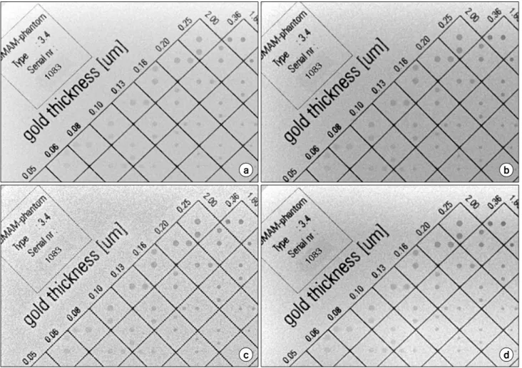
Fig. 2 shows an original and processed images processed by the proposed method of applying gamma correction, MUSICA and CLHAE to wavelet basis and sigmoid function to wavelet coefficients. From the result shown, on any of contrast en- hancement algorithm used on wavelet basis along with co- efficient stretching, can enhance the contrast of an original.
2. Comparative study
1) Image profiles: In order to compare the performance of the proposed method with other methods which are applied directly upon original images, the intensity profile of each processed image was acquired and analysed. Fig. 3 refers to comparative line profile on the gold plate of same position in CDMAM phantom with the direct application of CLAHE and the proposed method from the original image. It has been measured in order to the compare the local signal variation which represents contrast as well as noise of the signal.
Referring to the figure, the level of noise amplification by the contrast stretching is lower in the proposed method than the
2 2
b a
b
a S
CNR S
σ σ +
= −
Fig. 2. Comparison of images of original (a) and proposed method where gamma correction (b), MUSICA (c), CLAHE (d) were applied on the wavelet basis with sigmoid function applied on wavelet coefficients.
Fig. 3. Image Profile of CLHAE applied directly upon original image (a) and CLAHE applied on wavelet basis with sigmoid enhancement on wavelet coefficient (b).
direct application on the original image. As stated above, the amplification of noise component was suppressed by assigning the transition point from linear to sigmoid function when wavelet coefficient was enhanced. This result suggests that the proposed method can enhance the contrast as well as suppress the amplification of noise which takes place on direct applica- tion of the contrast enhancement algorithms.
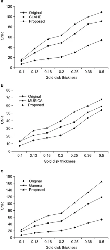
2) Comparative measure on contrast to noise ratio (CNR): CNR refers to the ability of an image to distinguish between contrasts of acquired image and the inherent noise in the image. It is defined
(9)
where, Sa and Sb are signal intensities for region a and b.
Fig. 4. Comparative measure of contrast to noise ratio between original, directly applied and proposed method of CLAHE (a), MUSICA (b), and Gamma Correction (c).
2 2
b
a σ
σ + refers the noise, which is usually measured from the region a and b, i.e, the measured standard deviation of the re- gion a and b. Fig. 4 shows the measured CNRs of CDMAM phantom images of various gold disk thicknesses where origi- nal, direct and proposed methods which were acquired as shown in Fig. 2 were compared. From the result shown in the figures, application of the conventional contrast enhancement methods on the wavelet basis and sigmoid enhancement func- tion on wavelet coefficients is better than the direct application on original image in terms of CNR.
3. Applications
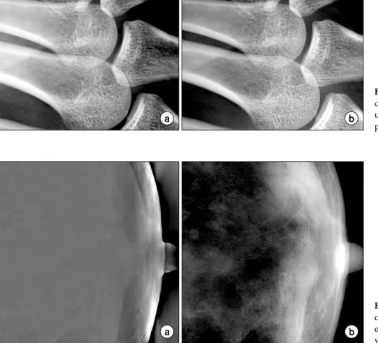
Fig. 5 shows the magnified view of finger joints. Compared with the direct application of CLAHE, sharpness is further en- hanced in the proposed method. The proposed method was al- so applied to mammographic images and result was compared with the previous contrast enhancement using wavelet trans- form (Fig. 6).8) Referring to the figure, the proposed method has shown better in terms of image quality than the previous method of contrast enhancement using wavelet transform.
DISCUSSION
Contrast as well as detail of an image could be enhanced by applying conventional contrast enhancement method and sig- moid magnitude stretching function on wavelet basis and co- efficients respectively. The method is better than the direct ap- plication of conventional method in terms of CNR as well as the interpretation of its detail.
Maximum decomposition level for applying proposed meth- od was two in the case of 1024×1024 sized 12 bit images.
Manipulation from further level would cause artefacts due to Gibbs phenomena. Applying the proposed method from one decomposition level was good enough to express the contrast enhancement as well as detail. Since the contrast enhancement algorithm is being applied on the low pass filtered residual (wavelet basis), the manipulation on wavelet basis without ap- plying magnitude stretching function (sigmoid function) on co- efficients would cause blurring on edges. The amount of stretching should be set adaptively according to the character- istic of the image. In the case of images where gray level in-
Fig. 6. Comparative view of result on mammographic image process- ed by conventional method using wavelet (a) and proposed method.
Fig. 5. Magnified finger joint detail of CLAHE applied directly upon original image (a) and proposed method (b).
tensities are generally uniform, assigning high weights on wavelet coefficients would cause amplification of noise component.
Through by comparing the line profile of CDMAM phantom and CNR obtained above, the proposed method could enhance the contrast as well as suppress the amplification of noise which takes place on direct application of contrast enhance- ment algorithms. In addition, proposed method has shown bet- ter in terms of image quality than previous method to enhance contrast by using wavelet transform.
Evaluation was done only upon on gray level images.
Flexibility of the proposed method to colour images remains to be determined. Also, the method is restricted to 2J×2J sized images due to the nature of wavelet transform definition.
CONCLUSION
The contrast enhancement as well as the detail enhancement
of an image could be simultaneously achieved by enhancing wavelet basis and wavelet coefficients at the same time. Since wavelet basis is low pass filtered components, applying the contrast enhancement algorithm on the wavelet caused a less amount of noise amplification than the direct application to an original image. Since wavelet coefficients contain high pass filtered components, applying the sigmoid function to stretch the magnitude of coefficients would result in detail enhance- ment. The method is flexible, and thus be applicable to vari- ous fields especially in radiology, where contrast as well as detail is of importance.
REFERENCES
1. Sakellaropoulos P, Costaridou L, Panayiotakis G: A wavelet-based spatially adaptive method for mammographic contrast enhancement. Physics in Medicine and Biology 48:
787-803 (2003)
2. Bronnikov AV, Duifhuis G: Wavelet-based image enhance-
웨이브렛과 기저 계수를 이용한 X-ray 영상의 대조도 향상기법
*가톨릭대학교 의과대학 의공학교실, †부산대학교 기계공학부
박천주*ㆍ김도일*ㆍ장도윤*ㆍ윤한빈†ㆍ최보영*ㆍ김호경†ㆍ이형구*
본 연구에서는 이산 웨이브렛 도메인에서 기저계수와 웨이브렛 계수의 변환을 이용하여 X-ray 이미지의 대조도를 향상 시키는 방법을 제안하였다. 기저 함수의 변환은 기존에 나와있는 contrast limited adaptive histogram equalization (CLAHE) 와 multi-scale image contrast amplification (MUSICA)와 같은 보편적인 영상의 대조도 향상 기법을 적용하였으며 대조도 가 향상된 저해상도 기저 계수의 역변환으로 인한 Blurring 현상을 방지하고 또한 이미지의 선명도를 향상시키기 위하여 웨이브렛 계수에 sigmoid function을 적용하였다. 본 알고리즘에 대한 성능 평가를 위하여 contrast detail mammography (CDMAM) 팬텀의 영상을 획득하여 기존에 사용한 대조도 향상 기법들과 contrast to noise ratio (CNR) 및 line profile에 대 한 비교평가를 실시 하였고 그 결과 기존의 대조도 향상기법을 웨이브렛 도메인에서 적용하는 것이 뛰어나다는 것을 알 수 있었다. 본 영상 대조도 향상기법은 영상의 대조도를 증가시키는 동시에 잡음의 증폭을 효율적으로 억제할 수 있다.
따라서 본 연구는 의료 영상뿐 만이 아닌 대조도와 선명도가 중요시되는 여려 분야에 적용될 수 있으리라 기대된다.
중심단어: X-ray 영상 향상 기법, 웨이브렛 변환, 대조도 향상 기법 ment in x-ray imaging and tomography. Applied Optics 37:
4437-4448 (1998)
3. Gonzalez RC, Woods RE: Digital Image Processing, Second Edition, Prentice Hall, 372-378
4. Stephen MP, Karel Z: Adaptive Histogram Equalization and Its Variations. Computer Vision Graphics and Image Processing 39:355-368 (1987)
5. Vuylsteke P, Schoeters E: Multiscale Image Contrast Ampli- fication (MUSICATM). SPIE Vol. 2167 Image Processing 551-560 (1994)
6. Laine A, Fan J, Yang W: Wavelets for contrast enhance- ment of digital mammography. Engineering in Medicine and Biology, IEEE Vol. 14:536-550 (1995)
7. Zhang C, Wang X, Wang J, Zhang H: Contrast Enhance- ment for Image Based on Discrete Stationary Wavelet Trans- form. SPIE, Vol. 6043, 60430Y, 1-8 (2005)
8. Dippel S, Stahl M, Wiemker R, Blaffert T: Multiscale Contrast Enhancement for Radiographies: Laplacian Pyramid Versus Fast Wavelet Transform. SPIE, Vol. 6043, 60430Y, 1-8 (2005)