272
Open Access
Carotid Intima-Media Thickness, Not Carotid Plaque, is Associated With Large Territory Cerebral Infarction in Patients With Ischemic Stroke
Hyun Ju Yoon, MD1, Myung Ho Jeong, MD1, Kye Hun Kim, MD1, Younggeun Ahn, MD1, Jeong Gwan Cho, MD1, Jong Chun Park, MD1, Jung Chaee Kang, MD1 and Jang Ho Bae, MD2
1The Heart Research Center of Chonnam National University Hospital, Gwangju,
2Department of Cardiology, Konyang University College of Medicine, Daejeon, Korea
ABSTRACT
Background and Objectives: Carotid intima-media thickness (IMT) has been associated with an increased risk of ischemic stroke. To better understand this association, we evaluated the relationships of vascular risk factors, in- cluding carotid IMT and carotid plaque, and large territory cerebral infarction and small vessel stroke. Subjects and Methods: A total of 502 patients with acute ischemic stroke were divided into two groups according to neu- rologic examinations and imaging studies; 1) a large territory infarction group (group I: n=126, 64.4±11 years, 78 males) and 2) a small vessel stroke group (group II: n=376, 62.5±11 years, 242 males). We evaluated associations between (a) territory and non-territory strokes and (b) age, sex, potential vascular risk factors, carotid image and cardiac function (by echocardiography). Results: We did not find significant between group differences of age, sex, diabetes, previous history of ischemic stroke, plaque (presence, site and size of carotid plaque), and velocity of ca- rotid blood flow and left ventricle ejection fraction. However, group I had a higher incidence of hypertension (p=0.006), smoking (p=0.003), and dyslipidemia (p=0.001). Group I had thicker carotid IMT than group II (right carotid: 0.81±0.21 mm vs. 0.76±0.19 mm, p=0.035; left carotid: 0.88±0.23 mm vs. 0.80±0.20 mm, p=0.014) and a higher e/e’ level (12.08 vs. 9.66, p<0.001). Dyslipidemia, thicker carotid IMT and elevated E/E’ ratios were significant independent predictors for large territory infarction in patients with ischemic stroke. Conclusion: Caro- tid IMT is significantly increased in patients with large territory infarction compared with those with small vessel stroke. (Korean Circ J 2010;40:272-276)
KEY WORDS: Cerebral infarction; Stroke; Carotid arteries.
Introduction
The carotid arteries are responsible for providing blo- od flow to the brain. If the carotid artery is blocked, or if a piece of carotid artery plaque dislodges and travels
to the brain, the individual can suffer a stroke. This can lead to paralysis, loss of ability to speak, blindness, loss of independence, and even death. Stroke is currently a leading cause of disability and the major leading cause of death in developing contries.1) A large portion of st- roke cases result from cerebral ischemia; the remainder is from hemorrhage. Large territory cerebral infarction tends to be the result of ischemic insult more than small vessel lacunar infarction.
In earlier studies, carotid atherosclerosis was confirm- ed by autopsy or by angiography.2-5) However, over the last decade, the development of noninvasive ultrasound techniques has made the visualization and measurement of the layers of the arterial wall and plaques possible in large population samples.6-9) It has been suggested that the intima-media thickness (IMT) of the common carotid artery (CCA) is one of the most sensitive markers for the
Received: August 23, 2009
Revision Received: November 23, 2009 Accepted: December 25, 2009
Correspondence: Myung Ho Jeong, MD,The Heart Research Center of Ch- onnam National University Hospital, 167 Jaebong-ro, Dong-gu, Gwang- ju 501-757, Korea
Tel: 82-62-220-6243, Fax: 82-62-228-7174 E-mail: [email protected]
This is an Open Access article distributed under the terms of the Creative Commons Attribution Non-Commercial License (http://creativecommons.
org/licenses/by-nc/3.0) which permits unrestricted non-commercial use, distribution, and reproduction in any medium, provided the original work is properly cited.
cc
earliest stages of atherosclerosis.10)
Increases in carotid IMT have been associated with increased risk of ischemic stroke.11) We did this study to evaluate the relationship of vascular risk factors, includ- ing carotid IMT and carotid plaque, with large territory cerebral infarction, one of the common subtypes of is- chemic stroke and small vessel stroke.
Subjects and Methods
Study population
A total of 502 patients with acute ischemic stroke were enrolled in our study between April 2007 to Septemter 2008.
Neurologists and radiologists diagnosed ischemic st- roke by neurologic examination and imaging study. If patients had any embolic source such as infective endo- carditis and intracardiac thrombi, they were excluded.
Patients with ischemic stroke were divided into two gr- oups according to infarct territory: Group I was defined by large territory infarctions (n=126, 64.4±11 years, 78 males). Group II was defined by small vessel strokes including lacunar infarction (n=376, 62.5±11 years, 242 males). We evaluated the association of territory and non-territory strokes with age, sex, potential vascular risk factors, carotid image and cardiac function (by echocar- diography).
Definition of hypertension, diabetes, dyslipidemia and territory infarction
Subjects were considered to have hypertension if their blood pressure was ≥140/≥90 mmHg as recommend- ed by the Joint National Committee (JNC) VII,12) or if they were on treatment for hypertension. The American Diabetes Association criteria13) were used to define dia- betes mellitus (DM). We considered a subject to have DM when the fasting plasma glucose levels were ≥126 mg/dL in 2 consecutive assessments or if they were on treatment for DM. Dyslipidemia was diagnosed according to the 2004 update of National Cholesterol Education Program guidelines.14)
According to these guidelines, we included in the st- udy patients with a level of low density lipoprotein-ch- olesterol ≥160 mg/d, a level of high density lipopro- tein-cholesterol ≤40 mg/dL and a level of triglycerides
≥150 mg/dL.15) Large territory cerebral infarction was defined if a major cerebral artery territory infarction was shown by a neurologic examination and an imag- ing study. This included anterior, middle and posterior cerebral infarction. Lacunar, cortical or subcortical in- farction were classified as small vessel stroke.
Laboratory tests
Blood sampling for serum lipid profiles and glucose were obtained after at least 14 hours of fasting.
Measurement of carotid intima-media thickness and carotid flow velocity
Carotid B-mode ultrasound was performed on both common carotid arteries and internal carotid arteries using a 10 MHz linear probe (VIVID 7, GE, USA). Im- ages were interpreted at the last centimeter of the CCA prior to the carotid bulb. We first described the presence or absence of plaques or of atheromas, which were de- fined as a focal widening relative to adjacent segments protruding into the lumen more than 1.5 mm, with or without calcifications. On a longitudinal two-dimensio- nal ultrasound image of the carotid artery, the anterior (near) and posterior (far) walls of the carotid artery ap- pear as two bright white lines separated by a hypoecho- genic space. End-diastolic images were frozen, and the far wall IMT was identified as the region between the lu- men-intima interface and the media-adventitia inter- face.16) Peak systolic and end diastolic carotid flow velo- city was measured by pulse wave Doppler on the CCA and the internal carotid artery (ICA).17)
Measurement of aortic intima-media thickness Transesophageal echocardiography was done using an Acuson 128 XP or Sequoia C256 ultrasonograph (Sie- mens, Mountain View, CA, USA) equipped with a 3.5- to 7.0-MHz multiplane probe. To ensure imaging of the entire thoracic aorta, the probe was rotated posteriorly and advanced to the distal esophagus and withdrawn slowly to scan the descending aorta and aortic arch. The probe was then rotated and advanced again to image the ascending aorta. The maximal value of the aortic IMT was collected for each patient.18) Transesophageal echo- cardiography was performed by experienced investigators who had no knowledge of the results of our other analyses.
Statistical analysis
Data are reported as mean±SD. In univariate ana- lysis, risk factors for different end-points were analyzed using the Chi-square test for discrete variables and St- udent’s t-test for continuous variables. Multiple logistic regression analysis was used to determine a model with independent predictive factors. A p<0.05 was consider- ed statistically significant. The software for statistical an- alysis was Statistical Package for the Social Sciences 13.0.
Results
Baseline characteristics
We did not find any significant between group differ- ences for age, sex, diabetes, or previous history of ische- mic stroke. However, group I patients with large territory cerebral infarct had a significantly higher percentage of hypertension (p=0.006), smoking (p=0.003), and dys- lipidemia (p=0.001) (Table 1). Also, group I had thick- er carotid IMT values than did group II (right CCA:
0.81±0.21 mm vs. 0.76±0.19 mm, p=0.035; left CCA:
0.88±0.23 mm vs. 0.80±0.20 mm, p=0.014) despite the absence of significant differences between the two groups for the presence of carotid plaque, the site of ca- rotid plaque, the size of carotid plaque and the velocity of carotid blood flow (Table 2 and 3). Regarding echo- cardiographic parameters, group I showed higher E/E’
ratios-which indicate diastolic dysfunction-than did gr- oup II, whereas there was no significant between group difference for left ventricle ejection fraction, which in- dicates systolic dysfunction (Table 4).
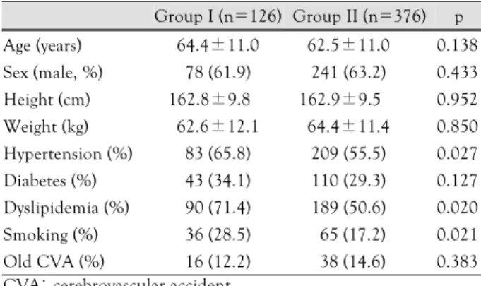
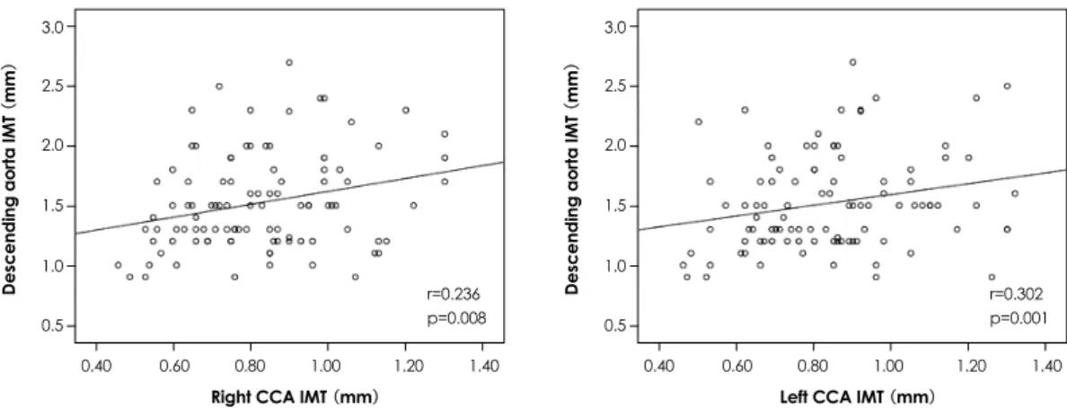
Correlation of carotid and aortic atherosclerosis We found a positive and significant correlations be- tween carotid IMT and descending thoracic aortic IMT (right CCA r=0.261, p=0.009; left CCA r=0.232, p=
0.021) (Fig. 1). But, there were no significant correl- ations between descending thoracic aorta IMT and in- farction territory. Descending thoracic aorta plaques were also not associated with infarct subgroup.
Independent predictors of territory infarction By multivariate regression analysis, dyslipidemia, th- icker carotid IMT and elevated E/E’ were significant in- dependent predictors of large territory infarction in pa- tients with ischemic stroke (Table 5).
Discussion
In this study, we compared CCA IMT and plaque for the risk assessment of respective stroke subtypes. Caro-
Table 1. Baseline clinical characteristics
Group I (n=126) Group II (n=376) p Age (years) 64.4±11.0 62.5±11.0 0.138 Sex (male, %) 78 (61.9) 241 (63.2) 0.433 Height (cm) 162.8±9.8 162.9±9.5 0.952 Weight (kg) 62.6±12.1 64.4±11.4 0.850 Hypertension (%) 83 (65.8) 209 (55.5) 0.027 Diabetes (%) 43 (34.1) 110 (29.3) 0.127 Dyslipidemia (%) 90 (71.4) 189 (50.6) 0.020 Smoking (%) 36 (28.5) 65 (17.2) 0.021 Old CVA (%) 16 (12.2) 38 (14.6) 0.383 CVA: cerebrovascular accident
Table 2. Comparison of IMT and plaque in patients with ischemic stroke
Group I (n=126)
Group II (n=376) p Right IMT (mm) 0.82±0.21 0.76±0.19 0.005
Plaque (%) 27 (36.9) 83 (34.1) 0.386 Plaque site (bulb, %) 24 (85.7) 59 (70.2) 0.082 Plaque size (mm) 2.77±0.88 2.37±0.71 0.319 Left IMT (mm) 0.88±0.23 0.79±0.20 0.001 Plaque (%) 25 (34.2) 78 (32.2) 0.434 Plaque site (bulb, %) 18 (81.8) 57 (76) 0.400 Plaque size (mm) 1.5±1.20 1.46±1.21 0.588 Descending aorta IMT 1.47±0.39 1.53±0.41 0.498 IMT: intima-media thickness
Table 3. Comparison of carotid blood flow velocity in the patients with ischemic stroke
Group I (n=126)
Group II (n=376) p Right CCA PS (m/sec) .045.3±20.0 .050.3±27.0 0.068
CCA ED 12.9±7.2 14.4±7.4 0.034
ICA PS 46.2±27.2 51.8±31.2 0.086
ICA ED 16.6±11.1 18.5±12.3 0.152
ICA/CCA 1.01±0.8 1.12±1.3 0.381 Left CCA PS (m/sec) 48.3±24.1 53.1±27.0 0.087
CCA ED 13.4±7.5 15.7±9.7 0.017
ICA PS 44.1±24.5 49.2±34.6 0.160
ICA ED 16.4±9.5 18.4±1.5 0.203
ICA/CCA 0.92±0.9 0.88±0.5 0.093 CCA: common carotid artery, ICA: internal carotid artery, PS: peak systolic velocity, ED: end diastolic velocity
Table 4. Comparison of echocardiographic parameters in patients with ischemic stroke
Group I (n=126)
Group II
(n=376) p
LVEDD (mm) 47.9±5.6 48.8±4.5 0.056
LVESD (mm) 31.3±4.9 31.6±4.6 0.637
IVS (mm) 9.45±1.4 9.46±1.5 0.940
LVPW (mm) 9.26±1.3 9.32±1.4 0.698
EF (%) 63.8±7.0 64.0±6.9 0.853
AOD (mm) 32.5±3.9 32.8±5.6 0.531
LAD (mm) 39.9±7.7 37.6±6.5 0.001
E (m/s) 0.63±0.3 0.58±2.4 0.080
A (m/s) 0.69±0.1 0.71±0.2 0.437
E/A 0.77±0.3 0.81±0.3 0.230
DT (msec) 238.9±156 204.0±68 0.001
E’ (m/s) 0.061±0.1 0.071±0.1 0.495
A’ (m/s) 0.092±0.1 0.099±0.1 0.980
S’ (m/s) 0.079±0.1 0.082±0.1 0.744
E/E’ 11.41±8.3 9.67±4.0 0.002 LVEDD: left ventricular end diastolic dimension, LVESD: left ven- tricular end systolic dimension, IVS: interventricular septal thick- ness, LVPW: left ventricular posterior wall thickness, EF: ejection fraction, AOD: aortic diameter, LAD: left atrial diameter, E: mitral inflow E velocity, A: mitral inflow A contraction velocity, DT: de- celeration time, E’: mitral septal annular velocity, A’: mitral septal an- nulus A’ velocity, S’: mitral septal annulus systolic velocity
Table 5. Independent predictors of large territory infarction in pa- tients with ischemic stroke
Hazard ratio Confidence interval p Hypertension 1.359 0.869-2.127 0.179 Dyslipidemia 2.066 1.312-3.254 0.002
Smoking 1.673 1.014-2.761 0.051
CCA IMT 4.710 01.754-12.645 0.002
E/E’ 1.039 1.004-1.074 0.027
CCA: common carotid artery, IMT: intima-media thickness, E/E’:
mitral inflow E velocity/mitral septal annular velocity
tid IMT was significantly increased in patients with large territory infarction compare with those with small ves- sel stroke. Various noninvasive imaging has been recom- mended by the American Heart Association for evalu- ation of risk in primary prevention.19) Carotid IMT is well known as a noninvasive surrogate marker of coro- nary atherosclerosis. IMT is increased in patients who are at risk for cardiovascular disease and in those pa- tients with atherosclerotic disease.20-23) O’Leary et al.24) reported that an increased carotid IMT is associated with a higher risk of stroke and acute myocardial infarc- tion in an elderly population and it is also a more po- werful predictor of cardiovascular disease than the con- ventional risk factors for atherosclerosis. Similarly, Tou- boul et al.25) demonstrated a greater CCA IMT in pa- tients with all major cerebral infarction subtypes com- pared with controls. In contrast, Nagai et al.26) reported a greater plaque score only for atherothrombotic and lacunar infarction patients compared to nonstroke pa- tients. Consequently, the implications of carotid athero- sclerosis remain to be established for each stroke subtype.
Certain known risk factors exist for the development of carotid artery atherosclerosis. These include smoking, diabetes, high blood pressure, high cholesterol, family history of cardiac or vascular disease and advanced age.
The present results indicated that hypertension, smok- ing and dyslipidemia are more common in patients with large territory cerebral infarct.
Some previous investigators reported that carotid pla- que was superior to carotid IMT for stratification of car- diovascular risk, and that carotid IMT was not a signi- ficant predictor for myocardial infarction and stroke.27)28) However, carotid IMT was different from carotid plaque as a predictor for developing stroke subtypes in this study.
Also, IMT of the descending thoracic aorta showed a strong correlation with coronary atherosclerosis and was a good predictor for stroke and peripheral arterial dise- ase in some reports.29) In our study, descending thor- acic aorta IMT was correlated with carotid IMT as in the previous study, whereas they did not find a significant
correlation with infarction subtype. Therefore, we th- ought that the atherosclerosis of the descending thoracic aorta was related to ischemic stroke, but this variable did not differentiate infarct subtype. Based on our findings, carotid IMT evaluation was more sensitive than other surrogate markers such as carotid plaque or descend- ing aorta IMT for differentiating stroke subtypes in pa- tients with ischemic stroke.
Additionally, diastolic dysfunction was more common in patients with large territory cerebral infarcts. Many investigators insist that diastolic dysfunction is not a disease but a step of the aging process resulting from de- creasing left ventricle compliance.30)31) Left ventricular systolic function tends to be preserved compared with diastolic function until old age. Although age differences were not significant between groups, the large territory infarction group showed higher E/E’ than did the small vessel infarction group. This finding suggests a large in- farction effect on diastolic function rather than on systo- lic function in patients with ischemic stroke.
The current study has several limitations, both in the diagnosis of stroke subtypes and in the transferability of conclusions to the general population. Because this study had a cross-sectional designed, whether it can be appli- ed to all individuals with stroke is not clear. Large pro- spective studies are still necessary to establish a link be- tween these measures and future risk of stroke subtypes.
Acknowledgments
This study was supported by a grant from the Korea Healthcare technology R&D project (A084869), Ministry for Health, Welfare
& Family Affairs, Republic of Korea, and the Cardiovascular Re- search Foundation, Asia.
REFERENCES
1) Wolf PA, D’Agostino RB. Epidemiology of stroke. In: Barnett HJ, Mohr JP, Stein BM, Yatsu FM editors. Stroke. 3rd ed. New York:
Churchill Livingstone;1998. p.3-28.
2) Young W, Gofman JW, Tandy R, Malamud N, Waters ES. The qu- antitation of atherosclerosis: III. the extent of correlation of de- grees of atherosclerosis within and between the coronary and ce- rebral vascular beds. Am J Cardiol 1960;6:300-8.
3.0 2.5 2.0 1.5 1.0 0.5
Descending aorta IMT (mm)
0.40 0.60 0.80 1.00 1.20 1.40 Right CCA IMT (mm)
Fig. 1. Correlation of descending thoracic aortic intima-media thickness (IMT) and common carotid artery (CCA) IMT.
r=0.236 p=0.008
3.0 2.5 2.0 1.5 1.0 0.5
Descending aorta IMT (mm)
0.40 0.60 0.80 1.00 1.20 1.40 Left CCA IMT (mm)
r=0.302 p=0.001
3) Mitchell JR, Schwartz CJ. Relationship between arterial disease in different sites: a study of the aorta and coronary, carotid and iliac arteries. Br Med J 1962;1:1293-301.
4) Solberg LA, Eggen DA. Localization and sequence of develop- ment of atherosclerotic lesions in the carotid and vertebral ar- teries. Circulation 1971;43:711-24.
5) Hertzer NR, Young JR, Bevan EG, et al. Coronary angiography in 506 patients with extracranial cerebrovascular disease. Arch Intern Med 1985;145:849-52.
6) Handa N, Masayasu M, Hiroaki M, et al. Ultrasound evaluation of early carotid atherosclerosis. Stroke 1990;21:1567-72.
7) Geroulakos G, O’Gorman DJ, Kalodiki E, Sheridan DJ, Nicol- aides AN. The carotid intima-media thickness as a marker of the presence of severe asymptomatic coronary artery disease. Eur Heart J 1994;15:781-5.
8) Gnasso A, Irace C, Mattioli PL, Pujia A. Carotid intima-media thickness and coronary heart disease risk factors. Atherosclero- sis 1996;119:7-15.
9) Lorenz MW, Kegler S, Steinmetz H, Markus HS, Sitzer M. Ca- rotid intima-media thickening indicates a higher vascular risk ac- ross a wide age range: prospective data from the carotid athero- sclerosis progression study (CAPS). Stroke 2006;37:87-92.
10) Howard G, Sharrett AR, Heiss G, et al. Carotid artery intima- media thickness distribution in general populations as evaluated by B-mode ultrasound. Stroke 1993;24:1297-304.
11) Salonen R, Seppanen K, Rauramaa R, Salonen JT. Prevalence of carotid atherosclerosis and serum cholesterol levels in eastern Finland. Arteriosclerosis 1988;8:788-92.
12) Chobanian AV, Bakris GL, Black HR, et al. The Seventh Report of the Joint National Committee on Prevention, Detection, Ev- aluation, and Treatment of High Blood Pressure: the JNC 7 Re- port. JAMA 2003;289:2560-72.
13) Expert Committee on the Diagnosis and the Classification of Diabetes Mellitus. Report of the expert committee on the diagnosis and the classification of diabetes mellitus. Diabetes Care 1997;20:
1183-97.
14) Grundy SM, Cleeman JI, Merz CN, et al. Implications of recent clinical trials for the National Cholesterol Education Program Ad- ult Treatment Panel III guidelines. Circulation 2004;110:227-39.
15) Hatzitoliosa AI, Athyrosb VG, Karagiannisb A, et al. Implemen- tation of strategy for the management of overt dyslipidemia: the IMPROVE-dyslipidemia study. Int J Cardiol 2009;134:322-9.
16) Park KR, Kim KY, Yoon SM, Bae JH, Seong IW. Correlation between intima-media thickness in carotid artery and the extent of coronary atherosclerosis. Korean Circ J 2003;33:401-8.
17) Withers CE, Gosink BB, Keightley AM, et al. Duplex carotid so- nography: peak systolic velocity in quantifying internal carotid artery stenosis. J Ultrasound Med 1990;9:345-9.
18) Bae JH, Bassenge E, Park KR, Kim KY, Schwemmer M. Signi- ficance of the intima-media thickness of the thoracic aorta in pa- tients with coronary atherosclerosis. Clin Cardiol 2003;26: 574-8.
19) Smith SC Jr, Greenland P, Grundy SM. Prevention conference: V.
beyond secondary prevention: identifying the high-risk patient for primary prevention. Circulation 2000;101:111-6.
20) Grobbee DE, Bots ML. Carotid artery intima-media thickness as an indicator of generalized atherosclerosis. J Intern Med 1994;
236:567-73.
21) Pignoli P, Tremoli E, Poli A, Oreste P, Paoletti R. Intimal plus medial thickness of the arterial wall: a direct measurement with ultrasound imaging. Circulation 1986;74:1399-406.
22) Hulthe J, Wikstrand J, Emanuelsson H, Wiklund O, de Feyter PJ, Wendelhag I. Atherosclerotic changes in the carotid artery bulb as measured by B-mode ultrasound are associated with the ex- tent of coronary atherosclerosis. Stroke 1997;28:1189-94.
23) Bae JH, Seung KB, Jung HO, et al. Analysis of Korean carotid intima-media thickness in Korean healthy subjects and patients with risk factors: Korea multi-center epidemiological study. Kor- ean Circ J 2005;35:513-24.
24) O’Leary DH, Polak JF, Kronmal RA, et al. Thickening of the ca- rotid wall: a marker for atherosclerosis in the elderly? Stroke 1996;27:224-31.
25) Touboul PJ, Elbaz A, Koller C, et al. Common carotid artery in- tima-media thickness and brain infarction. Circulation 2000;102:
313-8.
26) Nagai Y, Kitagawa K, Sakaguchi M, et al. Significance of earlier carotid atherosclerosis for stroke subtypes. Stroke 2001;32:1780-5.
27) Ebrahim S, Papacosta O, Whincup P, et al. Carotid plaque, intima- media thickness, cardiovascular risk factors, and prevalent cardio- vascular disease in men and women: the British Regional Heart Study. Stroke 1999;30:841-50.
28) Bots ML, Hoes AW, Koudstaal PJ, Hofman A, Gobbee DE. Com- mon carotid intima-media thickness and risk of stroke and myo- cardial infarction: the Rotterdam Study. Circulation 1997;96:
1432-7.
29) Yoon HJ, Hyun DW, Kwon TG, Kim KH, Bae JH. Prognostic significance of descending thoracic aorta intima-media thickness in patients with coronary atherosclerosis. Korean Circ J 2007;37:
365-72.
30) Masugata H, Senda S, Yoshikawa K, et al. Relationships between echocardiographic findings, pulse wave velocity, and carotid ath- erosclerosis in type 2 diabetic patients. Hypertens Res 2005;28:
965-71.
31) Salmasi A, Alimo A, Jepson E, Dancy M. Age-associated ch- anges in left ventricular diastolic function are related to increas- ing left ventricular mass. Am J Hypertens 2003;16:473-7.