865
The Pathophysiology and Diagnostic Approaches for
Diastolic Left Ventricular Dysfunction: A Clinical Perspective
Jong-Won Ha, MD, PhD1 and Jae K. Oh, MD2
1Cardiology Division, Yonsei University College of Medicine, Seoul, Korea
2Division of Cardiovascular Diseases, Mayo Clinic, Rochester, MN
ABSTRACT
Heart failure with a normal ejection fraction is interchangeably termed diastolic heart failure. This condition is often unrecognized and it does have diagnostic, prognostic and therapeutic implications that are distinct from those con- ditions with systolic dysfunction. It is clinically important to understand and assess the diastolic function to reliably manage the patients suffering with heart failure. With the results of randomized trials for this distinct clinical syn- drome, as well as the probability of better diagnostic testing in the future, physicians will in a better position not only to diagnose diastolic dysfunction or heart failure, but also to manage it more effectively. In this review, the physiology of the diastole and how to evaluate the abnormalities of the diastolic function will be discussed. (Korean Circulation J 2005;35:865-876)
KEY WORDS:Pathophysiology;Diastolic dysfunction;Heart failure.
Epidemiology
Heart failure is the most common hospital diagnosis for patients 65 or older at the time of discharge, and it is a major cardiovascular disorder that’s still increasing in prevalence. In the early 20th century, 4% of the po- pulation reached 65 years of age; however, by the year 2010, it is predicted that 35% of the population will be older than 65 years of age.1) Cardiovascular disease is the most common cause of morbidity and mortality for the elderly. Investigators have traditionally focused on sys- tolic functional abnormalities to explain the signs and symptoms of heart failure. However, it has become in- creasingly evident that diastolic functional abnormalities also play a major role for precipitating heart failure and determining the prognosis.2-5)
Four epidemiological investigations have demonstra- ted that nearly half of the congestive heart failure sub- jects in the community have normal systolic function.6-9) Some researchers have argued that apparent “diastolic heart failure” may actually be undetected transient sys- tolic dysfunction that is caused by an acute afterload mismatch that’s induced by hypertension or this is cau- sed by ischemia. In a recent study of 38 consecutive
patients who had acute pulmonary edema and marked systolic hypertension, their heart failure was due to exacerbation of the diastolic dysfunction via hyperten- sion, and not by transient systolic dysfunction or mitral regurgitation.10) However, this condition, which commonly referred to as diastolic dysfunction, is often unrecognized and it does have diagnostic, prognostic and therapeutic implications that are distinct from those patients with systolic dysfunction. Thus, it is clinically important to understand and assess the diastolic func- tion to reliably manage those patients with heart fai- lure. In this review, we will discuss the physiology of the diastole and how to evaluate abnormalities of diastolic function.
Physiology of Diastole
Abnormalities of ventricular relaxation and filling contribute to heart failure because these features result in an abnormally high ventricular diastolic pressure in relation to the diastolic volume. The most important factors that determine the relation between pressure and volume during ventricular diastole are the rate and extent of myocardial relaxation and the generation of restoring forces. Both are important determinants of early diastolic filling, and the passive pressure- volume relationship of the ventricle determines the mid-late diastolic filling after the relaxation is comple- ted.11-14)
Correspondence:Jong-Won Ha, MD, PhD, Cardiology Division Yonsei Cardiovascular Hospital Yonsei University College of Medicine, 134 Shin- chon-dong, Seodaemun-gu, Seoul 120-752, Korea
Tel: 82-2-2228-8460, Fax: 82-2-393-2041 E-mail: [email protected]
Myocardial relaxation
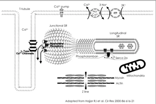
The normal cardiac contraction/relaxation cycle re- quires a precise, transient rise and fall in the level of intracellular calcium ions. The sarcoplasmic reticulum (SR) has a central role in orchestrating the movement of calcium during each contraction and relaxation.15)16) Contraction of the cardiac muscle is initiated by the cellular action potential that causes opening of the L- type sarcolemmal calcium channels through which the calcium ions enter the cytosol. Influx of calcium ions through the L-type channels results in the release of large amounts of calcium ions from the adjacent SR through the ryanodine receptor channels, and this process is ter- med calcium-induced calcium release. These calcium ions bind to troponin C that ultimately disinhibits the interaction of actin and myosin with the resultant cross- bridge formation(Fig. 1).11)12) Myocardial relaxation is primarily accomplished by the removal of calcium ions from troponin C by an enzyme located in SR, which is called SR calcium ATPase or SERCA2, and the sarcolem- mal sodium-calcium exchanger. In humans, approxima- tely 75% of calcium ions are removed by SERCA2 and 25% are removed by the Na/Ca exchanger.17) The acti- vity of SERCA2 is modulated by phospholamban, ano- ther SR protein located near SERCA2. The calcium ion uptake by SERCA2 is enhanced via phosphorylation by protein kinase A and other kinases.18) Failure of the normal mechanisms for the reuptake and extrusion of calcium ions that are released during contraction can result in slowing of relaxation or an inability to return the cytosolic calcium concentration to normal diastolic levels. The latter causes a diastolic calcium overload and incomplete relaxation with excessive diastolic tension or stiffening.19-21) In an experimental model of senescence,
it has been demonstrated that there is a decrease of the SR Ca2+ uptake during relaxation, and this is associated with a decrease in the content and activity of SERCA2.22) More recently, SERCA2 protein levels were found to be significantly decreased in senescent human myocar- dium.23) This decrease in SERCA2 levels was associated with impaired myocardial function at the study’s baseline and further deterioration occurred during hypoxic con- ditions.23) Thus, a decrease in the SERCA2 content and the associated decrease in the SR Ca2+ uptake have been suggested to play a major role in the diastolic dysfunc- tion. The vulnerability of calcium reuptake is a contri- buting factor for the abnormal left ventricular relaxation that’s been noted early in cardiac disease states, and this is despite the normal systolic function.
Myocardial relaxation is also modulated by factors at the extracellular and organ levels. Endocardial and vas- cular endothelial modulators have been proposed as being important factors in the physiology of myocardial relaxation.11)24) The vascular endothelium also appears to play an important role in these responses. In an experiment with papillary muscle preparation, Brutsaert et al. have found that denudation of the endocardium resulted in earlier and rapid relaxation in conjunction with a very modest reduction of the developed force.11) Although its effects are less clear in normal hearts, an- giotensin II has been shown to slow myocardial relaxa- tion in hypertrophied hearts.24) Other locally released substances that may influence myocardial relaxation in- clude endothelin-1, natriuretic peptides, prostaglandins and adenyl purines. The physiologic and pathophysio- logic significance of these modulators of relaxation remains to be elucidated.
It’s interesting that the relevance of caffeine for fa-
Adapted from Hajjar RJ et al. Cir Res 2000;86:616-21 Na+
Ca2+
Ca2+
T-tubule Ca2+ pump Ca2+ 2K+
Junctional SR
Longitudinal SR
Phospholamban Serca 2a
Mitochondria Myosin
Actin Z line
3 Na+
Fig. 1. The Ca2+ handling proteins involved in Ca2+ movement (adapted from Hajjar RJ et al. Cir Res 2000;86:616-21). SR: sarcoplasmic reticulum.
cilitating diastolic dysfunction in the intact heart was demonstrated in the experimental canine model of de- mand ischemia.25) It was also suggested that caffeine favorably affected myocardial relaxation without altering contractility.26)
Restoring forces
Restoring forces are generated when the ventricle con- tracts to an end-systolic volume(ESV) that is less than its equilibrium volume(Vo), which is the volume in the fully relaxed state when the transmural pressure is zero, thereby storing energy by compressing the elastic ele- ments in the myocardium.13)24) The physical properties of the normal ventricular chamber allow potential energy to be stored during systole in the form of a lower ven- tricular pressure. This potential energy is converted to kinetic energy in the form of elastic recoil with the resultant suction of blood from the left atrium into the ventricle early during diastole.27) Other factors, especially complex shape changes during systole, may also play a role in causing suction during the subsequent diastole.28) During systole the left ventricle demonstrates a counter clockwise twist extending from the apex to the base, and in early diastole the left ventricle untwists clockwise to release the stored energy and this creates a suction force not unlike the release of a compressed spring. Suction is most important under conditions of stress when con- tractility is high and the ESV is small, and when filling must be enhanced such as during exercise. Conversely, when contractility is impaired, the ESV is larger and the suction is reduced or lost as a mechanism of filling.29) The passive pressure-volume relation of the ventricle
Laplace’s law provides for the relationship between pressure and the chamber geometry when a stress is
applied to the ventricle. In the simplest case of a sphere, Laplace’s law states that pressure is proportional to the radius of the chamber divided by its wall thickness. Thus, a chamber with thicker walls requires a larger distending pressure to achieve a given volume. The intrinsic stif- fness of the tissue in the ventricular wall is the other key determinant of the distending force that’s required to passively fill the chamber. The elastic properties are most responsible for wall stiffness at low filling pres- sures.30) At higher distending pressures, the myocardial connective tissue matrix assumes a key role. The intrin- sic passive stiffness of any tissue, including the myocar- dium, can be quantified as the change in stress required for producing a given change in stretch(or strain). Bio- logic tissues have a curvilinear passive stress-strain re- lationship. The amount of blood contained within the vessels in the heart wall is also a determinant of the stiffness of the myocardial tissue, which contributes to the pressure-volume relationship(PVR).31) As a result, a greater distending pressure is required to fill the cham- ber when the myocardial blood volume increases. The PVR of the passive ventricle is curvilinear, paralleling the passive stress-strain relationship. At any point on this curve, the ratio of the change in pressure to the change in volume is represented as the operating chamber stiff- ness; its inverse is the operating compliance. Like the stress-strain relationship, the PVR can be converted into a linear relationship by plotting the operating chamber stiffness versus pressure(Fig. 2). The slope of this rela- tion is the chamber-stiffness constant. The parietal pe- ricardium and right ventricle form external constraints to filling, and this influences the PVR as well. The pa- rietal pericardium has a very compliant PVR at low vo- lumes, but then it makes a sharp transition to a very steep relationship. Increases in the right ventricular dia-
Adapted from Zile MR et al. Prog Cardiovasc Dis. 2005;47:307-13 Left ventricular volume
Left ventricular pressure
Systolic
heart failure Normal Diastolic
heart failure
Left ventricular volume
Left ventricular pressure
Systolic
heart failure Normal Diastolic
heart failure
Fig. 2. The left ventricular end-diastolic pressure-volume relationship describes the diastolic properties of the left ventricle. The slope of the relation- ship indicates the passive chamber stiffness. Shifting the pressure-volume curve leftward indicates a stiffer ventricle, whereas a rightward shift indica- tes greater compliance (adapted from Zile MR et al. Prog Cardiovasc Dis. 2005;47:307-13).
stolic volume and pressure influence the left ventricular PVR similarly, albeit more modestly because the right ventricular changes are transmitted to the left ventricle via the ventricular septum. This phenomenon is termed diastolic-ventricular interaction and it is based on the fact that the total cardiac volume within the pericardial sac remains constant; hence, overfilling of the right ventricle will result in reduced filling of the left ventri- cle and vice versa. This is manifested most dramatically in constrictive pericarditis. The ventricular and pericar- dial effects on the left ventricular PVR are often linked.
Diagnostic Evaluation by Echocardiography
Doppler echocardiography has been successfully app- lied in the past decade to assess diastolic filling, and it has become a reliable, reproducible and practical no- ninvasive method for the identification and longitudinal follow-up of those patients with diastolic dysfunction.32) Mitral valve inflow
Assessment of transmitral blood flow velocities has served as the backbone for evaluating the diastolic func- tion via Doppler echocardiography since its first des- cription by Kitabatake in 1982.33) The velocity curve is influenced by several parameters, including the preload, afterload, contractile state, heart rate, myocardial rela- xation and the left ventricular compliance. Theoretical models, computer simulation and experimental animal models have shown that the left atrial pressure, the rate of isovolumic ventricular relaxation(τ), the end-systo- lic volume and the left ventricular minimal pressure are
the major determinants of transmitral Doppler fil- ling.34-38) Impairment of left ventricular relaxation, which is the earliest manifestation of diastolic dysfunction, re- sults in a prolongation of the isovolumic relaxation time and a reduction in the early transmitral flow velocity(E) with prolongation of the E-wave deceleration time and an augmented A velocity. In contrast, increasing filling pressures result in shortening of the isovolumic relaxa- tion time, an increased early transmitral gradient and consequently, a high early transmitral flow velocity, shor- tening of the deceleration time and a reduction in the atrial flow velocity. Since the mitral flow Doppler profile depends on both of the above parameters, progressive elevation of the left atrial pressure in the ventricles with reduced isovolumic relaxation will reverse the classic pat- tern of impaired myocardial relaxation towards a “nor- mal” appearing profile; i.e., the so called pseudonormal pattern.
Transmitral flow velocity curves show a progression over time, and this demonstrates the natural progression of the underlying myocardial dysfunction. In a normal middle-aged subject, the mitral flow velocity curve con- sists of an E/A ratio slightly >1.0 and a deceleration time of -200 ms. In the early stages of diastolic dysfunc- tion, which is referred to as mild diastolic dysfunction, impaired(delayed) relaxation of the left ventricle pre- dominates and this results in the most typical mitral flow velocity profile. At this stage, there is little if any increase in the rest left ventricular diastolic pressure or the mean left atrial pressure. Increased filling pressure may develop with exercise because of shortening of the diastolic filling period. With progression of disease, the filling pressure in the particular left atrial pressure starts
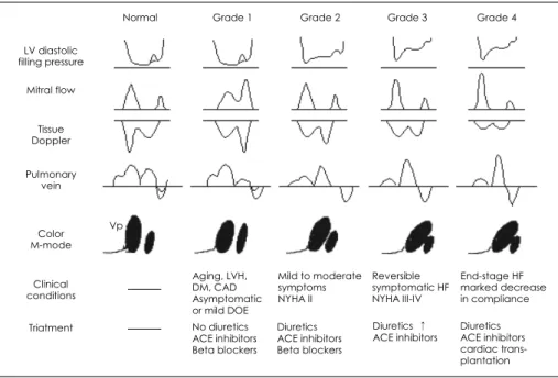
Normal Grade 1 Grade 2 Grade 3 Grade 4
LV diastolic filling pressure
Mitral flow
Tissue Doppler
Pulmonary vein
Color M-mode
Clinical conditions
Triatment
Aging, LVH, DM, CAD Asymptomatic or mild DOE No diuretics ACE inhibitors Beta blockers
Mild to moderate symptoms NYHA II Diuretics ACE inhibitors Beta blockers
Reversible symptomatic HF NYHA III-IV Diuretics ↑ ACE inhibitors
End-stage HF marked decrease in compliance Diuretics ACE inhibitors cardiac trans- plantation Vp
Fig. 3. Diagram of a proposed grading system for diastolic dysfunction, as based on the progression of disease patterns in the patients with cardiac disease. LV: left ventricle, LVH: left ventricular hypertrophy, DM: diabetes mellitus, CAD: coronary atery disease, DOE: dyspnea on exertion, ACE: angiotensin converting enzyme, NYHA: New York Heart Association, HF: heart failure, Vp: early diastolic propagation velocity.
to increase at rest; thus, this increases the driving pres- sure across the mitral valve. This has the effect of nor- malizing the E velocity and the DT of the E velocity.
This phase represents moderate diastolic dysfunction.
In more advanced disease, as the effective operative compliance decreases, the left atrial pressure becomes even higher and this produces a restrictive filling pattern that’s characterized by a tall E velocity, a shortened DT and a small A velocity; this all indicative of severe dias- tolic dysfunction. In patients with left ventricular sys- tolic dysfunction, a restricted filling pattern has been associated with a worse functional class and exercise in- tolerance.39) A short deceleration time(<140 ms) is indi- cative of a poor prognosis that’s independent of the degree of systolic dysfunction.40-42) On the basis of this progression of disease patterns, a grading system has been proposed for the severity of diastolic dysfunction as assessed with Doppler echocardiography (Fig. 3).43) Using a scale of I to IV, grade I identifies a patient with an abnormal relaxation pattern and minimal or no symp- toms of heart failure at rest. The patients with grade I diastolic dysfunction may develop dyspnea with moderate or greater exertion or they may develop symptoms of heart failure if the contribution from atrial contraction is lost, as occurs with the development of atrial fibrilla- tion. With grade II diastolic dysfunction, there is a pseu- donormalization pattern on the mitral flow velocity curves and increased filling pressures at rest; this pro- duces symptoms with mild to moderate exertion. Patients with grade III diastolic dysfunction have a restrictive filling pattern on the mitral flow velocity curves, a severe increase in the filling pressures and they expe- rience symptoms with minimal exertion or at rest. The treatment of heart failure or such bedside maneuvers that result in a decrease preload(an upright sitting pos- ture or a Valsalva maneuver) may produce changes in the mitral flow velocity curve so that for a patient with grade III diastolic dysfunction, the velocity profile may revert to grade II or even grade I diastolic dysfunction, which is indicative of a good prognosis. On the other hand, some patients with severe abnormalities of ven- tricular compliance and end-stage heart disease maintain a severely restrictive pattern even after optimal medical therapy. These patients have the poorest prognosis and they are classified as having grade IV(irreversible) dia- stolic dysfunction.44)
Because of the opposing effects on these variables according to the relaxation and filling pressures, it is difficult to evaluate the relaxation properties of the left ventricle when the left atrial pressure is unknown. Mo- reover, the relationships between the deceleration time or E/A ratio and the filling pressures are not as strong in those patients with normal systolic function.45)46) Therefore, it is desirable to have additional variables to complement the mitral inflow velocity during the eva-
luation of diastolic function.
Additional echocardiographic parameters can assess ventricular relaxation and the filling pressure since these are the two most prominent components of diastolic dysfunction. Impaired ventricular relaxation indicates the presence of diastolic dysfunction, and the level of fil- ling pressure elevation indicates the extent of the dias- tolic dysfunction. Ventricular relaxation is assessed via the mitral annular velocity recording by performing tissue Doppler echocardiography(TDE), and the mitral inflow propagation velocity is assessed by color M-mode and less directly by the preload reduction. The left ventri- cular filling pressure is estimated by the mitral inflow deceleration time(especially when there is left ventri- cular systolic dysfunction), pulmonary vein flow velocity analysis, comparison of the flow duration(the mitral A wave vs the pulmonary venous atrial flow reversal), and the ratio of the transmitral flow velocity to the mitral annular velocity, which will be discussed later. Demons- ration of impaired relaxation and/or increased filling pressures by any of the above modalities signifies the presence and severity of diastolic dysfunction. In addi- tion, the two-dimensional echocardiographic features such as increased left ventricular wall thickness, left atrial enlargement and ventricular interaction reflect the pre- sence of diastolic dysfunction.
Tissue Doppler echocardiography
TDE is a recent Doppler application that allows di- rect measurement of myocardial velocities. TDE uses low-velocity and high-amplitude Doppler signals, in con- trast to the mitral inflow, which uses high-velocity and low-amplitude signals.47) TDE is the easiest and most re- producible method to evaluate myocardial relaxation by measuring the mitral annulus velocity during diastole.
The mitral annulus velocity reflects shortening and leng- thening of the left ventricular myocardial fibers along a longitudinal plane(from the base to the apex plane).
With prolonged myocardial relaxation, the ratio of the mitral annulus motion during atrial systole to the total diastolic annular motion is increased. It has been shown that the early diastolic mitral annulus velocity(E’), as determined by TDE, is relatively independent of the pre- load, especially in those patients showing reduced myo- cardial relaxation, and it is useful in differentiating a pseudonormal from a normal mitral inflow velocity pat- tern.48)49) As the left ventricular filling pressure increases, the mitral E velocity becomes progressively higher whe- reas the E’ velocity remains reduced. Therefore, the assessment of mitral annular velocity by employing TDE may be useful for the evaluation of diastolic dysfunc- tion, and TDE has overcome some of the inherent dif- ficulties in assessing diastolic function with utilizing transmitral inflow velocities alone. Nagueh et al.49) have recently shown that when the mitral E velocity was cor-
rected for the influence of myocardial relaxation(i.e. the E/E’ ratio), it was found to be well correlated with the mean pulmonary capillary wedge pressure(PCWP). In their study, an E/E’(lateral annulus) ratio of >10 detected a mean PCWP >15 mm Hg with a sensitivity of 97% and specificity of 78%. Ommen et al. have as- sessed the association between E’(septal annulus) and the left ventricular filling pressures in 100 consecutive patients who were referred for cardiac catheterization, and they found that an E/E’ ratio >15 identified an increased left ventricular filling pressure(Fig. 4).46) Na- gueh et al.50) have also showed that E’ remained unchan- ged with an increased transmitral gradient in those sub- jects with diastolic dysfunction, whereas it is increased in the subjects with a normal tau.
A very exciting observation was recently published showing that myocardial contraction and relaxation ve- locities, as assessed by TDE, were reduced in the patients with familial hypertrophic cardiomyopathy mutations (the positive genotype), but they were without the left ventricular hypertrophy phenotype.51) Early detection of mutation-positive subjects may allow therapeutic inter- ventions to prevent the later development of left ventri- cular hypertrophy, which is a major determinant of mortality and morbidity in these patients. Although much work is still required to link the genetic defects in the cardiac sarcomere with a clinical, noninvasive measure of global left ventricular function, this preli- minary observation has introduced a possible future application of this technique that will contribute to the better understanding and management of the patients suffering with hypertrophic cardiomyopathy or prema- ture diastolic dysfunction.
It was recently shown that E’, as measured by TDE, is reduced in the patients suffering with restrictive car- diomyopathy(RCM), whereas it is relatively normal or even accentuated in the patients suffering with constric- tive pericarditis(CP).52-54) Recording of E’ by using TDE is another useful means to diagnose CP when the mitral
inflow velocity reveals a restrictive filling pattern with- out sufficient respiratory variation. When the mitral inflow velocity shows a high E, a shortened deceleration time and no respiratory variation, then the diagnostic possibilities are normal(in healthy young individuals), restriction or constriction; however, E’, as measured by TDE and the hepatic vein flow velocity, in addition to the 2-dimensional echocardiographic findings, should be able to distinguish them.55) Therefore, the recording of E’ via TDE should be an essential part of the echo- cardiographic Doppler evaluation for all the patients suffering with heart failure, especially when CP is sus- pected.
Color flow propagation
The propagation of flow into the left ventricular cavity during early diastole by the color M-mode has been shown to correlate with the invasively measured time constant of relaxation, τ.56)57) A chamber with normal relaxation will demonstrate rapid flow propagation from the inflow to the left ventricular apex, while a slowly relaxing ventricle will demonstrate prolonged flow pro- pagation. It has also been shown that the propagation velocity is relatively load independent.58) However, there is not yet a consistent method for measuring propaga- tion velocity, and the various reported techniques are not interchangeable. In addition, the influence and chal- lenge of a rapid heart rate along with the fusion of the early and late diastolic flow is not yet fully understood.
The flow propagation velocity may erroneously appear to be normal in the patients suffering with diastolic dys- function and a preserved left ventricular systolic func- tion. This is particularly so when the left ventricular size is small and the heart rate is fast. Therefore, in small ventricles, this index may not accurately reflect the relaxation properties of the left ventricle.
Pulmonary venous flow velocity
Pulmonary vein flow patterns have been used to com- plement the mitral inflow velocity profiles. A normal pulmonary venous flow velocity curve consists of sys- tolic forward flow, diastolic forward flow and a reversal flow during atrial contraction. The systolic forward flow is influenced by the left atrial relaxation, the left atrial compliance, the mean left atrial pressure, the descent of the annulus toward the left ventricular apex, the right ventricular contraction and the presence of significant mitral regurgitation. The diastolic forward flow occurs at the time when there is an open conduit between the pulmonary vein, the left atrium and the left ventricle.
At the time of atrial contraction, there continues to be an open conduit between the pulmonary vein, the left atrium and the left ventricle. There is forward flow into the left ventricle, with blood also traveling in a retro- grade fashion into the pulmonary veins. When the left
0 5 10 15 20 25 30 35 40
MLVDP
E/E’<8 E/E’ 8-15 E/E’>15 Fig. 4. The mean LV diastolic pressure versus groups defined by values of the septal E/E’. An E/Ea ratio of 15 is highly specific for an eleva- ted LA pressure, whereas a ratio of 8 is very specific for normal to low filling pressures. LA: left atrium, MLVDP: mean left ventricular dias- tolic pressure.46)
atrial pressure is normal, most flow into the left atrium occurs during systole. However, when the left atrial pressure increases, the systolic forward flow decreases and flow occurs predominantly during diastole, resul- ting in a reversal of the systolic(S)/diastolic(D) ratio with blunting of the pulmonary venous S wave and an increase in the amplitude and duration of the atrial re- versal(AR) wave. This pattern has been used to distin- guish normal from “pseudonormal” transmitral Doppler filling. However, in those healthy young adults and ath- letes for whom the atrial contribution to the left ven- tricular filling is minimal and the left atrium behaves more as a “passive” conduit, blunting of the S wave is also commonly seen. The AR wave is usually shorter in amplitude and duration in normal subjects,59) but it may also be reduced in some patients suffering with mo- derate or severe diastolic dysfunction, which is possibly caused by atrial mechanical failure.60) A recent study that investigated the correlation between the deceleration time of the diastolic pulmonary venous flow and of the early filling mitral flow, and the pulmonary capillary wedge pressure in patients suffering with acute myocar- dial infarction showed that the deceleration time of the diastolic pulmonary venous flow correlated even better than did the deceleration time of the mitral E velocity with the pulmonary capillary wedge pressure.61) Howe- ver, the most challenging aspect in practice is the overall feasibility of obtaining interpretable pulmonary venous flow signals. Several studies have documented that this assessment is possible in only 64-73% of pa- tients. Moreover, even for the patients with adequate pulmonary venous flow signals, the ability of the pul- monary venous flow parameters to detect elevated left ventricular filling pressures is inconclusive or it’s some- times confusing. Additionally, similar to the mitral in- flow velocity, its accuracy for predicting left ventricular filling pressures is dependent upon the presence of si- nus rhythm.
Preload manipulation
Preload manipulation can also be used to comple-
ment the mitral inflow pattern. A reduction in preload by having the patient sit, by performing a Valsalva ma- neuver or by administering sublingual nitroglycerin may be able to unmask the underlying impaired relaxation of the LV(Fig. 5, 6), and so decrease the E/A ratio to less than 1.0. Conversely, an increase in preload by per- forming a leg raise may result in deterioration of the diastolic function and the LV filling pattern. Fig. 7 shows the transmitral inflow velocity tracing obtained by pul- sed-wave Doppler echocardiography in a 71-year-old man with restrictive cardiomyopathy. The mitral flow pattern at baseline(left panel) showed that the early filling (the E wave) is lower than the filling during atrial con- traction with a prolonged deceleration time, which is consistent with a pattern of impaired relaxation(grade I). After elevating both legs for 3 minutes(the middle panel), the mitral flow pattern changed with promi- nent mid-diastolic filling, an increased E/A ratio and a shortened deceleration time of the E wave(grade II).
Fig. 5. Pulsed-wave Doppler echocardiography of the transmitral inflow in a 62-year-old man with ischemic cardiomyopathy. A: triphasic mitral in- flow velocity with prominent mid-diastolic filling (indicated by arrow). B: a Valsalva maneuver unmasked a delayed relaxation pattern and the mid- diastolic filling disappeared during the Valsalva maneuver. E denotes the peak velocity of early filling; A is the peak velocity of late filling during atrial contraction.
Baseline
A
Valsalva
B
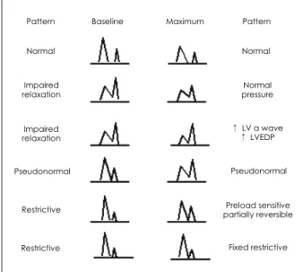
Mitral inflow filling pattern: response to valsalva maneuver
Pattern Baseline Maximum Pattern
Normal Normal
Normal pressure Impaired
relaxation
Impaired relaxation
↑ LV a wave
↑ LVEDP
Pseudonormal Pseudonormal
Restrictive Preload sensitive
partially reversible
Restrictive Fixed restrictive
Fig. 6. Various responses of the mitral inflow to a Valsalva maneuver.
LV: left ventricle, LVEDP: left ventricular end-diastolic pressure.
The mitral inflow pattern returned to baseline during performance of a Valsalva maneuver. However, similar to the pulmonary venous flow pattern, the ability to assess the response to a Valsalva maneuver is limited by the technical feasibility. The fusion of the E and A wave during the relative tachycardia of the Valsalva maneu- ver may hinder any accurate measurement.
Flow duration during atrial contraction
The comparison of flow duration between mitral in- flow and pulmonary venous flow during atrial contrac- tion is helpful for determining an elevated left ventricular end-diastolic pressure. The reversal of flow into the pulmonary vein during atrial contraction tends to in- crease in velocity and duration(relative to mitral A wave duration) with the worsening diastolic properties. This is the result of the worsening relative compliance of the left ventricle compared with the pulmonary venous conduit. Upon atrial contraction, a poorly compliant ventricle will manifest a steep end diastolic pressure rise for a small volume of transmitral flow, and this results in rapid and early equalization of the left ventricular pressure and the left atrial pressure, and cessation of forward flow will prematurely occur. Meanwhile, flow will continue into the pulmonary venous circuit. An elevated left ventricular end-diastolic pressure can be detected with high specificity if the atrial reversal flow duration is >30 msec of the mitral A duration.59)62) Index of myocardial performance(Tei index)
The integrated assessment of the systolic and diasto- lic left ventricular function would be ideal for the eva- luation of heart failure. An index of myocardial per- formance(IMP) has been devised to incorporate both the systolic and diastolic time intervals for expressing global performance, and this is referred to as the Tei index. This is estimated as the sum of the isovolumic contraction time and the isovolumic relaxation time divided by the ejection time. It can be easily obtained
and it’s been shown to be reproducible.63) Both systolic and diastolic dysfunction results in abnormality of myo- cardial relaxation, which prolongs the isovolumic rela- xation period. IMP has been demonstrated to predict morbidity and mortality in dilated cardiomyopathy,64) cardiac amyloidosis,65) primary pulmonary hyperten- sion,66) and myocardial infarction.67) Whether this index is useful for diagnosing and predicting the clinical out- come of primary diastolic dysfunction needs to be vali- dated with future studies.
The interval between the onset of mitral E and the annular early diastolic velocity(E’) as determined by tissue Doppler echocardiography
The different responses of the E and E’ velocities to an increase in preload illustrate the different mechani- sms generating the E and Ea velocities: when the LV myocardial relaxation is normal, E begins with the LV diastolic suction that’s induced by rapid relaxation, and this results in a simultaneous onset of E and E’. Howe- ver, if myocardial relaxation is impaired, early diastolic filling is initiated by the left atrial pressure at the time of the mitral valve opening, and the E’ velocity starts later as a result of the delayed myocardial relaxation.
Garcia and colleagues have shown that the onset of E’
occurred 7.5±3.5 ms after the peak mitral inflow ve- locity in 7 patients suffering with restrictive cardiomyo- pathy, whereas the E’ started 22±19 ms earlier than did the E in the normal group.52) Subsequently, the interval between the onset of the mitral E and E’, as determi- ned by TDE(TE-E’), has been shown to correlate with the time constant of the LV relaxation(τ); this was de- monstrated by Hasegawa and associates in their rather elegant animal experiment.68) With worsening of heart failure by inducing rapid pacing, E’ progressively decrea- sed in velocity and it was delayed in onset. Rivas-Gotz et al.69) demonstrated that the pulmonary capillary wedge pressure was closely related to IVRT/TE-E’., and an IVRT/
TE-Ea<2 was found to have a sensitivity of 91% and a
Valsalva
Baseline Leg up
Fig. 7. Transmitral inflow velocities obtained by pulsed-wave Doppler echocardiography in a 71-year-old man suffering with restrictive cardiomyo- pathy. The mitral flow pattern at baseline (left panel) showed that the early filling (the E wave) was lower than the filling during atrial contraction with a prolonged deceleration time, which was consistent with a pattern of impaired relaxation. After elevating both legs for 3 minutes (middle panel), the mitral flow pattern was changed with prominent mid-diastolic filling, an increased E/A ratio and a shortened deceleration time of the E wave. The mitral inflow pattern returned to baseline during a Valsalva maneuver. The abbreviations are the same as in Fig. 5.
specificity of 89% for detecting a PCWP >15 mm Hg.
However, Sohn and associates could not find a delay in the onset of mitral annulus velocity compared with the onset of the mitral inflow over a wide range of τ(31 to 70 ms); therefore, they found no correlation between TE-E’ and τ.70) They also pointed out that the equation IVRT/TE-E’ could not be applied with a zero denomina- tor when the onset of E and E’ was simultaneous. A potential limitation of clinically applying this ratio is the necessity for measuring the cardiac time intervals at dif- ferent locations and different cardiac cycles.71) An ap- proach to simultaneously record the onset of E and E’
may allow this approach to be more attractive for clinical use. Nevertheless, this new parameter, TE-E’ or IVRT/
TE-E’, can be useful to assess the LV filling pressure, and especially when E/E’ is indeterminate. Further clinical investigations and experience will be needed to deter- mine the reliability and the role of IVRT/TE-E’ for asses- sing the filling pressure.
Assessment of left ventricular torsion
During systole, the left ventricular apex rotates coun- terclockwise(as viewed from the apex), whereas the base rotates clockwise, and this creates a torsional deforma- tion originating in the dynamic interaction of the oppo- sitely-wound epicardial and endocardial myocardial fiber helices.72) Left ventricular torsion is an important aspect of such cardiac biomechanics as ejection and suction.
However, this has been difficult to measure. It has been recently shown that speckle tracking imaging can assess left ventricular torsional deformation.73) Assessment of left ven- tricular torsion may help understand the relationship between molecular changes and the left ventricular per- formance, and it may provide new concepts for the supe- rior management of patients suffering with heart failure.
Evaluation of left atrium
The left atrial size and function is frequently altered by various cardiac disorders; however, this has not re- ceived as much attention as it should have. This is now receiving its proper attention as it is now commonly realized that the left atrium is invariably dilated in the patients with left ventricular diastolic dysfunction. The left atrium is commonly enlarged in the patients suf- fering with mitral stenosis due to flow obstruction at the mitral valve level during diastole. During diastolic dysfunction, flow resistance occurs at the level of the left ventricle, and this results in left atrial enlargement.
Therefore, the left atrial size is a faithful mirror of the elevated left ventricular and left atrial filling pressures in the absence of any mitral valve disease. The left atrial volume has been shown to provide a more accurate assessment of the left atrial size than does the M-mode left atrial dimension. The left atrial volume usually gives indirect evidence for the increased left atrial pressure
that’s observed over an extended period of time rather than tracking acute changes in the left atrial pressure that are akin to the glycosylated hemoglobin level in the patients suffering with diabetes mellitus. Diastolic dys- function is associated decreased passive left atrial emp- tying; this results in a larger left atrial volume at the onset of atrial systole, which helps to maintain left atrial ejection. With the increase in the left atrial pressures, atrial stretch and enlargement of the chamber occur, and this leads to remodeling of the left atrial structure along with changes of the physiologic properties and electrical milieu of the left atrium; all this finally cul- minates in the development of atrial fibrillation. Tsang et al. have shown that the left atrial volume appears to be a strong predictor of incident atrial fibrillation, and this is incremental to the clinical risk factors.74)
Future Directions in the Assessment of Diastolic Function
Patients with significant heart disease may have en- tirely normal diastolic hemodynamics, as assessed in the resting state. Because most cardiac symptoms are preci- pitated by exertion, it may also be quite important to assess hemodynamic performance during some form of stress. The lack of a relation between the left ventricu- lar filling pressure and peak oxygen consumption has been reported and in a similar fashion, there appears to be almost no relation between the left ventricular function, as measured by the rest ejection fraction, and the resting cardiac output with the exercise capacity.
Thus, an evaluation both at rest and during exercise should enable physicians to assess the cardiovascular re- serve and the relationship between specific symptoms and any hemodynamic impairment. Furthermore, the physiologic information so obtained is often valuable for differentiating patients with cardiac causes of exercise intolerance from the patients with non-cardiac causes of exercise intolerance, and this may also be helpful in prescribing specific medical therapy and estimating the
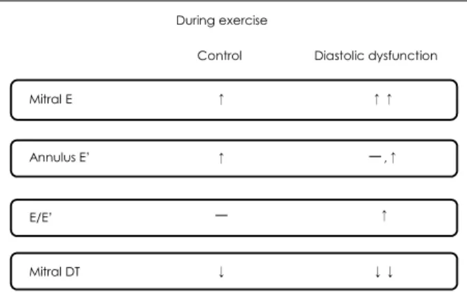
During exercise
Control Diastolic dysfunction
Mitral E
Annulus E’
E/E’
Mitral DT
↑
-
↓
↑↑
↑ -, ↑
↓↓
↑
Fig. 8. Changes of the mitral inflow E and mitral annular E’ velocities for diastolic dysfunction during exercise. DT: decalevation time.
prognosis. It is also conceivable that those patients with exertional dyspnea that can not be explained by the resting parameters of the left ventricular systolic and diastolic functions would have steep left ventricular pressure-volume relations during exercise as compared to those patients who have no symptoms of exertional dyspnea despite their similar degrees of left ventricular systolic and diastolic functions at rest.
A standard method for assessing the diastolic func- tional reserve, which can be defined as the capacity of the ventricle to accommodate the diastolic filling that’s necessary for increased demand as imposed by exercise without resulting in any marked increase in filling pres- sures. Therefore, for those patients suspected of having diastolic dysfunction with exertional dyspnea and they also have normal ventricular systolic and diastolic func- tions, exercise could unmask the diastolic abnormalities that were not evident under rest conditions.75) Patients with severe exercise limitation may be found to have a relatively preserved diastolic function even during exer- cise; this points to other etiologies such as pulmonary disease or deconditioning as the cause of the patient’s symptom. Alternatively, there may be patients who have a relatively normal or mildly impaired diastolic func- tion at rest, but they have the development of significant deterioration of the diastolic function during exercise.
In this setting, it is important to assess the diastolic function during stress or exercise with the provocation of symptoms. The development of a “diastolic stress test”
would be helpful for achieving better assessment and management of the patients with dyspnea(Fig. 8).76)77)
Diastolic dysfunction represents a growing clinical challenge to develop novel therapeutic approaches. Im- provements in our understanding of the molecular pa- thogenesis of diastolic dysfunction and its incorporation into the diagnostic and therapeutic approaches will enhance patient management in this disease popula- tion. Novel approaches that will evolve in the future will undoubtedly improve our evaluation and treatment of this entity. This makes it even more important to as- sess the diastolic function and its functional reserve.
Despite the high prevalence, substantial morbidity and significant mortality of diastolic heart failure, there has been no prospective, randomized, blinded pharmaco- logic trial data for guiding clinical decisions. Fortunately, large outcome studies are now ongoing to investigate the role of angiotensin converting enzyme inhibitors, angio- tensin receptor blockers and beta-blockers for patients suffering with diastolic heart failure or heart failure with preserved left ventricular systolic function. With the re- sults of these trials, as well as the better diagnostic tes- ting that will be available in the future, physicians should be in a better position to not only to diagnose diastolic dysfunction or heart failure, but also to manage it more effectively.
REFERENCES
1) Foot DK, Lewis RP, Pearson TA, Beller GA. Demographics and cardiology, 1950-2050. J Am Coll Cardiol 2000;35:1067-81.
2) Packer M. Abnormalities of diastolic function as a potential cause of exercise intolerance in chronic heart failure. Circulation 1990;81(Suppl III):III78-86.
3) Gaasch WH, Levine HJ, Quinones MA, Alexander JK. Left ven- tricular compliance: mechanisms and clinical implications. Am J Cardiol 1976;38:645-53.
4) Grossman W, McLaurin LP. Diastolic properties of the left ven- tricle. Ann Intern Med 1976;84:316-26.
5) Grossman W, Barry WH. Diastolic pressure-volume relations in the diseased heart. Fed Proc 1980;39:148-55.
6) Vasan RS, Larson MG, Benjamin EJ, Evans JC, Reiss C, Levy D.
Congestive heart failure in subjects with normal versus reduced left ventricular ejection fraction: prevalence and mortality in a population-based cohort. J Am Coll Cardiol 1999;33:1948-55.
7) Mosterd A, Hoes AW, de Bruyne MC, et al. Prevalence of heart failure and left ventricular dysfunction in the general population.
Eur Heart J 1999;20:447-55.
8) Kupari M, Lindroos M, Iivanainen AM, Heikkila J, Tilvis R.
Congestive heart failure in old age: prevalence, mechanisms and 4-year prognosis in the Helsinki Aging Study. J Intern Med 1997;
241:387-94.
9) Senni M, Tribouilloy CM, Rodeheffer RJ, et al. Congestive heart failure in the community: a study of all incident cases in Olmsted County, Minnesota, in 1991. Circulation 1998;98:2282-9.
10) Gandhi SK, Powers JC, Nomeir AM, et al. The pathogenesis of acute pulmonary edema associated with hypertension. N Engl J Med 2001;344:17-22.
11) Brutsaert DL, Meulemans AL, Sipido KR, Sys SU. Effects of damaging the endocardial surface on the mechanical perfor- mance of isolated cardiac muscle. Circ Res 1988;62:358-66.
12) Weiss JL, Frederiksen JW, Weisfeldt ML. Hemodynamic determi- nants of the time-course of fall in canine left ventricular pressure.
J Clin Invest 1976;58:751-60.
13) Yellin EL, Hori M, Yoram C, Sonnenblick EH, Gabbay S, Frater RW. Left ventricular relaxation in the filling and nonfilling intact canine heart. Am J Physiol 1986;250:H620-9.
14) Gaasch WH. Passive Elastic Properties of the Left Ventricle.
Philadelphia: Lea & Febiger; 1994.
15) Wahr PA, Michele DE, Metzger JM. Parvalbumin gene transfer corrects diastolic dysfunction in diseased cardiac myocytes. Proc Natl Acad Sci USA 1999;96:11982-5.
16) Ebashi S. Excitation-contraction coupling and the mechanism of muscle contraction. Annu Rev Physiol 1991;53:1-16.
17) Hasenfuss G. Calcium pump overexpession and myocardial func- tion: implications for gene therapy of myocardial failure. Circ Res 1998;83:966-8.
18) Koss KL, Kranias EG. Phospholamban: a prominent regulator of myocardial contractility. Circ Res 1996;79:1059-63.
19) Taffet GE, Michael LA, Tate CA. Exercise training improves lu- sitropy by isoproterenol in papillary muscles from aged rats. J Appl Physiol 1996;81:1488-94.
20) Takagishi Y, Rothery S, Issberner J, Levi A, Severs NJ. Spatial distribution of dihydropyridine receptors in the plasma mem- brane of guinea pig cardiac myocytes investigated by correlative confocal microscopy and label-fracture electron microscopy. J Electron Microsc 1997;46:165-70.
21) Tate CA, Helgason T, Hyek MF, et al. SERCA2a and mitochond- rial cytochrome oxidase expression are increased in hearts of exercise-trained old rats. Am J Physiol 1996;271:H68-72.
22) Taffet GE, Pham TT, Bick DL, Entman ML, Pownall HJ, Bick RJ.
The calcium uptake of the rat heart sarcoplasmic reticulum is altered by dietary lipid. J Membr Biol 1993;131:35-42.
23) Cain BS, Meldrum DR, Joo KS, et al. Human SERCA2a levels correlate inversely with age in senescent human myocardium. J Am Coll Cardiol 1998;32:458-67.
24) Shah AM, Grocott-Mason RM, Pepper CB, et al. The cardiac en- dothelium: cardioactive mediators. Prog Cardiovasc Dis 1996;39:
263-84.
25) Paulus WJ, Serizawa T, Grossman W. Altered left ventricular diastolic properties during pacing-induced ischemia in dogs with coronary stenoses: potentiation by caffeine. Circ Res 1982;50:
218-27.
26) Leite-Moreira AF, Correia-Pinto J, Gillebert TC. Load depen- dence of left ventricular contraction and relaxation: effects of caffeine. Basic Res Cardiol 1999;94:284-93.
27) Hori M, Yellin EL, Sonnenblick EH. Left ventricular diastolic suction as a mechanism of ventricular filling. Jpn Circ J 1982;
46:124-9.
28) Ingels NB Jr, Hansen DE, Daughters GT 2nd, Stinson EB, Alder- man EL, Miller DC. Relation between longitudinal, circumferen- tial, and oblique shortening and torsional deformation in the left ventricle of the transplanted heart. Circ Res 1989;64:915-27.
29) Solomon SB, Nikolic SD, Glantz SA, Yellin EL. Left ventricular diastolic function of remodeled myocardium in dogs with pacing induced heart failure. Am J Physiol 1998;274:H945-54.
30) Bell SP, Fabian J, LeWinter MM. Effects of dobutamine on left ventricular restoring forces. Am J Physiol 1998;275:H190-4.
31) Apstein CS, Grossman W. Opposite initial effects of supply and demand ischemia on left ventricular diastolic compliance: the ischemia-diastolic paradox. J Mol Cell Cardiol 1987;19:119-28.
32) Oh JK, Appleton CP, Hatle LK, Nishimura RA, Seward JB, Tajik AJ. The noninvasive assessment of left ventriclar diastolic func- tion with two-dimensional and Doppler echocardiography. J Am Soc Echocardiogr 1997;10:246-70.
33) Kitabatake A, Inoue M, Asao M, et al. Transmitral blood flow reflecting diastolic behavior of the left ventricle in health and di- sease: a study by pulsed Doppler technique. Jpn Circ J 1982;46:
92-102.
34) Nishimura RA, Abel MD, Hatle LK, Tajik AJ. Assessment of diastolic function of the heart: background and current applica- tions of Doppler echocardiography: part II. clinical studies. Mayo Clin Proc 1989;64:181-204.
35) Thomas JD, Weyman AE. Echo Doppler evaluation of left ven- tricular diastolic function: physics and physiology. Circulation 1991;84:977-90.
36) Choong CY, Herrmann HC, Weyman AE, Fifer MA. Preload dependence of Doppler-derived indices of left ventricular diasto- lic function in humans. J Am Coll Cardiol 1987;10:800-18.
37) Ishida Y, Mesinder JS, Tsujioka K, et al. Left ventricular filling dynamics: influence of left ventricular relaxation and left atrial pressure. Circulation 1986;74:187-96.
38) Stoddard MF, Pearson AC, Kern MJ, Ratcliff J, Mrosek G, La- bovitz AJ. Influence of alteration in preload on the pattern of left ventricular diastolic filling assessed by Doppler echocardiography in humans. Circulation 1989;79:1226-36.
39) Xie GY, Berk MR, Smith MD, DeMaria AN. Relation of Dop- pler transmitral flow patterns to functional status in congestive heart failure. Am Heart J 1996;131:766-71.
40) Rihal CS, Nishimura RA, Hatle LK, Bailey KR, Tajik AJ. Systo- lic and diastolic dysfunction in patients with clinical diagnosis of dilated cardiomyopathy: relation to symptoms and prognosis. Cir- culation 1994;90:2772-9.
41) Xie GY, Berk MR, Smith MD, Gurley JC, DeMaria AN. Pro- gnostic value of Doppler transmitral flow patterns in patients with
congestive heart failure. J Am Coll Cardiol 1994;24:132-9.
42) Klein AL, Hatle LK, Taliercio CP, et al. Prognostic significance of Doppler measures of diastolic function in cardiac amyloido- sis: a Doppler echocardiography study. Circulation 1991;83:
808-16.
43) Nishimura RA, Tajik AJ. Evaluation of diastolic filling of left ven- tricle in health and disease: Doppler echocardiography is the clinician’s Rosetta Stone. J Am Coll Cardiol 1997;30:8-18.
44) Pinamonti B, Zecchin M, di Lenarda A, Gregori D, Sinagra G, Camerini F. Persistence of restrictive left ventricular filling pat- tern in dilated cardiomyopathy: an ominous prognostic sign. J Am Coll Cardiol 1997;29:604-12.
45) Yamamoto K, Nishimura RA, Chaliki HP, Appleton CP, Holmes DR Jr, Redfield MM. Determination of left ventricular filling pressure by Doppler echocardiography in patients with coronary artery disease: critical role of left ventricular systolic function. J Am Coll Cardiol 1997;30:1819-26.
46) Ommen SR, Nishimura RA, Appleton CP, et al. Clinical utility of Doppler echocardiography and tissue Doppler imaging in the estimation of left ventricular filling pressures: a comparative simultaneous Doppler-catheterization study. Circulation 2000;102:
1788-94.
47) Kim KS. The usefulness of Doppler tissue image in evaluation of left ventricular systolic and diastolic dysfunction. Korean Circ J 2002;32:99-105.
48) Sohn DW, Chai IH, Lee DJ, et al. Assessment of mitral annulus velocity by Doppler tissue imaging in the evaluation of left ven- tricular diastolic function. J Am Coll Cardiol 1997;30:474-80.
49) Nagueh SF, Middleton KJ, Kopelen HA, Zoghbi WA, Quinones MA. Doppler tissue imaging: a noninvasive technique for eva- luation of left ventricular relaxation and estimation of filling pressures. J Am Coll Cardiol 1997;30:1527-33.
50) Nagueh SF, Sun H, Kopelen HA, Middleton KJ, Khoury DS.
Hemodynamic determinants of the mitral annulus diastolic velo- cities by tissue Doppler. J Am Coll Cardiol 2001;37:278-85.
51) Nagueh SF, Bachinski LL, Meyer D, et al. Tissue Doppler ima- ging consistently detects myocardial abnormalities in patients with hypertrophic cardiomyopathy and provides a novel means for an early diagnosis before and independently of hypertrophy. Circu- lation 2001;104:128-30.
52) Garcia MJ, Rodriguez L, Ares M, Griffin BP, Thomas JD, Klein AL. Differentiation of constrictive pericarditis from restrictive car- diomyopathy: assessment of left ventricular diastolic velocities in longitudinal axis by Doppler tissue imaging. J Am Coll Car- diol 1996;27:108-14.
53) Ha JW, Ommen SR, Tajik AJ, et al. Differentiation of constric- tive pericarditis from restrictive cardiomyopathy using mitral an- nular velocity by tissue Doppler echocardiography. Am J Cardiol 2004;94:316-9.
54) Ha JW, Oh JK, Ling LH, Nishimura RA, Seward JB, Tajik AJ.
Annulus paradoxus: transmitral flow velocity to mitral annular velocity ratio is inversely proportional to pulmonary capillary wedge pressure in patients with constrictive pericarditis. Circu- lation 2001;104:976-8.
55) Ha JW, Oh JK, Ommen SR, Ling LH, Tajik AJ. Diagnostic va- lue of mitral annular velocity for constrictive pericarditis in the absence of respiratory variation in mitral inflow velocity. J Am Soc Echocardiogr 2002;15:1468-71.
56) Brun P, Tribouilloy C, Duval AM, et al. Left ventricular flow propagation during early filling is related to wall relaxation: a color M-mode Doppler analysis. J Am Coll Cardiol 1992;20:
420-32.
57) Garcia MJ, Smedira NG, Greenberg NL, et al. Color M-mode Doppler flow propagation velocity is a preload insensitive index
of left ventricular relaxation: animal and human validation. J Am Coll Cardiol 2000;35:201-8.
58) Moller J, Poulsen S, Sondergaard E, Egstrup K. Preload depen- dence of color M-mode Doppler flow propagation velocity in con- trols and in patients with left ventricular dysfunction. J Am Soc Echocardiogr 2000;13:902-9.
59) Rossvoll O, Hatle LK. Pulmonary vein flow velocities recorded by transthoracic Doppler ultrasound: relation to left ventricular diastolic pressures. J Am Coll Cardiol 1993;21:1687-96.
60) Appleton CP, Hatle LK, Popp RL. Relation of transmitral flow elocity patterns to left ventricular diastolic function: new insights from a combined hemodynamic and Doppler echocardiographic study. J Am Coll Cardiol 1988;12:426-40.
61) Yamamuro A, Yoshida K, Hozumi T, et al. Noninvasive evaluation of pulmonary capillary wedge pressure in patients with acute myocardial infarction by deceleration time of pulmonary venous flow velocity in diastole. J Am Coll Cardiol 1999;34:90-4.
62) Kimura K, Murata K, Tanaka N, et al. The importance of pulmo- nary venous flow measurement for evaluating left ventricular end- diastolic pressure in patients with coronary artery disease in the early stage of diastolic dysfunction. J Am Soc Echocardiogr 2001;
14:987-93.
63) Bruch C, Schmermund A, Marin D, et al. Tei-index in patients with mild-to-moderate congestive heart failure. Eur Heart J 2000;
21:1888-95.
64) Dujardin KS, Tei C, Yeo TC, Hodge DO, Rossi A, Seward JB.
Prognostic value of a Doppler myocardial performance index combining systolic and diastolic performance in idiopathic dila- ted cardiomyopathy. Am J Cardiol 1998;82:1071-6.
65) Tei C, Dujardin KS, Hodge DO, Kyle RA, Tajik AJ, Seward JB.
Doppler index combining systolic and diastolic myocardial per- formance: clinical value in cardiac amyloidosis. J Am Coll Cardiol 1996;28:658-64.
66) Yeo TC, Dujardin KS, Tei C, Mahoney DW, McGoon MD, Se- ward JB. Value of a Doppler-derived myocardial performance index combining systolic and diastolic time intervals in predicting outcome in primary pulmonary hypertension. Am J Cardiol 1998;
81:1157-61.
67) Poulsen SH, Jensen SE, Nielsen JC, Moller JE, Egstrup K. Serial changes and prognostic implications of a Doppler-derived index of combined left ventricular systolic and diastolic myocardial performance in acute myocardial infarction. Am J Cardiol 2000;
85:19-25.
68) Hasegawa H, Little WC, Ohno M, et al. Diastolic mitral annular velocity during the development of heart failure. J Am Coll Car- diol 2003;41:1590-7.
69) Rivas-Gotz C, Khoury D, Manolios M, Rao L, Kopelen HA, Nagueh SF. Time interval between onset of mitral inflow and on- set of early diastolic velocity by tissue Doppler: a novel index of left ventricular relaxation. J Am Coll Cardiol 2003;42:1463-70.
70) Sohn DW, Kim YJ, Park YB, Choi YS. Clinical validity of mea- suring time difference between onset of mitral inflow and onset of early diastolic mitral annulus velocity in the evaluation of left ventricular diastolic function. J Am Coll Cardiol 2004;43:2097-101.
71) Oh JK. Echocardiography as a noninvasive Swan-Ganz catheter.
Circulation 2005;111:3192-4.
72) Buckberg GD, Weisfeldt ML, Ballester M, et al. Left ventricular form and function: scientific priorities and strategic planning for development of new views of disease. Circulation 2004;110:
e333-6.
73) Notomi Y, Lysyansky P, Setser RM, et al. Measurement of ven- tricular torsion by two-dimensional ultrasound speckle tracking imaging. J Am Coll Cardiol 2005;45:2034-41.
74) Tsang TS, Barnes ME, Bailey KR, et al. Left atrial volume: im- portant risk marker of incident atrial fibrillation in 1655 older men and women. Mayo Clin Proc 2001;76:467-75.
75) Ha JW, Oh JK. Echocardiographic evaluation of chronic dysp- nea: cardiovascular Imaging. In: A handbook for Clinical Prac- tice; 2005. p.164-74.
76) Ha JW, Lulic F, Bailey KR, et al. Effects of treadmill exercise on mitral inflow and annular velocities in healthy adults. Am J Car- diol 2003;91:114-5.
77) Ha JW, Oh JK, Pellikka PA, et al. Diastolic stress echocar- diography: a novel noninvasive diagnostic test for diastolic dys- function using supine bicycle exercise Doppler echocardiography.
J Am Soc Echocardiogr 2005;18:63-8.