DOI 10.3349/ymj.2008.49.1.19
Suppression of apoptosis is one of the hallmarks of carcino- genesis. Tumor cells endure apoptotic pressure by overex- pressing several antiapoptotic proteins, and FLICE inhibitory protein (FLIP) is one of the important antiapoptotic proteins that have been shown to be overexpressed in various primary tumor cells. FLIP has two death-effector domains in tandem, mimicking the prodomain of procaspase-8. It is recruited to Fadd in death-inducing signaling complex, thereby preventing the activation of procaspase-8. To date, three isoforms of human cytosolic FLIP (c-FLIP) and six viral homologs (v-FLIP) have been identified. Recently, the crystal structure of v-FLIP MC159 was determined for the first time as an atomic-detail FLIP structure, which revealed that two death effector domains are packed tightly against each other mainly through conserved hydrophobic interactions. The overexpres- sion of c-FLIP in tumor cells has been shown to be the determinant of the tumor’s resistance to death ligands such as FasL and TRAIL. It has also been shown that the down- regulation of c-FLIP results in sensitizing resistant tumor cells.
Therefore, the agents directly targeting c-FLIP at mRNA and protein levels are expected to be developed in near future and tested for the potential as a new class of anti-cancer drugs.
Kew Words: FLICE inhibitory protein, death-inducing signaling complex, Fas, apoptosis, cancer
APOPTOSIS AND CANCER
Apoptosis is a programmed way of cell death which has been characterized by shrinking of cells, condensation of nuclei, and internucleosomal degradation of DNA.
1,2Within 24 hours after this program is switched on, the apoptotic cell divides
into small blobs and is finally engulfed by neighboring cells.
3Since Dr. Stanley Korsmeyer had shown that apoptosis program is suppressed in B-cell lymphoma and its suppression enhances the development of B-cell lymphoma, thousands of studies have been accumulated to support the idea that the acquired resistance to apoptosis is a hallmark of most or perhaps all types of cancer.
4Moreover, a significant part of the benefits achieved by chemotheraphy relies on the induc- tion of apoptosis in tumor cells,
5and cancers with alterations in proteins involved in apoptosis signaling are often resistant to chemotheraphy.
6Therefore, drugs designed to restore the apoptosis program might be effective against tumor cells.
For selectivity, such drugs might induce cell death of only tumor cells because, unlike normal cells, they are under apoptotic stress and highly depen- dent on aberrations of the apoptosis signaling pathways to stay alive.
6For these reasons, apoptosis has been a very attractive phenomenon for the researchers who seek new strategies to fight against cancer.
Antiapoptotic proteins overexpressed in tumor cells have been recognized as targeting points for anti-cancer therapeutic interventions, and their inhibitors at the levels of mRNA and protein have been developed, which are mostly antisense oligonucleotides and small molecule inhibitors.
6-8Those drug candidate compounds are now mostly in the preclinical and early clinical stages. FLIP is an another important antiapoptotic protein overexpressed in various types of tumor cells,
9but the agents directly targeting it have not yet been reportedly developed.
8In this review, recent progress on FLIP research and its potential as an anti-cancer therapeutic target will be discussed.
FLIP as an Anti-Cancer Therapeutic Target
Jin Kuk Yang
Department of Chemistry, School of Natural Sciences, Soongsil University, Seoul, Korea.
Received October 23, 2007 Accepted November 3, 2007
This work was supported by the Soongsil University Research Fund to JKY.
Reprint address: requests to Dr. Jin Kuk Yang, Department of Chemistry, School of Natural Sciences, Soongsil University, Seoul, Korea. Tel: 82-2-820-0433, Fax: 82-2-824-4383, E-mail: jinkukyang
@ssu.ac.kr
INITIATOR CASPASE ACTIVATION IN INTRINSIC AND EXTRINSIC PATHWAYS
The central executioner of apoptosis is a set of cysteine proteases called caspases that are initially synthesized as inactive zymogens called pro- caspases. Upon the induction of apoptosis, procaspase is cleaved into p18 and p10 to form the active enzyme, which is a heterotetramer containing two p18/p10 heterodimers and two active sites.
10Based on their order of activation, caspases are classified into two families: initiator caspases and effector caspases.
11Initiator caspases (also known as apical caspases; caspase-8 & -9) are activated through autocatalytic cleavage on their own activation platform formed in response to upstream death signals. For example, caspase-8 is activated in death-inducing signaling complex (DISC) whose major components are Fas and Fadd.
12-14In caspase-9, the proteolytic activation is accomplished in apoptosome composed of Apaf-1 and cytochrom c (Fig. 1).
11Effector caspases (also known as executioner caspases) are proteolytically activated by initiator caspases. Once activated, effecter caspases (caspase-3 & -7) degrade more than 280 cellular proteins identified so far and consequently execute the cell death process.
15Death signals to activate the initiator caspases can occur internally from cytotoxic insults such as DNA damage or can be given externally in a form of cytokine collectively called as death ligands including Fas ligand (FasL) and TRAIL.
7In
intrinsic pathway, DNA damage leads to the phosphorylation of p53, which then induces transcriptional activation of proapoptotic proteins such as Bax, Puma and Noxa.
16These proteins change the permeability of mytochondiral mem- brane, which results in the release of several proteins including cytochrome c. Cytochrome c in cytosol interacts with Apaf-1 and they form a heptameric complex called apoptosome where procaspase-9 is activated (Fig. 1).
11In extrinsic pathway, binding of the trimeric death ligand to the death receptor induces the oligomerization of the death receptor which leads to the formation of DISC where procaspase-8 is recruited and activated.
12-14Death receptors are a subfamily of the TNF receptor superfamily, and eight human death receptors have been identified; Fas (also known as Apo-1 and CD95), TNF-R1, DR-3 (Apo- 3, TRAMP, WSL-1, LARD), TRAIL-R1 (DR-4), TRAIL-R2 (DR-5), DR-6, EDA-R and NGF-R.
17All death receptors have a death domain in their cytosolic part where the downstream signaling protein binds. Fas has been the most studied death receptor to date. Binding of FasL induces the oligomerization of Fas, and Fas recruits its downstream cytosolic adaptor protein Fadd, which in turn recruits procaspase-8.
12-14In addi- tion to Fas, TRAIL-R1 and -R2 should be men- tioned as members of special interests because their common ligand TRAIL was shown to induce apoptosis selectively in tumor cells but not in normal cells, highlighting its potential therapeutic
Fig. 1. Apoptosis signaling and the caspase activation.
application in cancer treatment.
18,19Protein-protein interactions in the activation of initiator caspases are mediated by three similar domains, which are death domain (DD), death- effecter domain (DED), and caspase-recruiting domain (CARD). In DISC for caspase-8 activation, DD in the cytosolic part of Fas interacts with DD of Fadd, and DED of Fadd in turn with DED of procaspase-8. In case of caspase-9 activation, Apaf-1 recruits procaspase-9 through CARD- CARD interaction. These three domains comprise the death domain superfamily, and they com- monly adopt a simple globular fold of the charac- teristic hexahelical bundle in a Greek key topology.
Even though these three domains are very similar in structure, their interactions are highly specific so that they interact only in a homotypic way (i.e.
DD-DD, DED-DED, and CARD-CARD) and there is no established interaction across members at least to date.
20These homotypic interactions of DD, DED and CARD play an essential role in forming heteromultimeric platforms for the initia- tor caspase activation, i.e. DISC and apoptosome, as well as in recruiting the proenzyme of the initiator caspases to those platforms.
FLIP AS AN INHIBITOR OF RECEPTOR- MEDIATED APOPTOSIS
Procaspase-8 has two DEDs in the N-terminus of the catalytic domain, and Fadd has one DED
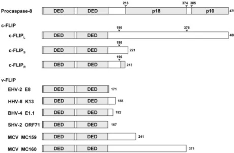
in the N-terminus. In 1997, other DED-containing proteins have been identified in several γ - herpesviruses and the poxvirus MCV.
21-23They have been collectively called viral FLICE inhibi- tory proteins (v-FLIP) because they show inhibitory effects on apoptosis signaling of caspase-8 which is also called FLICE. Soon after the identification of v-FLIP, their human cellular homologs (cellular-FLIP, c-FLIP) were also identified (Fig.
2).
24,25Although 13 distinct splice variants of c-FLIP mRNA have been described,
26only three have been detected at the protein level; c-FLIP
L, c-FLIP
S, and c-FLIP
R.
24-27The long isoform c-FLIP
Lhas a domain structure similar to procaspase-8:
two DED’s in N-terminus and caspase-like domain in C-terminus. However, its caspase-like domain lacks the catalytic cysteine residue con- served in caspases and consequently it is catalyti- cally inactive. All three isoforms of c-FLIP as well as v-FLIP have been reported to be recruited to DISC through its tandem DEDs interacting with DED of Fadd, and FLIP thereby excludes pro- caspase-8 from DISC. This may be the common mechanism of various FLIP’s to inhibit the caspase- 8 activation, although MC159 (v-FLIP from MCV) seems to act in a slightly different way.
28Although c-FLIP
Lhas been described in most reports as an inhibitor of caspase-8 activation in DISC, some other studies show that c-FLIP
Lcan activate the caspase-8.
29-33Recently, it has been proposed that c-FLIP
Lcan be either antiapoptotic or proapoptotic depending on its expression
Fig. 2. Various FLIPs and procaspase-8.
Arrows denote the sites cleaved by caspase-8 or procaspase-8. Shadow region in the C-terminus of c-FLIPR is its own unique 11-residue-long sequence which is not observed in other two isoforms, c-FLIPL and cFLIPS.
level,
34although all other FLIP’s have been reported to be solely anti-apoptotic. At low expression levels, which are probably found in most cells, c-FLIP
Lenhances the caspase-8 activation. At intermediate expression levels, found in some cell types such as monocytes/macrophages and certain tumors, c-FLIP
Lacts as an inhibitor of caspase-8 activation. At very high non-physiological concen- trations achieved by transient overexpression, c-FLIP
Lis cytotoxic by itself without the need for stimulation of Fas.
34Despite the dual role of c-FLIP
Lin caspase-8 activation, it should be emphasized that c-FLIP
Lacts as an antiapoptotic protein to inhibit caspase-8 activation at least in the range of physiological expression level in tumor cells.
NONAPOPTOTIC FUNCTIONS OF FLIP In addition to its activity in apoptosis signaling pathway, c-FLIP
Lhas been reported to activate NF- B pathway, especially in lymphocytes. κ
35-39NF- B activation by c-FLIP κ
Lrequires the cleav- ages of c-FLIP
Lat Asp-376 by fully processed mature caspase-8, i.e. p102-p182 heterotetramer, and/or at Asp-196 by procaspase-8.
37,38Intere- stingly, mature caspase-8 and procaspase-8 showed mutually exclusive proteolytic specificity on two cleavage sites in c-FLIP
L.
38These cleavages generate tandem-DED-containing N-terminal fragments of the molecular weight around 43 kDa and 22 kDa which are called p43-FLIP and p22-FLIP respec- tively. It has been shown that p22-FLIP can also be generated also from c-FLIP
S.
38Both the FLIP N-terminal fragments activate NF- B signaling via κ binding to the components of NF- B signaling κ pathway; p43-FLIP binds to TRAF237 and p22- FLIP directly binds to IKK complex.
38Notably, caspase-8 has also been reported to be essential for NF- B activation in T, B and NK cells. κ
40,41Involvement of c-FLIP
Land caspase-8 in NF- B κ activation may explain the dual role of c-FLIP
Lin caspase-8 activation discussed in the previous section.
42NF- B activation by c-FLIP κ
Land caspase-8 has been shown to play an important role in lymphocyte proliferation.
37,38In addition to NF- B signaling, c-FLIP κ
Lhas also been shown to activate Erk signaling pathway by binding to Raf-1.
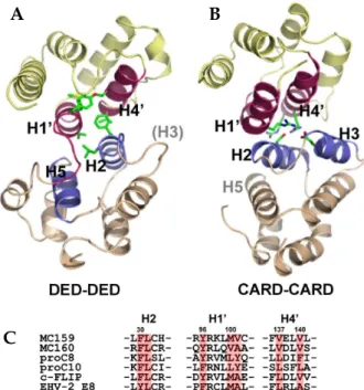
35,43FLIP STRUCTURE IN ATOMIC DETAIL The structure of a viral FLIP, MC159, has recently been revealed by X-ray crystallography for the first time as a FLIP structure (Fig. 3A).
28,44It shows that DED1 significantly deviates from the canonical death fold observed in Fadd DED
45and Fas DD.
46MC159 DED1 lacks the helix 3, and there is instead a short loop structure. The crystal structure also revealed that two DEDs pack tightly against each other through hydrophobic interac- tion. The atomic-detail DED-DED interaction mode observed in MC159, although intramole- cular, vividly contrasts to the previously reported intermolecular CARD-CARD interaction between procaspase-9 and Apaf-1 (Fig. 3A and 3B).
47Firstly, the CARD-CARD interface involves the helix 2/3 face (Apaf-1) and the helix 1/4 face (procaspase-9). In MC159 structure, however, the helix 2/5 face of DED1 meets the helix 1/4 face of DED2. Secondly, the interactions between two CARDs are mainly electrostatic. In contrast, the DED-DED interactions in MC159 are mainly hydrophobic. Interestingly with regards to the hydrophobic interface, the conserved hydrophobic patch in DED1 is buried in the interface, and the one in only DED2 is exposed. The hydrophobic patch is conserved in all known DEDs and has already been shown to be critical for the interac- tion of Fadd with other DED proteins such as procaspase-8.
45Other important observation is that the hydrophobic residues comprising this intramolecular DED-DED interface are conserved in other tandem-DED-containing proteins such as procaspase-8, -10, c-FLIP, and other v-FLIP’s (Fig.
3C). It implies that two DEDs in those proteins might also pack against each other in the same way as in MC159. However, it cannot rule out the possibility that two DEDs, in some circumstances, might get apart and the hydrophobic patch in DED1 could be exposed to play a role in the interaction with their binding partners.
RxDL sequence motif in the beginning of helix
6 is highly conserved among proteins containing
a DED or tandem DEDs.
48Mutation on the motif
in Fadd DED abolished its self-association which
is essential for its apoptosis signaling activity,
49and this motif is essential also for the antia-
poptotic activity of v-FLIP MC159.
50The crystal
structure of v-FLIP MC159 revealed that the arginine residue in the RxDL motif interacts with two acidic residues; an aspartate in the motif and a glutamate in helix 2 (Fig. 4). The three charged residues from helix 2 and 6 are highly conserved among DED proteins, and the sequence motif characteristic of DED was rephrased into E/D- RxDL motif. The triad of these three charged residues seems to play a structural role to hold the helix 2 and 6 together in correct positions, and consequently to help stabilize the entire DED fold.
The functional defect of the mutants on this motif reported previously is more likely a secondary effect caused by the structural disturbance. This idea is supported by recent study on Fadd DED.
Even though Fadd R72A mutant (corresponding to R in E/D-RxDL motif) shows the same circular dichroism spectra as the wild type, its NMR peaks exchange broaden and none of the DED resonances are visible.
51It implies that the Fadd R72A mutant has the same helical content as the wild type but its structural integrity of DED fold
is disturbed to some extent. Therefore, E/D-RxDL motif conserved in DEDs must play a structural role to help maintain the stability of DED fold.
TARGETING FLIP FOR CANCER THERAPY Elevated expression of c-FLIP has been found in various types of tumor cells which are often resistant to death-receptor-mediated apoptosis.
Those tumors include colorectal carcinoma,
52-55gastric carcinoma,
56-58pancreatic carcinoma,
59,60Hodgkin’s lymphoma,
61-63B cell chronic lymphocytic leukemia,
64,65melanoma,
24,66,67ovarian carcinoma,
68-70cervical carcinoma,
71bladder urothelial carcinoma,
72and prostate carcinoma.
73The expression of c-FLIP has been proven to be one of the major determinants of the resistance to death ligands such as FasL and TRAIL, and numerous reports have shown that down-regulation of c-FLIP results in sensitizing a various types of resistant tumor cells.
52,57,60,62,63,66,69,70,74-76Conversely, forced expression of c-FLIP renders cells resistant to Fas and/or TRAIL. These observations collectively imply that c-FLIP may be an attractive therapeutic target against at least the above mentioned kinds of tumors of which malignancy and resistance
Fig. 4. Conserved E/D-RxDL motif in DED fold.
Fig. 3. Structure of v-FLIP and comparison of DED-DED interface with CARD-CARD interface. (A) Structure of v-FLIP MC159 and the intramolecular DED-DED interaction. (B) CARD-CARD interaction observed between Apaf-1 and procaspase-9. (C) Hydrophobic residues in DED-DED interface are conserved among the proteins containing tandem DEDs.
A B
C
have been shown to be strongly dependent upon c-FLIP overexpression. In addition, v-FLIP K13 of human herpesvirus 8 (HHV8, also called Kaposi’s sarcoma-associated herpesvirus, KSHV) has also been shown to act as a tumor progression factor by inhibiting receptor-mediated apoptosis.
77To date, several kinds of small molecules have been known to lower c-FLIP expression and to sensitize the resistant tumor cell to death-receptor- mediated apoptosis.
9They include DNA-damaging agents (cisplatin and doxorubicin), RNA synthesis inhibitor (actinomycin D), protein synthesis inhi- bitor (cycloheximide), topoisomerase I inhibitors (camptothecin, 9-NC, topotecan), and histone deacetylase inhibitors (Trichostatin A).
9In addi- tion, the inhibitors of several kinases (MEK1/ 2, PKC and PI3K) also lower FLIP expression through blocking the corresponding signaling pathways for the transcriptional activation of FLIP. If it is considered that the above-mentioned tumors depend upon FLIP overexpression for the resistance to TRAIL,
18,78,79then the combination of TRAIL with these agents might be an attractive therapeutic strategy to kill those tumor cells.
However, it should be noted that the agents directly targeting FLIP at mRNA or protein levels has not yet been developed.
8Antiapoptotic proteins overexpressed in tumor cells have been recognized as attractive targets for anti-cancer therapeutic intervention. Compounds targeted to Bcl-2, IAP, and MDM2 at either mRNA or protein levels have been developed and are now in the stages of preclinical and early clinical trials.
6,8,80Various antisense oligonucleotides are targeted to their mRNA’s and small molecule inhibitors are designed to bind those proteins. The small molecule inhibitors are designed primarily based on their crystal structure in complex with their corresponding proapoptotic proteins; Bcl-XL complexed with Bad,
81XIAP complexed with Smac,
82,83and MDM2 complexed with p53.
84In development of the inhibitors of antiapoptotic proteins, FLIP seems to be left behind in com- parison to Bcl-2, IAP and MDM2. It is probably because FLIP was recognized more recently and the detailed information on its structure, especially in complex with Fadd, has not been available.
With regard to FLIP structure, it is notable that the crystal structure of v-FLIP MC159 has recently
been reported for the first time as an atomic-detail three-dimensional structure of FLIP.
28,44The antisense oligonucleotides and small molecule inhibitors directly targeting FLIP at the levels of mRNA and protein are expected to be developed in near future and tested for the potential as a new class of anti-cancer drugs.
REFERENCES
1. Kerr JF, Wyllie AH, Currie AR. Apoptosis: a basic biological phenomenon with wide-ranging implications in tissue kinetics. Br J Cancer 1972;26:239-57.
2. Thome M, Tschopp J. Regulation of lymphocyte proliferation and death by FLIP. Nat Rev Immunol 2001;1:50-8.
3. Wyllie AH, Kerr JF, Currie AR. Cell death: the significance of apoptosis. Int Rev Cytol 1980;68:251-306.
4. Hanahan D, Weinberg RA. The hallmarks of cancer.
Cell 2000;100:57-70.
5. Wajant H. Targeting the FLICE Inhibitory Protein (FLIP) in cancer therapy. Mol Interv 2003;3:124-7.
6. Fesik SW. Promoting apoptosis as a strategy for cancer drug discovery. Nat Rev Cancer 2005;5:876-85.
7. Debatin KM, Krammer PH. Death receptors in chemotherapy and cancer. Oncogene 2004;23:2950-66.
8. Reed JC, Pellecchia M. Apoptosis-based therapies for hematologic malignancies. Blood 2005;106:408-18.
9. Kataoka T. The caspase-8 modulator c-FLIP. Crit Rev Immunol 2005;25:31-58.
10. Hengartner MO. The biochemistry of apoptosis. Nature 2000;407:770-6.
11. Shi Y. Caspase activation: revisiting the induced proximity model. Cell 2004;117:855-8.
12. Muzio M, Chinnaiyan AM, Kischkel FC, O'Rourke K, Shevchenko A, Ni J, et al. FLICE, a novel FADD- homologous ICE/CED-3-like protease, is recruited to the CD95 (Fas/APO-1) death--inducing signaling complex. Cell 1996;85:817-27.
13. Chinnaiyan AM, Tepper CG, Seldin MF, O'Rourke K, Kischkel FC, Hellbardt S, et al. FADD/MORT1 is a common mediator of CD95 (Fas/APO-1) and tumor necrosis factor receptor-induced apoptosis. J Biol Chem 1996;271:4961-5.
14. Kischkel FC, Hellbardt S, Behrmann I, Germer M, Pawlita M, Krammer PH, et al. Cytotoxicity-dependent APO-1 (Fas/CD95)-associated proteins form a death- inducing signaling complex (DISC) with the receptor.
EMBO J 1995;14:5579-88.
15. Fischer U, Jänicke RU, Schulze-Osthoff K. Many cuts to ruin: a comprehensive update of caspase substrates.
Cell Death Differ 2003;10:76-100.
16. Roos WP, Kaina B. DNA damage-induced cell death by apoptosis. Trends Mol Med 2006;12:440-50.
17. French LE, Tschopp J. Protein-based therapeutic approaches targeting death receptors. Cell Death Differ 2003;10:117-23.
18. Wiley SR, Schooley K, Smolak PJ, Din WS, Huang CP, Nicholl JK, et al. Identification and characterization of a new member of the TNF family that induces apoptosis. Immunity 1995;3:673-82.
19. MacFarlane M. TRAIL-induced signalling and apoptosis.
Toxicol Lett 2003;139:89-97.
20. Lahm A, Paradisi A, Green DR, Melino G. Death fold domain interaction in apoptosis. Cell Death Differ 2003;10:10-2.
21. Thome M, Schneider P, Hofmann K, Fickenscher H, Meinl E, Neipel F, et al. Viral FLICE-inhibitory proteins (FLIPs) prevent apoptosis induced by death receptors.
Nature 1997;386:517-21.
22. Bertin J, Armstrong RC, Ottilie S, Martin DA, Wang Y, Banks S, et al. Death effector domain-containing herpesvirus and poxvirus proteins inhibit both Fas- and TNFR1-induced apoptosis. Proc Natl Acad Sci U S A 1997;94:1172-6.
23. Hu S, Vincenz C, Buller M, Dixit VM. A novel family of viral death effector domain-containing molecules that inhibit both CD-95- and tumor necrosis factor receptor-1-induced apoptosis. J Biol Chem 1997;272:
9621-4.
24. Irmler M, Thome M, Hahne M, Schneider P, Hofmann K, Steiner V, et al. Inhibition of death receptor signals by cellular FLIP. Nature 1997;388:190-5.
25. Hu S, Vincenz C, Ni J, Gentz R, Dixit VM. I-FLICE, a novel inhibitor of tumor necrosis factor receptor-1- and CD-95-induced apoptosis. J Biol Chem 1997;272:17255- 7.
26. Djerbi M, Darreh-Shori T, Zhivotovsky B, Grandien A.
Characterization of the human FLICE-inhibitory protein locus and comparison of the anti-apoptotic activity of four different flip isoforms. Scand J Immunol 2001;54:180-9.
27. Golks A, Brenner D, Fritsch C, Krammer PH, Lavrik IN. c-FLIPR, a new regulator of death receptor-induced apoptosis. J Biol Chem 2005;280:14507-13.
28. Yang JK, Wang L, Zheng L, Wan F, Ahmed M, Lenardo MJ, et al. Crystal structure of MC159 reveals molecular mechanism of DISC assembly and FLIP inhibition. Mol Cell 2005;20:939-49.
29. Yeh WC, Itie A, Elia AJ, Ng M, Shu HB, Wakeham A, et al. Requirement for Casper (c-FLIP) in regulation of death receptor-induced apoptosis and embryonic de- velopment. Immunity 2000;12:633-42.
30. Goltsev YV, Kovalenko AV, Arnold E, Varfolomeev EE, Brodianskii VM, Wallach D. CASH, a novel caspase homologue with death effector domains. J Biol Chem 1997;272:19641-4.
31. Han DK, Chaudhary PM, Wright ME, Friedman C, Trask BJ, Riedel RT, et al. MRIT, a novel death-effector domain-containing protein, interacts with caspases and BclXL and initiates cell death. Proc Natl Acad Sci U S A 1997;94:11333-8.
32. Inohara N, Koseki T, Hu Y, Chen S, Núñez G. CLARP, a death effector domain-containing protein interacts with caspase-8 and regulates apoptosis. Proc Natl Acad Sci U S A 1997;94:10717-22.
33. Shu HB, Halpin DR, Goeddel DV. Casper is a FADD- and caspase-related inducer of apoptosis. Immunity 1997;6:751-63.
34. Chang DW, Xing Z, Pan Y, Algeciras-Schimnich A, Barnhart BC, Yaish-Ohad S, et al. c-FLIP(L) is a dual function regulator for caspase-8 activation and CD95-mediated apoptosis. EMBO J 2002;21:3704-14.
35. Kataoka T, Budd RC, Holler N, Thome M, Martinon F, Irmler M, et al. The caspase-8 inhibitor FLIP promotes activation of NF-kappaB and Erk signaling pathways.
Curr Biol 2000;10:640-8.
36. Hu WH, Johnson H, Shu HB. Activation of NF-kappaB by FADD, Casper, and caspase-8. J Biol Chem 2000;275:
10838-44.
37. Kataoka T, Tschopp J. N-terminal fragment of c-FLIP(L) processed by caspase 8 specifically interacts with TRAF2 and induces activation of the NF-kappaB signaling pathway. Mol Cell Biol 2004;24:2627-36.
38. Golks A, Brenner D, Krammer PH, Lavrik IN. The c-FLIP-NH2 terminus (p22-FLIP) induces NF-kappaB activation. J Exp Med 2006;203:1295-305.
39. Chaudhary PM, Eby MT, Jasmin A, Kumar A, Liu L, Hood L. Activation of the NF-kappaB pathway by caspase 8 and its homologs. Oncogene 2000;19:4451-60.
40. Chun HJ, Zheng L, Ahmad M, Wang J, Speirs CK, Siegel RM, et al. Pleiotropic defects in lymphocyte activation caused by caspase-8 mutations lead to human immunodeficiency. Nature 2002;419:395-9.
41. Su H, Bidère N, Zheng L, Cubre A, Sakai K, Dale J, et al. Requirement for caspase-8 in NF-kappaB activation by antigen receptor. Science 2005;307:1465-8.
42. Peter ME. The flip side of FLIP. Biochem J 2004;382(Pt 2):e1-3.
43. Park SJ, Kim YY, Ju JW, Han BG, Park SI, Park BJ.
Alternative splicing variants of c-FLIP transduce the differential signal through the Raf or TRAF2 in TNF-induced cell proliferation. Biochem Biophys Res Commun 2001;289:1205-10.
44. Li FY, Jeffrey PD, Yu JW, Shi Y. Crystal structure of a viral FLIP: insights into FLIP-mediated inhibition of death receptor signaling. J Biol Chem 2006;281:2960-8.
45. Eberstadt M, Huang B, Chen Z, Meadows RP, Ng SC, Zheng L, et al. NMR structure and mutagenesis of the FADD (Mort1) death-effector domain. Nature 1998;392:
941-5.
46. Huang B, Eberstadt M, Olejniczak ET, Meadows RP, Fesik SW. NMR structure and mutagenesis of the Fas (APO-1/CD95) death domain. Nature 1996;384:638-41.
47. Qin H, Srinivasula SM, Wu G, Fernandes-Alnemri T, Alnemri ES, Shi Y. Structural basis of procaspase-9 recruitment by the apoptotic protease-activating factor 1. Nature 1999;399:549-57.
48. Tibbetts MD, Zheng L, Lenardo MJ. The death effector domain protein family: regulators of cellular
homeostasis. Nat Immunol 2003;4:404-9.
49. Muppidi JR, Lobito AA, Ramaswamy M, Yang JK, Wang L, Wu H, et al. Homotypic FADD interactions through a conserved RXDLL motif are required for death receptor-induced apoptosis. Cell Death Differ 2006;13:1641-50.
50. Garvey TL, Bertin J, Siegel RM, Wang GH, Lenardo MJ, Cohen JI. Binding of FADD and caspase-8 to molluscum contagiosum virus MC159 v-FLIP is not sufficient for its antiapoptotic function. J Virol 2002;76:697-706.
51. Carrington PE, Sandu C, Wei Y, Hill JM, Morisawa G, Huang T, et al. The structure of FADD and its mode of interaction with procaspase-8. Mol Cell 2006;22:599- 610.
52. Hernandez A, Wang QD, Schwartz SA, Evers BM.
Sensitization of human colon cancer cells to TRAIL- mediated apoptosis. J Gastrointest Surg 2001;5:56-65.
53. Ryu BK, Lee MG, Chi SG, Kim YW, Park JH. Increased expression of cFLIP(L) in colonic adenocarcinoma. J Pathol 2001;194:15-9.
54. Korkolopoulou P, Saetta AA, Levidou G, Gigelou F, Lazaris A, Thymara I, et al. c-FLIP expression in colorectal carcinomas: association with Fas/FasL ex- pression and prognostic implications. Histopathology 2007;51:150-6.
55. Ullenhag GJ, Mukherjee A, Watson NF, Al-Attar AH, Scholefield JH, Durrant LG. Overexpression of FLIPL is an independent marker of poor prognosis in colorectal cancer patients. Clin Cancer Res 2007;13:5070-5.
56. Lee SH, Kim HS, Kim SY, Lee YS, Park WS, Kim SH, et al. Increased expression of FLIP, an inhibitor of Fas- mediated apoptosis, in stomach cancer. APMIS 2003;
111:309-14.
57. Nam SY, Jung GA, Hur GC, Chung HY, Kim WH, Seol DW, et al. Upregulation of FLIP(S) by Akt, a possible inhibition mechanism of TRAIL-induced apoptosis in human gastric cancers. Cancer Sci 2003;94:1066-73.
58. Zhou XD, Yu JP, Liu J, Luo HS, Chen HX, Yu HG.
Overexpression of cellular FLICE-inhibitory protein (FLIP) in gastric adenocarcinoma. Clin Sci (Lond) 2004;
106:397-405.
59. Elnemr A, Ohta T, Yachie A, Kayahara M, Kitagawa H, Fujimura T, et al. Human pancreatic cancer cells disable function of Fas receptors at several levels in Fas signal transduction pathway. Int J Oncol 2001;18:311-6.
60. Mori T, Doi R, Toyoda E, Koizumi M, Ito D, Kami K, et al. Regulation of the resistance to TRAIL-induced apoptosis as a new strategy for pancreatic cancer.
Surgery 2005;138:71-7.
61. Thomas RK, Kallenborn A, Wickenhauser C, Schultze JL, Draube A, Vockerodt M, et al. Constitutive expres- sion of c-FLIP in Hodgkin and Reed-Sternberg cells.
Am J Pathol 2002;160:1521-8.
62. Dutton A, O'Neil JD, Milner AE, Reynolds GM, Starczynski J, Crocker J, et al. Expression of the cellular FLICE-inhibitory protein (c-FLIP) protects Hodgkin's lymphoma cells from autonomous Fas-mediated death.
Proc Natl Acad Sci U S A 2004;101:6611-6.
63. Mathas S, Lietz A, Anagnostopoulos I, Hummel F, Wiesner B, Janz M, et al. c-FLIP mediates resistance of Hodgkin/Reed-Sternberg cells to death receptor- induced apoptosis. J Exp Med 2004;199:1041-52.
64. Olsson A, Diaz T, Aguilar-Santelises M, Osterborg A, Celsing F, Jondal M, et al. Sensitization to TRAIL- induced apoptosis and modulation of FLICE-inhibitory protein in B chronic lymphocytic leukemia by actinomycin D. Leukemia 2001;15:1868-77.
65. MacFarlane M, Harper N, Snowden RT, Dyer MJ, Barnett GA, Pringle JH, et al. Mechanisms of resistance to TRAIL-induced apoptosis in primary B cell chronic lymphocytic leukaemia. Oncogene 2002;21:6809-18.
66. Griffith TS, Chin WA, Jackson GC, Lynch DH, Kubin MZ. Intracellular regulation of TRAIL-induced apoptosis in human melanoma cells. J Immunol 1998;161:2833-40.
67. Bullani RR, Huard B, Viard-Leveugle I, Byers HR, Irmler M, Saurat JH, et al. Selective expression of FLIP in malignant melanocytic skin lesions. J Invest Dermatol 2001;117:360-4.
68. Xiao CW, Yan X, Li Y, Reddy SA, Tsang BK. Resistance of human ovarian cancer cells to tumor necrosis factor alpha is a consequence of nuclear factor kappaB-mediated induction of Fas-associated death domain-like inter- leukin-1beta-converting enzyme-like inhibitory protein.
Endocrinology 2003;144:623-30.
69. Abedini MR, Qiu Q, Yan X, Tsang BK. Possible role of FLICE-like inhibitory protein (FLIP) in chemoresistant ovarian cancer cells in vitro. Oncogene 2004;23:6997- 7004.
70. Mezzanzanica D, Balladore E, Turatti F, Luison E, Alberti P, Bagnoli M, et al. CD95-mediated apoptosis is impaired at receptor level by cellular FLICE- inhibitory protein (long form) in wild-type p53 human ovarian carcinoma. Clin Cancer Res 2004;10:5202-14.
71. Wang W, Wang S, Song X, Sima N, Xu X, Luo A, et al. The relationship between c-FLIP expression and human papillomavirus E2 gene disruption in cervical carcinogenesis. Gynecol Oncol 2007;105:571-7.
72. Korkolopoulou P, Goudopoulou A, Voutsinas G, Thomas-Tsagli E, Kapralos P, Patsouris E, et al. c-FLIP expression in bladder urothelial carcinomas: its role in resistance to Fas-mediated apoptosis and clinico- pathologic correlations. Urology 2004;63:1198-204.
73. Zhang X, Jin TG, Yang H, DeWolf WC, Khosravi-Far R, Olumi AF. Persistent c-FLIP(L) expression is necessary and sufficient to maintain resistance to tumor necrosis factor-related apoptosis-inducing ligand- mediated apoptosis in prostate cancer. Cancer Res 2004;64:7086-91.
74. Siegmund D, Hadwiger P, Pfizenmaier K, Vornlocher HP, Wajant H. Selective inhibition of FLICE-like inhibitory protein expression with small interfering RNA oligonucleotides is sufficient to sensitize tumor cells for TRAIL-induced apoptosis. Mol Med 2002;
8:725-32.
75. Ricci MS, Jin Z, Dews M, Yu D, Thomas-Tikhonenko A, Dicker DT, et al. Direct repression of FLIP
expression by c-myc is a major determinant of TRAIL sensitivity. Mol Cell Biol 2004;24:8541-55.
76. Kim K, Fisher MJ, Xu SQ, el-Deiry WS. Molecular determinants of response to TRAIL in killing of normal and cancer cells. Clin Cancer Res 2000;6:335-46.
77. Djerbi M, Screpanti V, Catrina AI, Bogen B, Biberfeld P, Grandien A. The inhibitor of death receptor signaling, FLICE-inhibitory protein defines a new class of tumor progression factors. J Exp Med 1999;190:
1025-32.
78. Ashkenazi A, Pai RC, Fong S, Leung S, Lawrence DA, Marsters SA, et al. Safety and antitumor activity of recombinant soluble Apo2 ligand. J Clin Invest 1999;
104:155-62.
79. Walczak H, Miller RE, Ariail K, Gliniak B, Griffith TS, Kubin M, et al. Tumoricidal activity of tumor necrosis factor-related apoptosis-inducing ligand in vivo. Nat Med 1999;5:157-63.
80. Arkin M. Protein-protein interactions and cancer: small molecules going in for the kill. Curr Opin Chem Biol 2005;9:317-24.
81. Sattler M, Liang H, Nettesheim D, Meadows RP, Harlan JE, Eberstadt M, et al. Structure of Bcl-xL-Bak peptide complex: recognition between regulators of apoptosis. Science 1997;275:983-6.
82. Liu Z, Sun C, Olejniczak ET, Meadows RP, Betz SF, Oost T, et al. Structural basis for binding of Smac/
DIABLO to the XIAP BIR3 domain. Nature 2000;408:
1004-8.
83. Wu G, Chai J, Suber TL, Wu JW, Du C, Wang X, et al. Structural basis of IAP recognition by Smac/
DIABLO. Nature 2000;408:1008-12.
84. Kussie PH, Gorina S, Marechal V, Elenbaas B, Moreau J, Levine AJ, et al. Structure of the MDM2 oncoprotein bound to the p53 tumor suppressor transactivation domain. Science 1996;274:948-53.