Yonsei Med J http://www.eymj.org Volume 50 Number 4 August 2009 591
Pulmonary infarctions typically present as wedge-shaped peripheral parenchymal opacities on computed tomography (CT).1Rarely, however, they present as pul- monary nodules or mass.2,3An undiagnosed pulmonary nodule or mass should be evaluated by CT, positron emission tomography (PET) scan, and lung biopsy to confirm malignancy.4
We report herein a case of pulmonary infarction presenting an asymptomatic pulmonary nodule which was initially suspected of malignancy and was finally confirmed by video-assisted thoracoscopic surgery (VATS).
A 68-year-old man was referred for an undetermined pulmonary nodule which was revealed by a health check up. He had had hypertension for 10 years and was taking antihypertensive medications. He was a 30 pack-year ex-smoker. On admission, his blood pressure was 120 / 70 mmHg, pulse rate 76/minute, respiratory rate 18/minute, and body temperature 36.6˚C. He did not complain any chest symptoms. On physical examination, there were no remarkable findings.
Laboratory tests showed no abnormal findings. Sputum examinations for acid-fast bacilli were negative and for cytology were negative for malignancy.
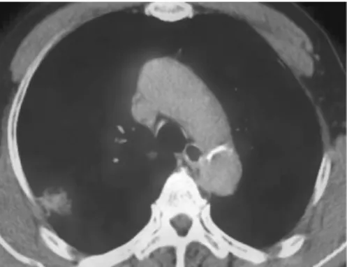
Chest radiography revealed a solitary pulmonary nodule (SPN) in the right upper lobe. Chest CT scan showed a solitary parenchymal opacity with subpleural base, convex borders and focal low attenuation area within density, and 3×2 cm in size in the right upper lobe of the lung. The margin was slightly lobulated, and enhanced by contrast media.
PET-CT scan revealed an abnormal hypermetabolic nodule with maximal standard uptake value of 1.0. CT-guided transthoracic needle aspiration was done
Case Report
DOI 10.3349/ymj.2009.50.4.591pISSN: 0513-5796, eISSN: 1976-2437 Yonsei Med J 50(4): 591-593, 2009
A Pulmonary Nodule due to Pulmonary Infarction Diagnosed by Video-Assisted Thoracoscopy
Jae Hwa Cho,
1Joung Taek Kim,
2Lucia Kim,
3Kyung Hee Lee,
4Jeong Seon Ryu,
1Seung Min Kwak,
1and Hong Lyeol Lee
1Departments of 1 Internal Medicine, 2 Thoracic Surgery, 3 Pathology, 4 Radiology, School of Medicine, Inha University, Incheon, Korea.
We report a pulmonary infarction in 68-year-old man who was referred for an asymptomatic pulmonary nodule in chest radiography. Computed tomography (CT), positron emission tomography (PET), and transthoracic needle aspiration suggested suspicion for malignancy. Video-assisted thoracoscopic surgery (VATS) was performed for histologic diagnosis. Our case is a pulmonary nodule due to pulmonary infarction diagnosed by VATS in Korea.
Key Words : Solitary pulmonary nodule, pulmonary infarction, video-assist thoracoscopic surgery
Received: June 13, 2007 Revised: January 4, 2008 Accepted: January 4, 2008
Corresponding author: Dr. Jae Hwa Cho, Department of Internal Medicine, School of Medicine, Inha University, 7-206 Sinheung- dong 3-ga, Jung-gu, Incheon 400-103, Korea.
Tel: 82-32-890-3490, Fax: 82-32-882-6578 E-mail: [email protected]
∙The authors have no financial conflicts of interest.
© Copyright:
Yonsei University College of Medicine 2009
INTRODUCTION
CASE REPORT
for further evaluation of a pulmonary nodule. Cytology showed a few atypical cells and reported suspicion for malignancy. Lung perfusion scan revealed focal perfusion defects in the right upper lobe. To confirm malignancy before radical lung resection, wedge resection was performed by VATS. On gross section, there was a relati- vely well demarcated mass, measuring 2.3×2.0 cm. The cut surface of mass was yellow, tan colored, necrotic and granular. No thrombi and inflammations around vessels were seen. The doppler ultrasound study of lower extre- mities revealed no evidence of deep vein thrombosis.
An asymptomatic pulmonary nodule in an elderly patient with 30 pack-year smoking should be evaluated by CT, PET-CT scan and lung aspiration cytology. These studies suspected lung malignancy, but wedge resection via VATS revealed localized pulmonary infarction.
Pulmonary infarctions result from pulmonary throm- boembolism, heart failure, vasculitis, infection, and malig- nancy.1,5In one study, undiagnosed SPNs or masses were found in 16 patients among 43 patients with pulmonary infarctions diagnosed by surgical lung biopsy.2Six patients had features suggestive for malignancy on CT scan, PET scan or transthoracic needle aspiration. Fifty-percent of
patients received oral anticoagulant therapy, 2 for chronic atrial fibrillation and 1 for deep vein thrombosis.2In our patient, there were no predisposing factors of venous throm- boembolism and no chest symptoms.
Radiologically, pulmonary infarctions appear parenchy- mal density with broad pleural base, convex borders, linear strands from apex of the infarct toward hilum, truncated apex, broad pleural base, and low attenuation areas within the lesion.6Pulmonary infarctions could be suggested by peripheral parenchymal opacities with characteristic findings on CT scan. PET-CT scan is usually used in initial and follow up staging of lung cancer. False-positive results can occur in acute inflammation or infectious conditions. Pulmonary embolisms have abnormally hypermetabolic nodules and small occult infarctions also have hypermetabolic nodules in the peripheral lung.7
The transthoracic needle aspiration cytology revealed false-positive results. Histologically, atypical reactive alveolar lining type II cells and metaplastic cells near the infarction can result in this false-positive cytology results.8,9 In cases of the peripheral lung nodules in subpleural regions, the possibility of pulmonary infarctions should be considered, even in the absence of evidence of venous thromboembolism. VATS should be performed to confirm the nature of nodules.
This study was supported by INHA University Research Grant (INHA).
1. He H, Stein MW, Zalta B, Haramati LB. Pulmonary infarction:
spectrum of findings on multidetector helical CT. J Thorac Imag- ing 2006;21:1-7.
2. George CJ, Tazelaar HD, Swensen SJ, Ryu JH. Clinicoradio- logical features of pulmonary infarctions mimicking lung cancer.
Mayo Clin Proc 2004;79:895-8.
3. Tsunezuka Y, Hiranuma C, Sato H, Kurumaya H. Pseudotumor due to pulmonary infarction diagnosed by video-assisted thoraco- scopic surgery. Ann Thorac Cardiovasc Surg 1999;5:402-4.
4. Jeong YJ, Yi CA, Lee KS. Solitary pulmonary nodules:
detection, characterization, and guidance for further diagnostic workup and treatment. AJR Am J Roentgenol 2007;188:57-68.
5. Tsao MS, Schraufnagel D, Wang NS. Pathogenesis of pulmonary infarction. Am J Med 1982;72:599-606.
6. Balakrishnan J, Meziane MA, Siegelman SS, Fishman EK.
Pulmonary infarction: CT appearance with pathologic correlation.
J Comput Assist Tomogr 1989;13:941-5.
7. Kamel EM, McKee TA, Calcagni ML, Schmidt S, Markl S, Castaldo S, et al. Occult lung infarction may induce false Jae Hwa Cho, et al.
Yonsei Med J http://www.eymj.org Volume 50 Number 4 August 2009 592
DISCUSSION
REFERENCES
Fig. 1. Chest CT scan revealed a broad pleural base, convex borders, and a low attenuation area within the density in right upper lobe of lung.
Fig. 2. PET-CT scan showed an abnormal hypermetabolic uptake in right upper lobe of lung.
ACKNOWLEDGEMENTS
SPN in Pulmonary Infarction
Yonsei Med J http://www.eymj.org Volume 50 Number 4 August 2009 593 interpre-tation of 18F-FDG PET in primary staging of pulmonary
malig-nancies. Eur J Nucl Med Mol Imaging 2005;32:641-6.
8. Kaminsky DA, Leiman G. False-positive sputum cytology in a case of pulmonary infarction. Respir Care 2004;49:186-8.
9. Scoggins WG, Smith RH, Frable WJ, O’Donohue WJ Jr. False- positive cytological diagnosis of lung carcinoma in patients with pulmonary infarcts. Ann Thorac Surg 1977;24:474-80.