대흉외지 2007;40:851-854 □ 증례보고 □
− 851 −
*울산병원 흉부외과
Department of Thoracic and Cardiovascular Surgery, Ulsan General Hospital
**울산병원 마취과
Department of Anesthesia, Ulsan General Hospital
논문접수일:2007년 8월 6일, 심사통과일:2007년 10월 19일
책임저자:김상익 (680-742) 울산시 남구 신정 5동 34-72, 울산병원 흉부외과 (Tel) 052-259-5026, (Fax) 052-259-5120, E-mail: [email protected] 본 논문의 저작권 및 전자매체의 지적소유권은 대한흉부외과학회에 있다.
동맥류를 동반한 양측성 관상동맥 -폐동맥루의 외과적 교정
1예 보고
김 상 익*ㆍ김 병 훈*ㆍ노 정 섭**
Surgical Treatment of Bilateral Coronary to Pulmonary Artery Fistulae with a Saccular Aneurysm
A case report
Sang-Ik Kim, M.D.*, Byung-Hun Kim, M.D.*, Jeong-Sup Noh, M.D.**
A 76-year-old woman with a history of chest pain and palpitation, was diagnosed with bilateral coronary to pulmo- nary artery fistulae with a concomitant saccular aneurysm, which is quite rare. Suture closure of the fistular ves- sels around the pulmonary artery root, the removal of a saccular aneurysm, and the transpulmonary closure of coronary to pulmonary artery fistulae were performed. The patient was well at 4 months after surgery.
(Korean J Thorac Cardiovasc Surg 2007;40:851-854) Key words: 1. Fistula
2. Aneurysm
3. Coronary artery fistula
증 례
76세 여자 환자로 2년간의 약물 치료에도 불구하고 호 전되지 않는 흉부 압박감, 운동 시 흉통, 전신 허약감, 점 점 더 자주 발생하는 심계 항진으로 수술을 받기 위해 내 원하였다. 과거력상 흉부 외상을 당한 적은 없었다. 2년 전에 흉부 압박감과 심계항진이 있어서 본원 응급실을 방 문했는데 심전도 검사에서 V
1∼V
6까지 T-파의 역전과 혈 액 검사에서 CPK-MB (5.6 ng/mL), troponin-I (9.2 ng/mL) 소견을 보였다. 관상동맥조영술 결과 좌전하행지 근위부 에서 동정맥루가 기시하여 주폐동맥의 좌측면으로 들어 가는데 동정맥루의 근위부에는 동맥류가 형성되어 있고 주폐동맥으로 들어가는 원위부는 확장된 소견을 보였다.
또 우측 관상동맥 입구에서 기시해서 주폐동맥의 우측면
으로 들어가는 동정맥루 혈관이 관찰되었다. 관상동맥에
서 주폐동맥으로 동맥혈의 steal로 인한 심장 허혈로 진단
을 내리고 코일 색전법(coil embolization)을 통한 치료를
시도했지만 동정맥루 내로 카테타 진입의 실패로 비수술
적 방법은 포기하고 수술을 권유했으나 거부를 해서 보존
적 치료만 이루어졌다. 내원 후 시행한 관상동맥조영술
사진(Fig. 1)과 흉부 단층촬영사진(Fig. 2)에서 좌측 동정맥
루의 동맥류 크기는 2년 전에 비해 조금 더 늘어나 있었
다. 심장초음파 검사에서 좌심실의 비정상적인 이완(rela-
xation) 소견을 보였다. 수술은 정중 흉골 절개를 시행하고
심낭을 수직으로 절개하였다. 주폐동맥 근부의 좌측면에
서 길이는 2 cm 정도이고 직경이 7 mm로 확장된 혈관이
대흉외지
2007;40:851-854
− 852 −
Fig. 2. Chest computed tomography demonstrates the origin of the left coronary-to-pulmonary artery fistula from the left anterior descending cor- onary artery (A, arrow) and a saccular aneurysm located behind the main pulmonary artery (B, arrow head).
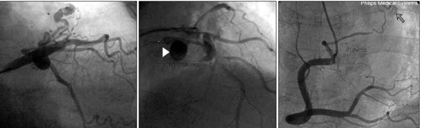
Fig. 1. Preoperative coronary angiographies show bilateral coronary to pulmonary artery fistulae with a saccular aneurysm (arrow head). Small arrows indicate tortuous arteriovenous fistulae originated from the coronary arteries and a brown arrow indicates the contour of the pulmonary valve cusp.
주폐동맥으로 들어가는 것이 관찰되었고 주변에서 thrill이 촉지되었다. 상행대동맥과 주폐동맥 사이를 양측으로 벌 린 후 심외막 주위의 지방 조직 속에 파묻혀있는 동정맥 루 혈관들을 찾기 위해 박리를 하였고 저혈압, 부정맥 등 이 나타나서 심폐기 가동 하에 수술을 진행하기로 했다.
무명동맥 근처의 상행대동맥에 동맥관(arterial cannula)을, 우심방이와 하대정맥 근처의 우심방에 정맥관(venous can- nula)을 삽입하고 심폐기를 가동시킨 후 우측 폐정맥의 입 구를 통해 좌심실 내로 벤트 카테타를 삽입하였다. 상행 대동맥을 겸자로 물고 심정지액(HTK solution)을 대동맥 근부에 투여하고 심정지를 시킨 후 주폐동맥의 sinotubular junction에서 1 cm 원위부를 횡으로 절개하였다. 주폐동맥
의 좌측 내면, sinotubular junction의 직 상부에 직경 4 mm
의 동정맥루의 입구가 보였고 6-0 인조사로 봉합을 하였
다. 우측 관상동맥에서 기시하는 동정맥루의 입구를 찾기
위해 주폐동맥의 내부를 조사했지만 찾지 못했다. 주폐동
맥을 완전 절단 후 근위부를 사방으로 견인하면서 주폐동
맥 근부의 외막과 주변 지방 조직을 박리하면서 주폐동맥
주변의 의심되는 작은 혈관들은 모두 제거하였고 주폐동
맥 뒤쪽 중앙 하부의 지방조직 속에 파묻혀있는 동맥류
(saccular 형태로 직경 1.5 cm 정도임)를 찾아서 절제하였
다. 절단된 주폐동맥을 6-0 인조사로 문합을 하고 thrill이
사라진 것을 확인 후 수술을 끝마쳤다. 병리 조직 검사에
서 동맥류의 중막에 퇴행성 변화와 internal elastic lamina
김상익 외
양측성 관상동맥-폐동맥루
− 853 −
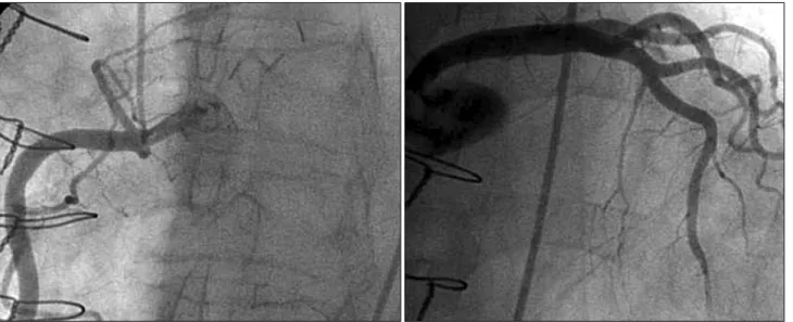
Fig. 3. Postoperative coronary angiography demonstrates the disappearance of bilateral coronary to pulmonary artery fistulae with a saccular aneurysm.
의 비후가 관찰되었다. 술 후 시행한 관상동맥조영술 검 사에서 양측의 동정맥루들은 모두 제거되었고(Fig. 3) 술 전 증상들은 모두 사라진 상태이며 현재 외래 추적 관찰 중이다.
고 찰
관상동맥조영술 검사 중에 선천성 기형인 관상동맥-폐 동맥루가 발견되는 경우는 0.1∼0.2%이고 양측성으로 발 생하면서 동정맥루 혈관에 동맥류가 만들어진 경우는 아 주 드물다[1,2]. 관상동맥-폐동맥루의 경우 20세 이하에서 는 무증상을 보이는 경향이 있고 나이가 들어서 증상이 나타날 수 있는데 관상동맥에서 폐동맥으로 동맥혈의 steal로 인한 심근 허혈 또는 경색으로 발생하는 흉통, 장 기간의 좌-우 단락으로 인한 호흡곤란 및 피로, 심계항진 (부정맥), 심내막염, 급사 등이 발생할 수 있다. 그러나 대 부분의 경우 무증상을 보인다[3]. 본 환자의 경우 좌측 동 정맥루 근위부의 동맥류(saccular type)와 주폐동맥으로 들 어가는 원위부 확장과 같은 혈관 변화를 볼 때 환자의 임 상 증상은 좌전하행지에서 기시한 동정맥루에 의한 동맥 혈의 steal때문에 발생된 것으로 사료된다. Akashi 등[4]은 관상동맥-폐동맥루를 보인 69세 여자 환자에서 동맥류의 자연 파열을 보고하였는데 본 환자의 경우 2년 전에 비해 동맥류가 조금 더 커진 양상을 보여서 파열 가능성이 있 다고 사료된다. Nakatani 등[5]은 55세 남자 환자에서 양측
성 관상동맥-폐동맥루가 4년을 경과하면서 거의 소실된 경우를 보고하였지만 대부분은 자연적으로 소실되지 않 는다. 증상이 없을 경우 보존적 치료를 하면서 규칙적으 로 관찰하면 되고 증상이 있을 경우 병변 또는 환자의 상 태에 따라 일부에서 coil 또는 device를 적용한 카테타 교 정술(transcatheter closure)[6]이 시술되고 있지만 주로 외과 적 교정술[7,8]로 치료되고 있다. 외과적 교정술의 기본 개 념은 동정맥루의 발생부위와 종착부위를 봉합하는 것인 데 특히 Huang 등[8]은 양측성 관상동맥-폐동맥루인 경우 는 술 후 재발 가능성 때문에 심폐기 가동과 심정지하에 수술 진행하고 잔존할 수 있는 동정맥루의 입구를 폐동맥 내부에서 확인할 것을 주장하였다. 결론적으로 흉통, 심계 항진 등 관상동맥에서 폐동맥으로 steal로 인한 증상을 보 인, 동맥류를 동반한 양측성의 관상동맥-폐동맥루 환자를 외과적 교정술로 좋은 결과를 얻었기에 문헌 고찰과 함께 보고한다.
참 고 문 헌
1. Darwazah AK, Hussein IH, Hawari MH. Congenital cir-
cumflex coronary arteriovenous fistula with aneurysmal termination in the pulmonary artery. Tex Heart Inst J
2005;32:56-9.2. Olearchyk AS, Runk DM, Alavi M, Grosso MA.
Congenital bilateral coronary-to-pulmonary artery fistulas.
Ann Thorac Surg 1997;64:233-5.
대흉외지
2007;40:851-854
− 854 −
3. Sapin P, Frantz E, Jain A, Nichols TC, Dehmer GJ.Coronary artery fistula; an abnormality affecting all age groups. Medicine 1990;69:101-3.
4. Akashi H, Tayama E, Tayama K, et al. Rupture of an
aneurysm from a coronary artery fistula: a case report.
Circ J 2003;67:551-3.
5. Nakatani S, Nanto S, Masuyama T, Tumai J, Kodama K.
Spontaneous near disappearance of bilateral coronary ar- tery-pulmonary artery fistulas. Chest 1991;99:1288-9.
6. Armsby LR, Keane JF, Sherwood MC, Forbess JM, Perry
SB, Lock JE. Management of coronary artery fistulae:
Patient selection and results of transcatheter closure. J
Am Coll Cardiol 2002;39:1026-32.7. Kim H, Park JK, Kim YH, et al. Bilateral coronary ar-
tery to pulmonary artery fistula: two case report. Korean
J Thorac Cardiovasc Surg 2007;40:225-7.8. Huang YK, Lei MH, Lu MS, Tseng CN, Chang JP, Chu JJ. Bilateral coronary-to-pulmonary artery fistulas. Ann Thorac Surg 2006;82:1886-8.