비용에서 CC Chemokines mRNA의 발현
중앙대학교 의과대학 이비인후과학교실,1 고려대학교 의과대학 이비인후과학교실2
황찬승1·이성욱1·홍영호1·김 훈1·김춘길1·정학현2
Expression of CC Chemokines mRNA in Nasal Polyps
Chan Seung Hwang, MD1, Seong Wook Lee, MD1, Young Ho Hong, MD1, Hoon Kim, MD1, Chun Gil Kim, MD1 and Hak Hyun Jung, MD2
1Department of Otolaryngology, College of Medicine, Chung-Ang University, Seoul,
2Department of Otolaryngology-Head & Neck Surgery, College of Medicine, Korea University, Seoul, Korea
- -
-- ABSTRACT ----
Background:Nasal polyposis can be defined as a chronic inflammatory disease of the paranasal sinus mu- cosa. The exact pathogenesis of nasal polyp is unknown, but inflammation is thought to be an important factor in the developement of nasal polyposis. Histologically, the stroma of nasal polyps consists of variable inflam- matory cellular infiltrates including eosinophils. Eosinophil is an important inflammatory cell. CC chemokines (RANTES, eotaxin, MCP-3, and MCP-4) are powerful chemotactic cytokines for eosinophils. Objective:To understand the events involved in eosinophil migration into inflammatory sites, we performed the analysis of CC chemokines mRNA in the nasal polyps, allergic inferior turbinate mucosas and normal inferior turbinate mucosas. Materials and Methods:Expression levels of CC chemokines mRNA were examined using RT- PCR in 20 nasal polyps, 7 allergic inferior turbinate mucosas and 6 normal inferior turbinate mucosas.
Results:The expression levels of CC chemokines mRNA were higher in nasal polyps than allergic inferior turbinate mucosas and normal inferior turbinate mucosas. The expression levels of RANTES and MCP-3 mRNA were higher than eotaxin and MCP-4 mRNA (p<0.01). The infiltrating eosinophils were correlated the expre- ssion levels of RANTES, eotaxin, MCP-3, and MCP-4 mRNA (p<0.001). Conclusions:These results suggest that inflammation is an important factor in the pathogenesis of nasal polyposis and CC chemokines (RANTES, eotaxin, MCP-3, and MCP-4) play a role in eosinophils migration into inflammatory sites. With the development of immunological reagents to detect the CC chemokines, it will be important to compare proteins and mRNA expressions in these tissue. ((((J Clinical Otolaryngol 1999;10:250-258))))
KEY WORDS:Nasal polyp·RANTES·Eotaxin·MCP-3·MCP-4.
서 론
비용은 비과학 영역의 흔한 질환으로 그 원인과 병인
은 아직 확실하게 규명되지 않았으나, 염증, 알레르기, 자율신경의 조절장애, 유전자 장애, 탄수화물 대사장애, 혈관운동의 불균형 등의 가설이 제시되고 있으며, 비용 의 형성과 성장에 있어서 염증반응이 현재까지 가장 중 요한 요인으로 생각되고 있다.1)
비용의 형성에서 염증반응은 여러 염증매개체와 cy- tokine이 관여한다. 비용에 침착된 염증세포, 비용의 구 성 성분인 상피세포, 섬유아세포, 혈관내피세포 등에서 논문접수일:1999년 10월 10일
심사완료일:1999년 10월 25일
교신저자:황찬승, 140-717 서울 용산구 한강로 3가 65 중앙대학교 의과대학 이비인후과학교실
전화:(02) 748-9575・전송:(02) 792-6642 E-mail:[email protected]
분비되는 cytokine은 상피세포의 분화를 촉진하고, 조혈 세포의 기능을 조절하며 염증세포의 생존기간을 연장시 킴으로써 비용의 성장에 관여한다.2)
염증부위에 염증세포의 침착은 염증반응에서 중요한 과 정이며 최근 분자생물학적 기법의 발달에 따라 염증세 포들에 대하여 화학주성을 갖는 chemokine과 이러한 chemokine이 작용할 수 있는 염증세포에 분포하는 ch- emokine수용체에 대한 관심이 집중되고 있다. Che- mokine은 특정세포에 대하여 화학주성을 갖는 cyto- kine으로 정의되며, 분자의 아미노산 구조에 따라 CXC, CC chemokine으로 분류된다. CXC chemokine은 두 개의 cystein기 사이에 다른 아미노산 구조를 포함한 것으로 주로 다형핵 백혈구의 화학주성에 관여하고, CC chemokine은 두 개의 cystein기를 가진 것으로 림프 구, 단핵구, 호산구 및 호염기구에 화학주성을 가지고 있 다.3) 특히 RANTES, eotaxin, MCP-3, MCP-4 등이 호산구의 화학주성에 관여한다.3-6)
본 연구의 목적은 비용, 알레르기 하비갑개 비점막, 정 상 하비갑개 비점막 조직에서 CC chemokines인 RA- NTES, eotaxin, MCP-3, MCP-4 mRNA을 역전사- 중합효소반응(reverse transcriptase-polymerase chain reaction:RT-PCR)을 이용하여 관찰하고 조직내 침 윤된 호산구 수와 상관관계를 알아봄으로써 선택적 호 산구 침윤의 기전에 대해 이해하고자 하였다.
재료 및 방법
연구재료
중앙대학교 이비인후과에서 부비동염 및 비용으로 진 단 받고 내시경하 부비동수술 또는 비용 절제술을 받은 20명의 환자에서 20측의 비용조직을 채취하였다. 수양 성 비루, 코막힘, 재채기 및 소양감 등의 비알레르기 4대 증상중 2가지 이상이 1년중 6개월 이상 지속되고, 전체 혈청 I gE가 100 IU/ml 이상이고, 피부반응검사상 1가 지 이상에서 강양성을 보인 알레르기성 비염 7명의 환 자에서 하비갑개 절제술을 통하여 하비갑개 비점막 조 직을 채취하였다. 대조군으로는 위와 같은 비알레르기 검사에서 음성반응을 보인 6명에서 하비갑개 비점막 조 직을 채취하였다. 채취된 조직은 일부를 광학현미경하에
호산구 침윤을 관찰하기 위하여 10% 포르말린 용액에 고정하였고, 일부는 분자생물학적 연구를 위하여 즉시 -70℃에 보관하였다.
연구방법
광학 현미경의 조직 표본 제작과 호산구의 관찰
비용 20측, 알레르기 하비갑개 비점막 7측, 정상 하비 갑개 비점막 6측에서 광학현미경 조직 표본을 제작하였 다. 조직은 10% 포르말린 용액에 24시간 고정한 다음 파라핀 포매과정을 거쳐 4~5 μm 두께로 박절하여 파 라핀 절편을 작성하였다. Hematoxylin-Eosin 염색을 시행하여 광학현미경 400 배율에서 한 시야당 호산구 수를 세고 한 절편당 10시야를 관찰하여 그 평균을 산술 하여 호산구 수를 측정하였다.
전체 RNA 추출
전체 RNA(total RNA)를 추출하기 위하여 냉동 보 관된 각각의 비점막 조직을 eppendorf tube에 옮긴 후 RNA를 침전시키기 위하여 TRIzol용액(Life Technol- ogies, Gaithersberg, MD USA) 1 ml를 첨가하여 gl- ass homogenizer로 균질화 시킨 후 chloroform 용액 (Sigma chem. Co., St. Louis, MO USA) 0.2 ml을 넣어 잘 혼합시켰다. 여기서 얻은 재료를 14,000 rpm, 4℃
에서 20분간 원심분리한 후 상층액을 다른 eppendorf tube에 옮기고 핵산 침전이 잘 되도록 isopropanol을 첨가하여 약하게 흔들어 혼합시켰다. 10분 후 14,000 rpm, 4℃에서 10분간 원심분리하여 상층액은 제거하고 남은 침전물에 DEPC로 처리된 증류수로 희석한 et- hyl alcohol 1 ml을 첨가하여 RNA 침전물을 세척하고 10,000 rpm, 4℃에서 5분간 원심분리하였다. 다시 상 층액을 제거하고 30분간 대기중에서 건조시킨 후 RNA 침전물을 녹이기 위하여 DEPC로 처리한 증류수 50 μl 를 첨가한 후 농도와 순수도를 측정하여 이용하였다.
RANTES, eotaxin, MCP-3, MCP-4, β-actin primer의 제작
중합효소반응을 위한 RANTES, eotaxin, MCP-3, MCP-4, β-actin mRNA의 primer는 각각 다음과 같
으며, 각각의 중합효소반응 산물의 크기는 각각 196 bp, 156 bp, 376 bp, 234 bp, 334 bp이다(Table 1).
Reverse transcriptase-polymerase chain reaction(RT- PCR)
전체 RNA 0.5 μg에 oligo dT primer와 MMLV 역 전사 효소(molohey murine leukemia virus reverse transcriptase, Life Technologies, Gaithersberg, MD USA)를 첨가하여 전령 RNA(mRNA)를 compleme- ntary DNA(cDNA)로 역전사 시키고, 그중 일부를 이 용하여 중합효소반응(PCR)을 시행하였다.
PCR에는 Gene Amp PCR system 2400(Perkin Elmer)을 사용하였으며, 95℃로 30초간 변성시킨 후 반 응조건은 95℃ 30초 , 58℃에서 30초, 72℃ 1분 30초 로 총 35cycle을 시행하였다. 양성 대조군으로 RAN- TES, eotaxin, MCP-3, MCP-4와 β-actin 각각이 pGEM-T Easy Vector(Promega, Medison, WI USA) 에 함유된 plasmid DNA 1 ng을 사용하였고, 음성대조 군으로 cDNA 대신 dH 2O를 사용하였다.
PCR로 증폭된 산물은 2% agarose gel에 ethidium bromide로 염색한 후 자외선 투사기로 관찰하고 폴라로 이드 카메라로 촬영하였다. 또한 분리된 중합효소반응 산물의 일부는 pGEM-T Easy vector에 연결한 후 automated sequencer로 염기서열을 관찰하였다. NIH image analysis software(version 1.60)을 이용하여 각 조직의 RANTES, eotaxin, MCP-3, MCP-4, β- actin band 면적을 측정한 후 RANTES, eotaxin, MCP-
3, MCP-4 band의 면적을 β-actin band의 면적으로 나 누어 각 조직의 RANTES, eotaxin, MCP-3, MCP-4 mRNA의 양을 산출하였다.
통계학적 분석
비용, 알레르기 하비갑개 비점막, 정상 하비갑개 비점 막 조직에서 호산구 수와 RANTES, eotaxin, MCP-3, MCP-4 mRNA 양을 비교하기 위하여 ANOVA test 를 이용하여 5% 유의수준에서 통계학적으로 분석하였 다. 병리조직 소견상의 호산구 수와 RANTES, eotaxin, MCP-3, MCP-4 mRNA 발현양과의 상관관계를 알기 위하여 Pearson 상관계수를 이용하여 분석하였다. 모든 통계는 SPSS 통계 소프트웨어를 이용하였으며 p<0.05 인 경우를 유의하다고 보았다.
결 과
광학현미경을 이용한 호산구 소견
조직내 호산구 수는 비용조직에서 12.27±10.0개, 알 레르기 하비갑개 비점막 조직에서 10.6±7.3개, 정상 하 비갑개 비점막 조직에서 5.15±1.6개로 비용조직에서 호산구가 가장 많이 관찰되었으나 각 군간의 호산구 수 는 통계학적으로 유의한 차이는 없었다(p>0.05, AN- OVA test)(Table 2).
호산구는 주로 상피층과 기저막 아래 고유층에서 관 찰되었고, 비용조직중 염증성 비용보다는 부종성 비용에 서 더 많은 호산구가 관찰되었으며 많은 림프구 침윤도 Table 1. Primer sequences and expected length of PCR products
Primers Oligonucleotide sequences Length of PCR products
RANTES Sense 5’CGCTGTCATCCTCATTGCTA3’ 196 bp
Antisense 5’CACACACTTGGCGGTTCTT3’
Eotaxin Sense 5’CCTGGCCAATAGGAAGATACC3’ 156 bp
Antisense 5’ACTTCATGGAATCCTGCACC3’
MCP-3 Sense 5’CAAGGAG(G,A)TCTGTGCTGACC3’ 376 bp
Antisense 5’GTAGAGAAGGGAGGAGCATC3’
MCP-4 Sense 5’GAGCTATGAGATCACCACCAG3’ 234 bp
Antisense 5’CAAAGCATAGAAGAGGAGGCCAG3’
β-actin Sense 5’ACCTGTACGCCAACACAGTG3’ 334 bp
Antisense 5’GCCATGCCAATCTCATCTT3’
관찰되었다(Fig. 1). 정상 하비갑개 비점막 조직에서는 염증세포나 호산구의 침윤을 잘 관찰할 수 없었다.
RANTES, eotaxin, MCP-3, MCP-4 mRNA의 발현 RANTES mRNA 발현양은 1.12±1.0, eotaxin mR- NA는 0.36±0.5, MCP-3 mRNA는 0.96±1.1, MCP-
4 mRNA는 0.31±0.3으로 비용 조직에서 가장 높았 으나 각 군간의 발현양의 차이는 통계학적으로 유의한 차이는 없었다(p>0.05, ANOVA test)(Table 2)(Figs.
2 and 3). 또한 RANTES, MCP-3 mRNA 발현양 은 모든 조직에서 eotaxin, MCP-4 mRNA 발현양에 비해 통계학적으로 유의하게 높았다(p<0.01, ANOVA Table 2. CC chemokine expression levels and numbers of infiltrated eosinophils
RANTES/β-actin Eotaxin/β-actin MCP-3/β-actin MCP-4/β-actin No of eosinophil*
Polyp (N=20) 1.12±1.0 0.36±0.5 0.96±1.1 0.31±0.3 12.27±10.0 AT† (N=7) 0.93±0.6 0 0.80±1.2 0.17±0.2 10.6 ± 7.3 NAT‡ (N=6) 0.41±0.3 0 0.40±0.2 0.14±0.1 5.15± 1.6 Total (N=33) 0.95±0.8 0.22±0.3 0.82±0.9 0.25±0.2 10.62± 7.9
*number of eosinophil/high power field (×400) †allergic inferior turbinate mucosa
‡non-allergic inferior turbinate mucosa
Fig. 1. A:Edematous, eosinophilic polyp. Note the abundance of eosinophils, a thickening of the basement mem- brane (arrows). And the loose stroma contains pseudocystic spaces filled with fluid (arrow heads)(H & E, ×200). B:A chronic inflammatory type of polyp. The surface respiratory epithelium has areas with cuboidal metaplasia but no goblet cell hyperplasia. The basement membrane does not show any pronounced hyalinization. The stroma consists of connective tissue with some dilated vessels and a moderate amount of lymphocytes (H & E, ×200). C:Allergic inferior turbinate mucosa. The stroma consisted of a few of eosinophils and lymphocytes (H & E, ×200). D:Normal inferior turbinate mucosa. Typical structure of the inferior turbinate is shown with pseudostratified columnar ciliated epithelium. There are few inflammatory cells in the stroma (H & E, ×200).
A A A
A BB BB
C C C
C DD DD
test)(Table 2).
RANTES mRNA 발현과 호산구의 상관관계
RANTES mRNA 발현양과 조직내 침윤된 호산구 수 는 상관관계가 있었다(p<0.001, r=0.818)(Fig. 4).
Eotaxin mRNA 발현과 호산구의 상관관계
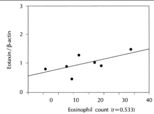
Eotaxin mRNA 발현은 7측의 비용조직에서만 관찰 되었으며 관찰된 eotaxin mRNA 발현양과 조직내 침윤 된 호산구 수는 상관관계가 있었다(p<0.001, r=0.533) (Fig. 5).
MCP-3 mRNA 발현과 호산구의 상관관계
MCP-3 mRNA 발현양과 조직내 침유된 호산구 수 는 상관관계가 있었다(p<0.001, r=0.865)(Fig. 6).
MCP-4 mRNA 발현과 호산구의 상관관계
MCP-4 mRNA 발현양과 조직내 침윤된 호산구 수
는 상관관계가 있었다(p<0.001, r=0.749)(Fig. 7).
고 찰
비용의 원인과 병인은 확실하게 규명되지는 않았지만 두가지 가설 즉, 염증이나 알레르기에 의하여 발생하는 것으로 생각되고 있다. 그러나 비용환자와 정상인 사이 에 피부반응 검사 결과가 차이가 없었으며, 특이 항체 IgE도 비용 환자에서 검출율이 높지 않고, 임상적으로 천식있는 환자에서 비용 발생율이 높지 않고, 비용조직 에서 많은 염증세포 축적이 관찰되는 것으로 보아 알레 르기보다는 염증이 비용의 원인과 병인에 중요한 역할 을 하는 것으로 생각된다.7)8) 비용의 형성에서 염증반응 은 여러 염증매개체와 cytokine이 관여한다. 비용에 침 착된 염증세포, 비용조직의 상피세포, 섬유아세포, 혈관 내피세포 등에서 분비되는 cytokine은 상피세포의 분화 를 촉진하고, 조혈세포의 기능을 조절하며 염증세포의 생 존기간을 연장시킴으로써 비용의 성장에 관여한다.2) Fig. 2. CC chemokines and β-actin analysis in the nasal polyps by RT-PCR. L indicates l00 bp ladder. P means posit- ive control clone including CC chemokines inserts in PGEM-T Easy plasmid vector and N means PCR amplification without template.
비용조직에서 관찰되는 염증세포로는 호산구, 림프구, 대식세포, 형질세포, 비만세포 등이 주로 관찰되며, 급성 염증질환에서 주로 관찰되는 다형핵 백혈구는 많이 관
찰되지 않고 특히 호산구가 많이 관찰되는 것으로 알려 져 있다.9)10) 호산구는 풍부한 cytotoxic protein, lipid mediators, oxygen metabolites, cytokines 등을 생성
Fig. 4. Relationship between RANTES mRNA expression levels and numbers of infiltrated eosinophils in the nasal polyps, allergic inferior turbinate mucosas and normal inferior turbinate mucosas (r=0.818, p<0.001).
Fig. 5. Relationship between eotaxin mRNA expression levels and numbers of infiltrated eosinophils in the nas- al polyps (r=0.533, p<0.001).
Fig. 3. CC chemokines and β-actin analysis in the allergic inferior turbinate musosas and normal inferior turbinate mucosas by RT-PCR. L indicates l00 bp ladder. P means positive control clone including CC chemokines inserts in PGEM-T Easy plasmid vector and N means PCR amplification without template. Symbol (*) means allergic inferior turbinate mucosas.
분비하며 이러한 물질들은 염증을 유발하게 된다.11) 만 성 염증질환과 관련이 있는 T림프구 또한 비용조직에서 많이 관찰되는데, 비용조직에서 CD8+(suppressor/cy- totoxic) 림프구가 CD4+(helper/inducer) 림프구보다 더 많이 관찰되고, 정상인의 하비갑개나 중비갑개 비점 막 조직에서 비용조직에서 보다 의미있게 CD4+ 림프구 가 많이 관찰되는 것으로 보아 T림프구 또한 비용형성 에 있어서 중요한 염증세포로 생각된다.12) 본 논문에서 도 비용조직에서 알레르기 비점막이나 정상 비점막 조 직에서 보다 호산구와 림프구 침착이 많았다.
염증세포의 염증부위 침착기전은 접착분자와 chem-
okine, chemokine 수용체 등이 관여하는 것으로 알려 져 있다.13) 염증세포의 염증부위로의 이동과정은 ro- lling, adhesion, diapedesis(transendothelial migra- tion), chemotaxis(migration to tissue) 등의 과정을 통하여 이루어지며 chemotaxis 과정에서 chemokine과 chemokine 수용체가 작용한다.14)
Chemokine은‘chemotactic cytokine’으로 1987년 처음으로 분류된 이래 특정 세포를 활성화시키며 infl- ammation mediator로서 기능을 갖고 있고, 특정 세포 에 대해 화학주성을 갖고 있기 때문에 최근 많이 연구 되고 있다.13) Chemokine의 구조는 특징적인 disulfide band로 연결된 4개의 cystein잔기로 이루어져 있고 처 음 2개의 cystein 위치에 따라 CXC, CC chemokine 으로 구분된다. 특히 CC chemokine중 RANTES, eo- taxin, MCP-3, MCP-4 등이 호산구의 화학주성에 주 로 관여한다.3)
RANTES는 T림프구, 대식세포, 섬유아세포, 혈소판, 기관지점막 상피세포, 비점막 상피세포 등에서 생성 분 비되고, TNF-α, IFN-γ, IL-β, 내독소(lipopolys- accharide) 등이 RANTES 발현을 상향조절(up-re- gulation)하고, IL-4, IL-13 등이 하향조절(down- regulation) 한다.15) RANTES는 기억세포(memory T cell), 단핵구 및 호산구에 화학주성을 가지며, 호산구와 T림프구를 활성화시키고 히스타민과 eosinophil cati- onic protein(ECP)의 분비에 의하여 급만성 알레르기 염증반응에 관여한다.16) RANTES의 호산구 순환(re- cruitment)유발기전에 대하여는 아직 확실하게 규명되 지는 않았지만 호산구에 존재하는 접착분자 Mac-1 발 현을 증가시키고, diapedesis 과정 후 호산구가 염증부 위로 이동하도록 직접적으로 자극함으로써 이루어진다.17) 이와 같이 RANTES는 호산구 침윤과 밀접한 연관성이 있는 것으로 보고되고 있으나,15)16) 최근의 보고에 의하 면 RANTES의 호산구에 대한 화학주성에 대해 의문이 제기되고 있다. 즉 재조합 인체 또는 기니픽 RAN-TES 유전자를 기니픽에 주사하면 호산구는 침윤되지 않고 거 대세포와 기억세포만이 침윤을 초래한다는 보고도 있
다.18)19) 그러나 이러한 재조합 유전자는 구성성분에 있
어서 RANTES의 형태를 전부 포함하지 않고 종(sp- ecies)간에도 차이가 있을 수 있다.
Fig. 6. Relationship between MCP-3 mRNA expression le- vels and numbers of infiltrated eosinophils in the nasal polyps, allergic inferior turbinate mucosas and normal inferior turbinate mucosas (r=0.865, p<0.001).
Fig. 7. Relationship between MCP-4 mRNA expression le- vels and numbers of infiltrated eosinophils in the nasal polyps, allergic inferior turbinate mucosas and normal in- ferior turbinate mucosas (r=0.749, p<0.001).
본 연구 결과에서 RANTES mRNA발현양은 조직내 침윤된 호산구 수와 상관관계가 있었으며, 이러한 소견 은 RANTES가 호산구 침윤에 밀접한 관계가 있음을 알 수 있다. 또한 조직내 호산구외 염증세포 침윤이 많은 비용조직에서 발현율이 가장 높고 알레르기 하비갑개 비 점막, 정상 하비갑개 비점막 순으로 발현되는 것으로 보 아 RANTES가 호산구외 다른 염증세포의 화학주성에 도 관여할 것으로 생각된다. 이에 대하여는 추후 면역조 직화학적 방법으로 규명하고자 한다.
Eotaxin은 기니픽에서 처음 발견되었으며 호흡점막의 상피세포, 내피세포, 섬유아세포, 호산구에서 생성 분비 되고 호중구, 단핵구, 림프구에 대해서는 화학주성을 갖 지 않고 호산구에 대해서만 선택적으로 화학주성을 갖는 chemokine으로 RANTES에 비해 호산구에 대해 강력 한 화학 주성을 갖는다.5) 또한 호산구에서 생성 분비된 eotaxin이 자가반응(autocrine reaction)기전으로 호산 구에 작용함으로써 호산구 침윤이 많은 알레르기 질환에 서 호산구 침윤을 더욱 촉진시킬 수 있다.20) 본 연구 결 과에서 eotaxin mRNA 발현은 7측의 비용조직에서만 관찰되었고, eotaxin mRNA 발현이 관찰된 조직내 침 윤된 호산구와 eotaxin mRNA 발현양과는 상관관계가 있었다. 이러한 소견은 염증반응시 조직내에서 eotaxin 이 RANTES에 비해 훨씬 적게 발현되고, 발현된 조직 수가 너무 적어 통계학적 의의를 말할 수는 없지만 호산 구 침윤과 연관성이 있을 것으로 생각된다.
MCP-3는 구조적으로 RANTES와 비슷하고 단핵구, T림프구, 호염기구, 호산구에 화학주성을 가지며 특히 호산구에 대해 강력한 화학주성을 나타내는 것으로 알려 져 있다.4) 본 연구결과에서 MCP-3 mRNA 발현양은 조직에 침윤된 호산구와 상관관계가 있었으며, 호산구외 염증세포 침윤이 많은 비용, 알레르기 하비갑개 비점막, 정상 하비갑개 비점막 순으로 높았다. 이러한 소견은 MCP-3은 호산구 침윤과 밀접한 관계가 있음을 알 수 있고 다른 염증 세포의 화학 주성에도 관여할 것으로 생 각한다.
MCP-4는 구조적이나 기능적으로 MCP-3이나 eo- taxin과 유사하며 단핵구, T림프구에 대해 화학주성이 있고 특히 호산구에 대해서는 eotaxin과 MCP-3만큼 강력한 화학주성을 갖는 것으로 알려져 있다.6) 본 연구
결과에서도 MCP-4 mRNA 발현양은 조직에 침윤된 호산구와 상관관계가 있어 호산구 침윤에 밀접한 연관 성이 있을 것으로 생각된다. 향후 CC chemokines에 대 한 면역학적 항체가 개발되어 면역조직화학 염색법으로 조직내에서 CC chemokines 발현을 관찰할 수 있다면 호산구 및 염증세포 침윤에 대한 기전을 보다 더 명확히 규명할 수 있을 것으로 생각된다.
요 약
이상의 결과를 요약하면 염증이 비용 원인과 병인에 중요한 요소로 생각되며 CC chemokines중 RANTES, eotaxin, MCP-3, MCP-4는 호산구 화학주성에 중요 한 역할을 할 것으로 생각된다. 특히 RANTES와 MCP- 3는 비용 조직과 알레르기 하비갑개 비점막 조직에서 가 장 많이 발현되는 것으로 보아 호산구와 관련된 염증질 환에서 매우 중요한 CC chemokines으로 생각한다.
중심 단어:비용・RANTES・Eotaxin・MCP-3・
MCP-4.
REFERENCES
1) Bernstein JM, Gorfien J, Noble J, Yankaskas JR. Nasal polyposis: Immunohistochemistry and bioelectrical find- ings (a hypothesis for the development of nasal polyp). J Allergy Clin Immunol 1997;99:165-75.
2) Murrol J, Xaubet A, Gaya A, Roca-ferrer J, Lopez E, Fe- rnandez JC, et al. Cytokine gene expression and release from epithelial cells. A comparison study between healthy nasal mucosa and nasal polyps. Clinical Experimental Allergy 1995;25:607-15.
3) Alam R. Chemokines in allergic inflammation. J Allergic Clin Immunol 1997;99:273-7.
4) Dahinden A, Geiser T, Brunner T, Tscharner v, Caput D, Ferrara P, et al. Monocyte chemotactic protein 3 is a most effective basophil- and eosinophil-activating chemokine.
J Exp Med 1994;179:751-6.
5) Ponath PD, Qin S, Post TW, Wang J, Wu L, Gerard NP, et al. Molecular cloning and characterization of a human eotaxin receptor expressed selectively on eosinophils. J Exp Med 1996;183:2437-48.
6) Uguccioni M, Loetscher P, Forssmann U, Dewald B, Li H, Lima HS, et al. Monocyte chemotactic protein 4 (MCP- 4), a novel structural and functional analogue of MCP-3 and eotaxin. J Exp Med 1996;183:2379-84.
7) Shatkin JS, Desupepehe KG, Thisted RA, Corey JP. Mu- cosal allergy in the absence of systemic allergy in nasal polyposis and rhinitis: a meta-analysis. Otolaryngol Head
Neck Surg 1994;111:553-6.
8) Settipane GA, Chafee FH. Nasal polyps in asthma and rh- initis: a review of 6,037 patient. J Allergy Clin Immunol 1977;59:17-21.
9) Jankowski R, Bene M, Haas F, Faure G, Simson C, Way- off M. Immunohistological characteristics of nasal polyps.
A comparison with healthy mucosa and chronic sinusitis.
Rhinology 1989;8:51-8.
10) Ogawa H. Atopic aspect of eosinophilic polyposis and a possible mechanism of eosinophil accumulation. Acta Ot- olaryngol (Stockh) Suppl 1986;430:12-7.
11) Gleich GJ. The eosinophil bronchial asthma. N Engl J Med 1990;323:1033-9.
12) Stoop AE, Heijden HAMD, Baan V, Bienwega J, et al.
Lymphocytes and nonlymphoid cells in human nasal po- lyps. J Allergy Clin Immunol 1991;87:470-5.
13) Kita H, Gleich GH. Chemokine active on eosinophils: po- tential roles in allergic inflammation. J Exp Med 1996;
183:2421-6.
14) Hernicks PAJ, Bloemen PGM, Nijkamp FP. Adhesion mo- lecules and the recruitment of eosinophils to the airways.
Research in Immunology 1997;148:18-28.
15) Terada N, Maesako KI, Hamano N, Houki G, Ikeda T. Eo- sinophil adhesion regulates RANTES production in nasal epithelial cell. J Immunol 1997;158:5464-70.
16) Su r S, Hirohito K, Gleich GJ, Chenier TC, Hunt LW. Eo- sinophil recruitment is associated with IL-5, but not with RANTES, 24hr after allergen challenge. J Allergy Clin Im- munol 1996;97:1272-8.
17) Alam R, Stafford S, Forsythe P, Harrison R, Faubion D, Brown MAL, et al. RANTES is a chemotactic and activa- ting factor for human eosinophil transendothelial zmigration induced by cytokine Ⅲ. Effect of the chemokine RANTES.
J Immunol 1994;153:2153-60.
18) C ampbell EM, Proudfoot AE, Yoshimura T. Recombinant guinea pig and human RANTES activate macrophage but not eosinophils in the guinea pig. J Immunol 1997;159:
1482-9.
19) Conti PARK, Reale M, Barbacane RC. Massive infiltra- tion of basophilic cells in inflammed tissue after injection of RANTES. Immunology Letters 1997;58:101-6.
20) Weller PF. Roles of eosinophils in allergy. Curr Opin Im- munol 1992;4:782-7.