ISSN: 2233-601X (Print) ISSN: 2093-6516 (Online)
− 291 −
Received: November 10, 2016, Revised: April 3, 2017, Accepted: April 7, 2017, Published online: August 5, 2017
Corresponding author: Jin-Ho Choi, Department of Thoracic and Cardiovascular Surgery, Dongguk University Ilsan Hospital, 27 Dongguk-ro, Ilsandong-gu, Goyang 10326, Korea
(Tel) 82-31-961-7285 (Fax) 82-31-961-7287 (E-mail) [email protected]
© The Korean Society for Thoracic and Cardiovascular Surgery. 2017. All right reserved.
This is an open access article distributed under the terms of the Creative Commons Attribution Non-Commercial License (http://creativecommons.org/
licenses/by-nc/4.0) which permits unrestricted non-commercial use, distribution, and reproduction in any medium, provided the original work is properly
cited.
Delayed Type III Endoleak Caused by Fabric Erosion after Endovascular Repair of an Abdominal Aortic Aneurysm
Jae Hang Lee, M.D., Eung-joong Kim, M.D., Jin-Ho Choi, M.D.
Department of Thoracic and Cardiovascular Surgery, Dongguk University Ilsan Hospital
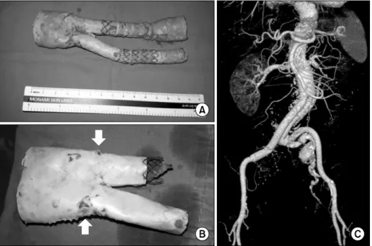
A 74-year-old patient presented with recurrent aneurysms in the infrarenal abdominal aorta and right com- mon iliac artery 6 years after endovascular aortic repair using endografts in the same location. The patient underwent an aorto-bi-iliac replacement with removal of the stent graft. Two holes measuring 2 mm each were found in the removed graft, and they appeared to have been caused by wear from continuous friction between the endograft and the aortic wall.
Key words: 1. Aneurysm 2. Aorta 3. Abdominal
4. Endovascular procedures 5. Endoleak
Case report
A 74-year-old female patient visited our hospital for the evaluation of a palpable abdominal mass. The patient had undergone successful endovascular aneu- rysm repair (EVAR) for an infrarenal abdominal aort- ic aneurysm (AAA) and a right common iliac artery (CIA) aneurysm 6 years ago, and had been lost to follow-up since 3 months after the procedure.
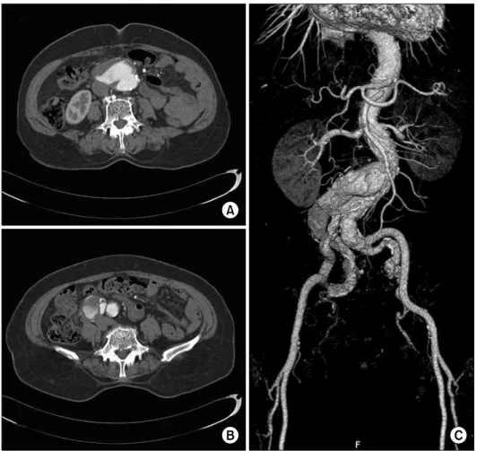
In computed tomographic angiography (CTA) be- fore the EVAR procedure, the patient had an aortic aneurysm; the maximal diameters of the infrarenal AAA and the right CIA aneurysm were 50 mm and 36 mm, respectively. Even though the aorta was tor- tuous with a proximal angle of 75
o, the proximal aortic diameter of 26 mm and proximal neck length of 33 mm seemed anatomically suitable for an EVAR procedure (Fig. 1). At that time, EVAR using the SEAL stent graft (S&G Biotech, Seongnam, Korea) was
performed under general anesthesia. A bifurcated proximal graft measuring 30 mm in diameter was used as the main body; on the right side, the right internal iliac artery was covered with a limb ex- tension using a 12-mm stent graft, and on the left side, a 20-mm bell bottom-shaped endograft was placed before the iliac bifurcation. After the surgery, no endoleak was observed on the CTA images either immediately or 2 months postoperatively. The patient did not visit the hospital again until 6 years later, when she presented with a palpable abdominal mass.
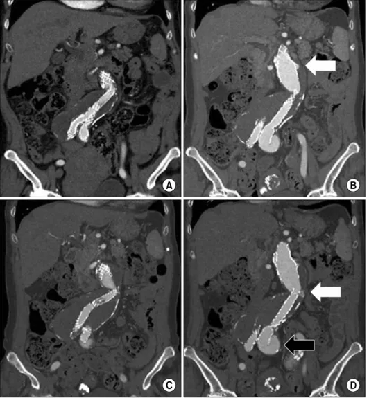
In the initial CTA images obtained during the more recent admission, type Ia and Ib endoleaks were ob- served, and a type III endoleak was also suspected (Fig. 2). The maximal diameters of the aneurysms were 61 mm for the AAA; 41 mm for the right CIA;
and 35 mm for the left CIA. This time, the patient underwent surgery rather than an endovascular in- tervention, and an aorto-bi-iliac bypass using a trans-
Korean J Thorac Cardiovasc Surg 2017;50:291-294 □ CASE REPORT □
https://doi.org/10.5090/kjtcs.2017.50.4.291